In vitro anti-cancer activity of Epipremnum aureum
Abstract
In the present study, in vitro anti-cancer activity of aerial parts of Epipremnum aureum extracts was performed using the MCF-7 breast cancer cell line. Soxhlet method was used with different solvents for extract preparation. The amount of apoptosis in MCF-7 cells was assessed using flow cytometry for each of the extracts. The chloroform and ethanol extracts had a considerable cytotoxic effect with IC50 values of 32.9 and 45.8 μg/mL respectively, while the conventional medication 5-fluorouracil produced an IC50 value of 19.2 μg/mL. The microscopic examination of the chloroform and ethanol extracts of E. aureum revealed the presence of apoptotic bodies, nuclear fragmentation, and tiny nuclei with strong chromatin condensation. These results suggest that chloroform extract of E. aureum is more effective against breast cancer.
Introduction
Breast cancer is regarded as the most dangerous form of cancer and is a leading cause of health issues among women globally (Akram et al., 2017). Currently, phar-maceutical medications, radiation therapy, and surgery are utilized in the management of breast cancer but a considerable number of circumstances involving these treatment options have proven unsuccessful because of drug resistance and toxicity.
The current landscape of cancer treatment relies on the utilization of plant-derived anti-cancer medicines (Wu et., 2021). Kramerial appacea (Al-Oqail, 2021), Ardisia crispa (Nordin et al., 2018), Mallotus phillippensis (Sakthidhasan et al., 2021) extracts and their secondary metabolites viz. isoflavones, coumestans, lignans, and prenyl flavonoids are widely acknowledged for their anti-cancer and antiproliferative activity.
Epipremnum aureum is a species in the arum family Araceae. It is a common household plant. There is no scientific study showing its anti-cancer effect on any organ. However, another plant of the same family, E. pinnatum has anti-cancer property using T-47D mammary carcinoma cells (Lan et al., 2007). Therefore, the primary goal of this work was to examine the anti-cancer effects of the different extracts of E. aureum using MCF-7 breast cancer cell line.
Materials and Methods
Chemicals
All ingredients for the current study i.e., acetone, chloroform, ethanol, ethyl acetate, petroleum ether, dimethyl sulfoxide (DMSO), 3-(4,5-dimethylthiazol-2-yl)2-5-diphenyltetrazolium bromide (MTT) were procured from Sigma Aldrich (India). MCF-7 cancer cell line was purchased from the National Centre for Cell Sciences, Pune.
Collection and authentication of plant
In the Ashta rural area of Sangli District, Maharashtra, India, the plant E. aureum was collected. The Botanical Survey of India, Pune, had authenticated and verified the plant (BSI/WRC/100-1/Tech./2020/117). The aerial parts were separated, cleaned, and dried for 3-4 weeks in the shade. The plant material was then subjected to crushing, resulting in coarsely powdered particles and placed in an airtight container for storage.
Preparation of extract
A soxhlet apparatus (locally made) was used to extract 500 g of plant aerial parts in turn using petroleum ether, chloroform, acetone, ethyl acetate, and ethanol in increasing order of their polarity. The solvent was then evaporated under decreased pressure by using a rotary evaporator (Labline PBU 6D; India) to produce the extracts. Finally, the yields were recorded (Abubakar et al., 2020).
Brine shrimp lethality assay
For each test substance, the initial cytotoxicity analysis is the brine shrimp fatality experiment. The brine shrimp species Artemia salina was raised in saltwater (natural brine water) with continuous light and aeration for 48 hours. The shrimps (naupliis) were collected after hatching and utilized in the experiment. Ten Nauplii were tallied and added to a test tube holding 5 mL of various concentrations (20, 50, 100, 200, and 500 μg/mL) of the petroleum ether, ethyl acetate, benzene, chloroform, and ethanol, taken in triplicate. The tube was then left to stand under a light source for 24 hours. To get the LC50, the percentage of death was computed for every extract and every concentration (Nerdy et al., 2021; ).
Box I: DAPI staining
Principle
A widely used nuclear and chromosome counterstain, DAPI (4′,6-diamidino-2-phenylindole) emits blue fluorescence upon binding to AT regions of DNA.
Requirements
CO2 incubator; DAPI; Dimethylformamide; Flat-bottom microplate (24-wells); Fluorescence microscope [Olympus FV4000, India]; Paraformaldehyde (4%); Phosphate buffer solution;
Procedure
Preparing solutions
Stock solution: Add deionized water (2 mL) or dimethylformamide (2 mL) to the entire contents of the DAPI vial to make a 14.3 mM (5 mg/mL).
Intermediate dilution: Add 2.1 µL of the DAPI stock solution to 100 µL phosphate buffer solution to make a 300 µM DAPI intermediate dilution
Working solution: Dilute the 300 µM DAPI intermediate dilution 1:1,000 in phosphate buffer solution as needed to make a 300 nM DAPI stain solution.
Labeling fixed cells
Step 1: After being inoculated in a 24-well flat-bottom microplate with cover slips, the cells were maintained at 37°C in a CO2 incubator for the whole night.
Step 2: Apply the extracts' IC50 values to the cell line after 24 hours.
Step 3: Following the incubation period, cells were fixed for 30 min with 4% paraformaldehyde and cleaned with phosphate buffer solution.
Step 4: After adding 20 µL of DAPI and letting it sit at room temperature in the dark for 5 min, the nuclear morphology was checked using a fluorescence microscope.
Note
DAPI has poor solubility in water, so sonicate as necessary to dissolve. The 5 mg/mL DAPI stock solution may be stored at 2–6°C for up to 6 months or at ≤–20°C for longer periods.
References
Lin et al., 2022
MTT assay
The MCF-7 breast cancer cell line was plated at a density of 1.0 × 10⁴ cells/well into a flat bottom 96-well plate and then grown at 37°C in a humidified 5%CO2 incubator. On the following day, the cells were subjected to different doses of the different solvent extracts at 10, 20, 40, 80, and 160 μg/mL. As controls, well-containing cells treated with DMSO (0.5%) as opposed to a test chemical was used. 5-Fluorouracil was employed as the norm. Following a 24-hour treatment with the test chemical, 10 μL of the MTT solution (0.5 mg/mL) was added to the wells, and the plates were incubated at 37°C for 4 hours. After removing the supernatant, 100 μL of DMSO was used to dissolve the violet formazan crystals, and a microplate ELISA Redder (Benspera E21, India) was used to quantify the amount of adsorption at 570 nm. Utilizing the dosage response inhibition curve, the IC50 values were determined.
The MTT assay is a method used to quantify the reduction of the yellow-colored MTT compound by the enzyme mitochondrial succinate dehydrogenase. The reduction of MTT resulted in the formation of insoluble and dark, purple-colored formazan, facilitated by the phytoconstituents found in the extracts of E. aureum. The vitality of cells was assessed by considering the amount of activity as an indicator, as the reduction of MTT is only observed in metabolically active cells (Ali et al., 2021; Turker and Bakar, 2023 for video clip).
Detection of apoptosis by annexin V/propidium iodide staining
Using an annexin V FITC/propidium iodide labeling kit, flow cytometry was used to analyze the induction of apoptosis in MCF-7 cells by various extracts. The extracts' IC50 values were applied to 1 × 10⁵ cells/mL over 24 hours. Following the duration of incubation, the cells were rinsed with PBS, suspended in 300 μL of binding solution that contained 1.5 μL of annexin V-FITC antibody and 1.5 μL of propidium iodide, and incubated for 5 min in the dark. Following staining, cells were subjected to flow cytometry analysis (Thermo Fisher Scientific, India), employing quadrant statistics to distinguish between groups of living, early apoptotic, late apoptotic, and necrotic cells (Kumar et al., 2018; Chazotte; Lin et al., 2022).
Statistical analysis
Data were shown as mean ± SD. For data analysis, three duplicates of the results were made. An analysis of variance (ANOVA) test that was conducted in one direction was used to determine how the conditions differed. The graph was shown using Graph Pad Prism 6.01 software (USA). A p-value of less than 0.05 was deemed significant.
Results
Yield
The plant aerial parts (500 g) showed higher yield with chloroform extract (6.4 g) followed by marginally less with ethanol extract (6.3 g) and then ethyl acetate extract (3.2 g), petroleum ether extract (1.0 g), benzene extract (0.9 g) and the least yield with ethyl acetate extract (0.7 g).
Brine shrimp lethality assay
The lowest mortality was noted at concentrations of 20 μg/mL while the highest mortalities were noted at 500 μg/mL. Results revealed chloroform extract showed high percent lethality (96.6%) followed by ethanol extract (90%) at 500 μg/mL concentration (Figure 1A,B).
Figure 1: Brine shrimp lethality and MTT cytotoxicity assay with determination of IC50 value for various extracts of E. aureum
MTT assay
To evaluate how effectively E. aureum inhibits the proliferation of human breast cancer cell lines, different extracts (petroleum ether, chloroform, acetone, ethyl acetate, and ethanol) of E. aureum were tested at different concentrations against MCF-7 cells. The percent inhibition also followed the same trend as the former result. The chloroform extract showed maximum inhibition with 61.7% followed by ethanol extract of 60.8% as compared to standard 5-fluorouracil (74.3%) at a concentration of 160 μg/mL.
The IC50 values of the extracts were determined to evaluate their effectiveness. 5-Florouracil was used to have an IC50 value of 19.2 μg/mL. Among five organic solvents extracted, chloroform and ethanolic extracts had shown significant antiproliferative effects on the MCF-7 cell lines. The chloroform extracts showed a significant cytotoxic effect against the MCF-7 cell line with an IC50 value of 32.9 μg/mL followed by ethanol extract of 45.8 μg/mL. In contrast, petroleum ether extract showed a moderate cytotoxic effect with an IC50 value of 91.8 μg/mL, followed by acetone extract at 106.8 μg/mL, whereas ethyl acetate extract exhibited weak cytotoxic effect against the MCF-7 cell line with an IC50 value of 145.1 μg/mL (Figure 1C, D).
Based on these findings, extracts exhibiting notable cytotoxic action were then stained with DAPI and annexin V/propidium iodide to determine apoptosis.
DAPI
The cells treated with the IC50 dose of ethanolic and chloroform extracts under the microscope displayed apoptotic bodies (small spherical fragments) and small nuclei with severe chromatin condensation, blebbing, and nuclear disintegration. On the other hand, the untreated cells (control) displayed uniformly faint blue staining and undamaged nuclei (Figure 2).
Figure 2: Microscopic analysis of chloroform and ethanol extracts of E. aureum
Detection of apoptosis
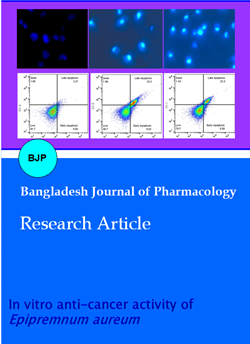
Figure 3 demonstrated the production of apoptosis using propidium iodide/annexin V staining, and the outcome indicated that chloroform extract showed a significant increase in apoptosis in MCF-7 cells. The late apoptotic cell population was increased by 33.3%, supporting findings.
Figure 3: Detection of apoptosis for chloroform and ethanol extracts of E. aureum
Discussion
The present study shows that higher yield occurs with chloroform and ethanol. These extracts show a high percentage of lethality using brine shrimp lethality assay. The chloroform extracts showed a significant cytotoxic effect against the MCF-7 cell lines. The microscopic findings are the presence of apoptotic bodies (small spherical fragments) and small nuclei with severe chromatin condensation, blebbing, and nuclear disintegration. The chloroform extract shows a significant increase in apoptosis in the MCF-7 cell lines.
Most of the organic compounds are extracted with 70 or 80% ethanol. It is somewhat difficult to conclude using ethanol extract. Therefore, emphasis will be given to the chloroform extract. Interestingly, chloroform extract shows a high percentage of lethality of brine shrimp which is confirmed by using MCF-7 cell lines.
E. aureum contains some secondary metabolites of which alkaloids, flavonoids, terpenoids, saponin glycosides, tannin, and phenolic compounds are the most prominent (Meshram and Srivastava, 2016). These were extracted using methanol. The study did not mention about the phytochemicals extracted by chloroform. The anti-cancer effect of chloroform extract may be due to the presence of terpenoids (Nuringtyas et al., 2014; Khang et al., 2020). Terpenoids mainly exert their anti-cancer effects by targeting various pathways, including mitochondrial death pathway, PI3K/Akt, and NF-KB pathways. Reactive oxygen species (ROS) play a critical role in the breast tumor microenvironment (Malla et al., 2021; Tai and Ascoli, 2011). Thus, the antioxidant enzymatic activity of a plant may be involved in the anti-cancer effect. The methanol extract of E. aureum shows a potent antioxidant effect (Meshram and Srivastava, 2016). The leaf, root, and stem of E. aureum were studied to find the extent of antioxidant activities. The decreasing order efficiency in the FRAP system is leaf > aerial root > stem. The chloroform extract of E. aureum was not studied. The chloroform extract of Vanda roxburghii shows an antioxidant effect (Uddin et al., 2015). To correlate the relationship between free radicle generation and the development of cancer, a study like the anti-cancer activity of chloroform extract of E. aureum leaf, root, and stem is necessary.
The E. pinnatum chloroform extract elicited both apop-totic and non-apoptotic programmed cell deaths impacted by the caspase-3 and c-myc genes' mRNA expre-ssion (Lan et al., 2007). So, chloroform extracts of E. aureum also induce apoptosis and inhibit cancer cell proliferation because it may contain polyphenols like quercetin and gallic acid (Khang et al., 2020). The results add to this body of evidence, demonstrating the efficacy of E. aureum chloroform extract in mitigating cancer progression. To further elucidate the mechanism of action underlying the cytotoxic effects of the chloroform extract, it was evaluated nuclear morphology using DAPI staining. Consistent with previous studies (Kumar et al., 2021, Eidet et al., 2014), it was observed small nuclei with intense chromatin condensation in cells treated with the chloroform extract. These morphological changes are characteristic of apoptotic cell death and provide additional evidence of the anti-cancer activity of the chloroform extract. The observed similarities in nuclear morphology between the study and previous reports further validate the efficacy of E. aureum chloroform extract in inducing apoptosis in cancer cells. Similar result was reported earlier where chloroform extract showed toxic to the breast cancer cell lines (Nuringtyas et al., 2014). This finding is consistent with previous research, where chloroform extracts have shown promising anti-cancer activity against MCF-7 cells.
Limitations of this study are as follows: a) thin-layer chromatography was not done which is necessary to identify the number of compounds in chloroform extract in comparison to other extracts; b) HPLC analysis of chloroform extract was not done; and c) animal model of breast cancer was not done.
Conclusion
This study suggests the anti-cancer potential of the aerial parts of E. aureum. Among the different extracts, chloroform extract showed effectiveness in combating breast cancer.
Ethical Issue
The cell line was maintained in Biocyte Institute of Research and Development, Sangli for conduct of cell line study as per the ethical guideline (NCCS/21-22/850).
Acknowledgement
Authors are thankful to the principal and management Appasaheb Birnale College of Pharmacy, Sangli for provided lab facility and chemicals for the present investigation.
References
Abubakar AR, Haque M. Preparation of medicinal plants: Basic extraction and fractionation procedures for experimental purposes. J Pharm Bioallied Sci. 2020; 12: 1-10.
Akram M, Iqbal M, Daniyal M, Khan AU. Awareness and current knowledge of breast cancer. Biol Res. 2017; 50: 1-23.
Ali A, Banerjee S, Kamaal S, Usman M, Das N, Afzal M, Alarifi A, Sepay N, Roy P, Ahmad M. Ligand substituent effect on the cytotoxicity activity of two new copper (ii) complexes bearing 8-hydroxyquinoline derivatives: Validated by MTT assay and apoptosis in MCF-7 cancer cell line (human breast cancer). RSC Adv. 2021; 11: 14362-73.
Al-Oqail MM. Anticancer efficacies of Kramerial appacea extracts against human breast cancer cell line (MCF-7): Role of oxidative stress and ROS generation. Saudi Pharm J. 2021; 29: 244-51.
Chazotte B. Labeling nuclear DNA using DAPI. Cold Spring Harb Protoc. 2011; 80-82.
Eidet JR, Pasovic L, Maria R, Jackson CJ, Utheim TP. Objective assessment of changes in nuclear morphology and cell distribution following induction of apoptosis. Diagn Pathol. 2014; 9: 92-100.
Khang DT, Dieu HD, Dung NK, Giang BT, Thao TL, Tien LT, Men TT, Loi HV, Tuan NT, Ay NV, Thuy NP. Anticancer and antioxidant of chloroform extracts from medical plants in the Mekong Delta, Vietnam. Asian J Plant Sci. 2020; 19: 398-405.
Kumar R, Saneja A, Panda AK. An annexin V-FITC-propidium iodide-based method for detecting apoptosis in a non-small cell lung cancer cell line. Methods Mol Biol. 2021; 2279: 213-23.
Lin S, Ke Z, Liu K, Zhu S, Li Z, Yin H, Chen Z. Identification of DAPI-stained normal, inflammatory, and carcinoma hepatic cells based on hyperspectral microscopy. Biomed Opt Express. 2022; 13: 2082-90.
Lan TM, Sulaiman SF, Muhammad TS. Anticancer medicinal plant, Epipremnum pinnatum (L.) Engl. chloroform extracts elicited both apoptotic and non-apoptotic cell deaths in T-47D mammary carcinoma cells. Curr Appl Sci Tech. 2007; 7: 24-43.
Malla R, Surepalli N, Farran B, Malhotra SV, Nagaraju GP. Reactive oxygen species (ROS): Critical roles in breast tumor microenvironment. Cri Rev Oncol Hematol. 2021; 160: 103285-96.
Meshram A, Srivastava N. Phytochemical screening and in vitro antioxidant potential of methanolic extract of Epipremnum aureum (Linden and Andre) GS Bunting. Int J Pharm Res Allied Sci. 2016 1; 5: 1-6.
Nerdy N, Lestari P, Sinaga JP, Ginting S, Zebua NF, Mierza V, Bakri TK. Brine shrimp (Artemiasalina Leach.) lethality test of ethanolic extract from green betel (Piper betle Linn.) and red betel (Piper crocatum Ruiz and Pav.) through the soxhletation method for cytotoxicity test. Maced J Med Sci. 2021; 9: 407-12.
Nordin ML, Abdul Kadir A, Zakaria ZA, Abdullah R, Abdullah MN. In vitro investigation of cytotoxic and antioxidative activities of Ardisia crispa against breast cancer cell lines, MCF-7 and MDA-MB-231. BMC Complement Altern Med. 2018; 18: 1-10.
Nuringtyas TR, Pratama Y, Galih G, Wahyuono S, Moeljo-pawiro S. Cytotoxicity of buah merah (Pandanus conoideus Lamk.) extract on breast cancer cell line (T47D). Indonesian J Biotech. 2014; 19: 71-78.
Sakthidhasan P, Kumar PS, Viswanathan MB. Cytotoxic potential of bioactive seed proteins from Mallotus philippensis against various cancer cell lines. Appl Nanosci. 2021; 12: 1-8.
Tai P, Ascoli M. Reactive oxygen species (ROS) play a critical role in the cAMP-induced activation of Ras and the phosphorylation of ERK1/2 in Leydig cells. Mol Endocrinol. 2011; 25: 885-93.
Turker NP, Bakar E. Effects of L-dopa and p-coumaric acid combination on oxidative stress, DNA damage, and mitochondrial apoptosis in neuroblastoma cells. Bangladesh J Pharmacol. 2023; 18: 49-57.
Uddin MN, Afrin R, Josim Uddin M, Uddin MJ, Alam AHMK, Rahman AA. Vanda roxburghii chloroform extract as a potential source of polyphenols with antioxidant and cholinesterase inhibitory activities: Identification of a strong phenolic antioxidant. BMC Complement Altern Med. 2015; 15: 195.
Wu TN, Chen HM, Shyur LF. Current advancements of plant-derived agents for triple-negative breast cancer therapy through deregulating cancer cell functions and reprogramming tumor microenvironment. Int J Mol Sci. 2021; 22: 13571.