ML098 enhances intracellular bacterial clearance in bladder epithelial cells by activating Rab7 and promoting lysosomal biogenesis
Abstract
The aim of this study was to investigate the effect of ML098 on the intracellular bacterial count of bladder epithelial cells infected with uropathogenic Escherichia coli. The number of intracellular bacteria were counted after cell lysis present in coated Luria-Bertani agar plates. Lysotracker red was used to detect the numbers of lysosomes in cells. The expression of lysosome associated membrane protein 1 within cells was detected by western blot and q-PCR methods. Rab7 pulldown activation assay was used to detect the expression of intracellular active Rab7. The results showed that ML098 (1 µM) could reduce the number of intracellular bacteria in bladder epithelial cells 6 hours after E. coli infection (p<0.05), increased the number of lysosomes in cells (p<0.05), and increased the expression of intracellular active Rab7 (p<0.05). Therefore, ML098 can promote intracellular bacterial clearance, possibly by increasing the expression of active Rab7 in cells, thereby increasing the number of lysosomes in bladder epithelial cells.
Introduction
Urinary tract infection is one of the most common infections in the world, causing 150 million cases annually, with one-fourth of patients facing recurrence, and some patients experiencing more than 6 relapses per year (Engel et al., 2024). Recurrent urinary tract infections not only affect the quality of life of patients, but also increase the possibility of the emergence of multidrug-resistant bacteria (Fisher et al., 2018).
Urinary tract pathogenic Escherichia coli is the main pathogen of urinary tract infection (Kitagawa et al., 2018), which can invade bladder epithelial cells, grow and reproduce inside the cells, and can reside in autophagy-related vesicles (Wang et al., 2019). Thus, avoiding the host's urine flushing and immune cell attacks and can seed urinary tract infection recurrence (Beebout et al., 2022). So, promoting the clearance of bacteria in bladder epithelial cells is of great significance for relieving urinary tract infection and reducing the recurrence (Yin et al., 2023). After invading bladder epithelial cells, E. coli can be recognized by intracellular autophagy, wrapped by autophagosomes, and then transported to lysosomes for clearance (Miao et al., 2015).
Rab7 is a small guanine nucleotide-binding protein hydrolase found on lysosomes, late endosomes, and multivesicular bodies (Bhattacharya et al., 2023). Rab7 has two forms: active Rab7 that binds to guanosine triphosphate (GTP) and inactive Rab7 that binds to guanosine diphosphate (GDP). Active Rab7 plays a crucial role in lysosomal biosynthesis and maintenance (Zhao et al., 2021).
To investigate whether increasing intracellular active Rab7 expression can enhance intracellular bacterial clearance, experiments were conducted using the Rab7 activator ML098, and it was explored whether ML098 can increase the number of intracellular lysosomes and promote bacterial clearance in HTB-9 cells.
Materials and Methods
Bacterial strains and growth conditions
The UTI89 (E. coli) strain was donated by the Central Laboratory of Nanjing University of Chinese Medicine. The strain was inoculated into Luria-Bertani liquid medium and then incubated at a constant temperature shaker at 10 x g and 37°C for 18 hours. Subsequently, it was centrifuged at 4°C and 1,000 x g for 10 min. After that, the bacterial concentration was adjusted to 4 × 10⁶ CFU/mL.
Grouping and bacterial infection of HTB-9 cells
Human transitional bladder epithelial cells (HTB-9, 5 x 10⁵ cells) were obtained from iCell (China, iCell-h232). HTB-9 cells were cultured in RPMI 1640 (Gibco, C11875500BT) supplemented with 10%fetal bovine serum (Bdbio, F801-050Hi) at 37°C in a 5%CO2 incubator. The cells were inoculated into 24-well plates and then randomly categorized into the control group, the E. coli group, and the E. coli + ML098 group. For the E. coli + ML098 group, ML098 (1 µM) (MCE, HY - 19800) was incorporated into its cell culture medium (Ghosh et al., 2020). In contrast, an equivalent volume of phosphate buffer solution was supplemented into the culture media of the other two groups as a control. 12 hours later, the culture media of the E. coli group and the E. coli + ML098 group were replaced with media containing E. coli (with the cell-to-bacteria ratio set as 1:100) (Li et al., 2024).
Lysotracker red staining
Lysotracker red DND-99 (Yeasen, 40739ES50) (1 µL) was added to 20 mL of complete culture medium and mixed well to obtain the lysotracker red working solution.
Preheated the lysotracker red working solution within an incubator at 37℃ for a hour. After 6 hours of cell infection, removed the culture medium, washed it 3 times with phosphate buffer solution, and then added 0.5 mL of preheated working solution. After 1 hour incubated, removed the working solution, added 0.5 mL fresh culture medium, and then observed and photographed under a fluorescence microscope (Chan et al., 2022). The red fluorescence intensity was detected by ImageJ software.
Box I: Intracellular bacterial count
Dilution of bacterial samples is needed to prevent the overcrowded growth. As a result, distinct individual colonies can be generated, which stand for the offspring of single bacteria, thereby facilitating the counting process.
Requirements
10%Fetal bovine serum (Bdbio, F801-050Hi); Gentamicin (Aladdin, G647699); HTB-9 cells; ImageJ software; Luria-Bertani agar plates; Methyl-D-mannoside (Aladdin, M425107); RPMI 1640 (Gibco, C11875500BT); Triton X-100 (Aladdin, M425107); UTI89 strain
Procedure
Step 1: HTB-9 cells (4 x 10⁵) were grown in RPMI 1640 medium supplemented with 10%fetal bovine serum that contained bacteria incubated at 37°C in a 5%CO2 incubator for 1 hour.
Step 2: Washed cells 3 times with phosphate buffer solution (pH 7.4).
Step 3: Gentamicin (100 µg/mL) was added to the complete culture medium and incubated for another 1 hour to eliminate extracellular bacteria.
Step 4: Washed cells 3 times with phosphate buffer solution.
Step 5: Methyl-D-mannoside (100 mM) and gentamicin (10 µg/mL) were added to the culture medium and grown within a 5%CO2 incubator at 37°C for 4 hours.
Step 6: At 2 and 6 hours after infection, 1 mL of PBS containing 0.1% Triton X-100 was added to each well and lysed for 5 min.
Step 7: Diluted the lysate at 0-fold, 10-fold and 100-fold and spread it on Luria-Bertani agar plates.
Step 8: After 12 hours, the Luria-Bertani agar plates were photographed by means of a digital camera. The photographs were subjected to analysis using ImageJ software to ascertain the number of intracellular bacteria.
Reference
Li et al., 2024
Western blot
HTB-9 cells (4 x 10⁵) were lysed in RIPA buffer with 1 mM phenylmethylsulfonyl fluoride (PMSF), protease inhibitors and phosphatase inhibitors for 30 min. The protein sample was determined using the BCA method (Ellis et al., 2024). After 10%SDS-PAGE electrophoresis separation, the gels were transferred to PVDF membranes. And then, the PVDF membranes were blocked with 5%fat-free milk for 1 hour. Then PVDF membranes were rinsed with TBST buffer 3 times for 5 min and probed with lysosome associated membrane protein 1 (Proteintech, 21997-1-AP, 1:1000), Rab7 (Proteintech, 55469-1-AP, 1:1000), anti-GAPDH (Biodragon, B1034) (1:5000), at 4℃ overnight. PVDF membranes were rinsed with PBS-T three times for 5 min, then incubated at room temperature for 1 hour in HRP conjugated secondary antibody (Proteintech, SA00001-2, 1:7500). Membranes were rinsed with PBS-T three times for 5 min, and then added ECL luminescent solution, imaged at Tanon scan imager. Grayscale values of all bands were detected (Pillai-Kastoori et al., 2020).
RT-qPCR
Extracted total RNA from cell samples using MolPure cell/tissue total RNA kit (Yeasen, 19221ES50), and then used HS RT SuperMix (Medicalbio, MR0110) to reverse transcribe RNA into cDNA. The SYBR qPCR master mix (Medicalbio, MR0321) was prepared in accordance with the provided instructions. The reaction mixture, consisting of 20 µL, comprised 0.4 µL each of upstream and downstream primers, 10 µL of SYBR Green master mix, 1 µL template cDNA, and 8.2 µL nuclease-free water. Data analyses were performed using the 2-ΔΔCt method with GAPDH serving as an internal control. The primers were synthesized by Sangon Biotech Corporation (Kretschmer-Kazemi et al., 2021). The sequences of the primers employed are as follows:
Lysosome associated membrane protein 1-F: TCTCAG-TGAACTACGACACCA,
Lysosome associated membrane protein 1-R: AGTGTA-TGTCCTCTTCCAAAAG,
Rab7-F: CTCATTATCGTCGGAGCCATTG, Rab7-R: AGTGTGGTCTGGTATTCCTCATA
GAPDH-F: TGTGGGCATCAATGGATTTGG, GAPDH-R: ACACCATGTATTCCGGGTCAAT
Rab7 pulldown activation assay
Sample preparation: The culture medium was removed and washed twice with 4℃ phosphate buffer solution, then added 1x assay/lysis buffer (Neweastbio, 30303) and waited for 15 min to lyse the cells. Centrifuged the cell lysate at 12,000 x g at 4°C for 10 min.
Affinity precipitation of activated G protein: Mixed 0.5 mL of supernatant with 0.5 mL of 1x assay/lysis buffer and added 1 µL of active Rab7 monoclonal antibody (Neweastbio, 26923). Thoroughly resuspend the protein A/G agarose (Neweastbio, 30301) bead slurry by vortex or titration. Sucked out 20 µL of suspended bead slurry and incubated at 4°C for 1 hour, then centrifuged for 1 min at 5,000 x g. The supernatant was discarded, then washed the beads 3 times with 0.5 mL of 1x assay/lysis buffer, centrifuged and discarded the supernatant. Resuspended the sample in 20 µL of 2x SDS-PAGE sample buffer. Boiled the sample for 5 min and centrifuged for 10 sec at 5,000 x g (Seaman et al., 2018).
Western blot analysis: The steps were the same as before.
Statistical analysis
Data are mean ± SEM. The analysis was conducted using Graphpad Prism 8 (Graphpad Software, USA). To determine whether there were statistically significant differences between the two groups, a t-test was employed. Significant results were determined when the p-value was less than 0.05.
Results
Effect on the count of intracellular bacteria
The results of the cell lysates plating experiment showed that two hours after bacterial infection, the number of intracellular bacteria in the E. coli group was 22883.3 ± 3787.0, and that in the E. coli + ML098 group was 22650 ± 4745.4. There was no significant difference in the number of intracellular bacteria between the E. coli group and the E. coli + ML098 group (p>0.05, Figure 1). Suggests that ML098 does not affect the early invasion ability of E. coli into HTB-9 cells. 6 hours after bacterial infection, the number of intracellular bacteria in the E. coli group was 24666.7 ± 3178.5, and that in the E. coli + ML098 group was 18216.7 ± 1979.3. The number of intracellular bacteria in the E. coli + ML098 group significantly decreased compared to the E. coli group (p<0.05, Figure 2).
Effect on the quantity of intracellular acidic lysosomes
To investigate whether ML098 can affect the number of lysosomes in HTB-9 cells, lysotracker red staining was conducted. The results of lysotracker red staining show-ed that the intracellular red fluorescence intensity of the control group was 280803.5 ± 58701.5, the E. coli group was 333322.8 ± 76931.1, and the E. coli + ML098 group was 913107.8 ± 182883.7. There was no significant difference in the number of intracellular acidic lysosomes in the HTB-9 cells between the E. coli group and the control group (p>0.05). Compared with the E. coli group, the number of intracellular acidic lysosomes increased in the E. coli + ML098 group (p<0.05).
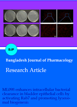
Figure 1: Effect of ML098 (1 µM) on the intracellular bacterial count of HTB-9 cells infected with E. coli. The number of intracellular bacteria in the E. coli group and E. coli + ML098 group 2 hours after infection (A, B). The number of intracellular bacteria between the E. coli group and E. coli + ML098 group 6 hours after infection (C, D). Compared to the E. coli group, ap<0.05; “ns” means statistically not significant
Figure 2: Effect of ML098 (1 µM) on the number of intracellular acidic lysosomes in HTB-9 cells infected with E. coli. The results of lysotracker red staining (A). The results of intensity of lysotracker red in the three groups (B). Compared to the E. coli group, ap<0.05; “ns” means statistically not significant
Effect on the expression of LAMP1 in HTB-9 cells
The expression of lysosome associated membrane protein 1 in E. coli group cells showed no significant difference compared to the control group (p>0.05, Figure 3). Compared with the E. coli group, the E. coli + ML098 group showed an increase in intracellular lysosome associated membrane protein 1 expression (p<0.05). The RT-qPCR results showed that the expression of intracellular lysosome associated membrane protein 1 mRNA in the E. coli group was no difference compared with the control group (p<0.05). And compared with the E. coli group, the expression of lysosome associated membrane protein 1 mRNA was significantly increased in the E. coli + ML098 group.
Figure 3: The effects of ML098 (1 µM) on the expression levels of lysosome associated membrane protein 1 (LAMP-1) and lysosome associated membrane protein 1 mRNA in HTB-9 cells infected with E. coli. The results of lysosome associated membrane protein 1 expression in three groups using the western blot method (A, B). Levels of LAMP-1 mRNA expression in three groups by RT-qPCR (C). Compared to the E. group, ap<0.05; “ns” means statistically not significant
Effect on the expression of Rab7 and active Rab7 in HTB-9 cells
To investigate the effect of ML098 on Rab7 and active Rab7 expression in bladder epithelial cells, western blot and GST-pull down assay were conducted. The results of western blot and RT-qPCR showed that there is no difference in the expression of Rab7 in the control, E. coli and E. coli + ML098 group (Figure 4). GST-pull down assay showed that the expression of intracellular active Rab7 in the E. coli group was increased compared with the control group (p<0.05) and the expression of intracellular active Rab7 in the E. coli + ML098 group was increased compared with the E. coli group (p<0.05).
Figure 4: Effect of ML098 (1 µM) on intracellular Rab7 levels and activity in HTB-9 cells infected with E. coli. The results of Rab7 expression in the three groups using the western blot method (A, B). The levels of active Rab7 in the three groups using the Rab7 pulldown activation assay (A, C). The results of the level of Rab7 mRNA in the three groups using RT-qPCR. Compared to the control group, ap<0.05. Compared to the E. coli group, bp<0.05; “ns” means statistically not significant
Discussion
The results of this study showed that ML098 does not affect E. coli UTI89's early invasion in HTB-9 cells but significantly reduced the number of bacteria in HTB-9 cells infected with E. coli 6 hours after infection. The red fluorescence intensity of lysosomes in the infected HTB-9 cells treated with ML098 was significantly higher, and the levels of lysosome associated membrane protein 1 and lysosome associated membrane protein 1 mRNA were significantly higher. ML098 treatment significantly increased the level of active Rab7. This suggests that ML098 may activate Rab7 to increase the number of lysosomes in HTB-9 cells and promote the clearance of E. coli in cells.
Many pathogenic microorganisms, including UPEC, Salmonella, Coronaviruses, and Mycobacterium tuberculosis, have evolved the ability to invade host cells, survive and reproduce inside the cells, and thereby evade clearance by the immune system (Xu et al., 2019). Previous studies have shown that promoting intracellular bacterial clearance can relieve urinary tract infections (Pang et al., 2022; Yin et al., 2023). During the resistance of bladder epithelial cells to urinary tract infection, the removal of intracellular bacteria by lysosomes is the key defense link, which can quickly reduce the intracellular bacterial load (Miao et al., 2015). Lysosomes are crucial organelles in cells, which are deeply involved in cell metabolism and autophagy. It efficiently engulfs and degrades damaged and aged organelles within cells, thereby maintaining the stability of the intracellular environment. In addition, lysosomes can also remove bacteria or viruses that invade cells and play an important role in the process of cell defense (Yim et al., 2020). Specifically, UPEC, the main pathogen of urinary tract infection, is recognized by autophagy after invading bladder epithelial cells. Autophagosome containing bacteria first binds to multivesicular bodies, and then transported to lysosomes, and finally removed by lysosomes. Therefore, regulating the function or quantity of lysosomes in bladder epithelial cells may be an effective strategy for the treatment of urinary tract infection. In the study of HeLa cells against coronaviruses, it was found that increasing the number of lysosomes could promote viral clearance (Ghosh et al., 2020). In the study of Porphyromonas gingivalis infection in oral epithelial cells, it was found that reducing the number of intracellular lysosomes was beneficial to the survival of the bacteria (Liu et al., 2023). Different from previous studies, this study involved different cell lines and pathogenic microorganisms.
Rab7 is a small GTPase localized in lysosomes, late endosomes and multivesicular bodies, which plays a key role in lysosomal biogenesis and maintenance (Wong et al., 2018). Active Rab7 can promote lysosome formation (Langemeyer et al., 2018). Knockout of Rab7 inhibited the maturation of late endosomes/multivesicular bodies, resulting in a decrease in the number of intracellular lysosomes (Vanlandingham et al., 2009). Rab7 exists in an active GTP-bound state and an inactive GDP-bound state (Cai et al., 2021; Zhao et al., 2021). Inhibition of Rab7 activity in HeLa cells significantly reduced the number of intracellular lysosomes, while enhancing the expression of active Rab7 increased the number of lysosomes (Ghosh et al., 2020). Rab7 knockout significantly reduced the ability of bladder epithelial cells to eliminate intracellular bacteria (Miao et al., 2015). Therefore, modulation of intracellular active Rab7 expression may be a potential target for the treatment of urinary tract infection.
As a RAB7 activator, ML098 is commonly used to regulate autophagy and apoptosis processes. ML098 alleviates ovarian aging-related oocyte quality decline by enhancing mitophagy (Jin et al., 2022), and ameliorates drug-related osteonecrosis of the jaw by enhancing autophagic flow and down-regulating apoptosis in a mouse model (Dong et al., 2023). For microbial studies, ML098 promoted coronavirus clearance by increasing the number of lysosomes in HeLa cells (Ghosh et al., 2020). Therefore, ML098 is considered as a potential drug for the treatment of intracellular infections. Different from previous studies, this study focused on investigating the bacterial clearance effect of Rab7 agonist ML098 in HTB-9 cells, aiming to evaluate the feasibility of Rab7 agonist ML098 as a potential drug for the treatment of urinary tract infections.
However, the study has some limitations. First, the study did not include any in vivo experiments to confirm the findings in a living organism, which is crucial for understanding the drug's efficacy and potential side effects in a real-world context. Second, while the study suggests that ML098 increases active Rab7 and lysosome biogenesis, the detailed molecular mechanisms underlying these effects were not fully elucidated.
Conclusion
ML098 can promote the clearance of intracellular E. coli UTI89 in infected bladder epithelial cells. This may be because ML098 enhances the expression of active Rab7 in bladder epithelial cells and increases the number of LAMP1 positive lysosomes in the cells.
Ethical Issue
Cell lines derived from expansion of primary cell cultures in vitro were not relevant material, as all of the original cells were divided and so the cell line had been created outside of the human body. The storage and use of cell lines created from primary human tissue, for research purposes, did not require an ethical approval.
Acknowledgements
Gratitude is extended to Mr. Youwei Huang for conducting part of the cell culture and western blotting experiments. Special thanks also go to Prof. Wei Jiang for providing guidance throughout the experiments.
References
Beebout CJ, Robertson GL, Reinfeld BI, Blee AM, Morales GH, Brannon JR, Chazin WJ, Rathmell WK, Rathmell JC, Gama V, Hadjifrangiskou M. Uropathogenic Escherichia coli subverts mitochondrial metabolism to enable intracellular bacterial pathogenesis in urinary tract infection. Nat Microbiol. 2022; 7: 1348-60.
Bhattacharya A, Mukherjee R, Kuncha SK, Brunstein ME, Rathore R, Junek S, Münch C, Dikic I. A lysosome mem-brane regeneration pathway depends on TBC1D15 and autophagic lysosomal reformation proteins. Nat Cell Biol. 2023; 25: 685-98.
Cai CZ, Yang C, Zhuang XX, Yuan NN, Wu MY, Tan JQ, Song JX, Cheung KH, Su H, Wang YT, Tang BS, Behrends C, Durairajan SSK, Yue Z, Li M, Lu JH. NRBF2 is a RAB7 effector required for autophagosome maturation and mediates the association of APP-CTFs with active form of RAB7 for degradation. Autophagy 2021; 17: 1112-30.
Chan H, Li Q, Wang X, Liu WY, Hu W, Zeng J, Xie C, Kwong TNY, Ho IHT, Liu X, Chen H, Yu J, Ko H, Chan RCY, Ip M, Gin T, Cheng ASL, Zhang L, Chan MTV, Wong SH, Wu WKK. Vitamin D3 and carbamazepine protect against Clostridioides difficile infection in mice by restoring macrophage lysosome acidification. Autophagy 2022; 18: 2050-67.
Dong X, He Y, An J, He L, Zheng Y, Wang X, Wang J, Chen S, Zhang Y. Increased apoptosis of gingival epithelium is associated with impaired autophagic flux in medication-related osteonecrosis of the jaw. Autophagy 2023; 19: 2899-911.
Ellis MJ, Lekka C, Holden KL, Tulmin H, Seedat F, O'Brien DP, Dhayal S, Zeissler ML, Knudsen JG, Kessler BM, Morgan NG, Todd JA, Richardson SJ, Stefana MI. Identification of high-performing antibodies for the reliable detection of Tau proteoforms by Western blotting and immunohistochemistry. Acta Neuropathol. 2024; 147: 87.
Engel DR, Wagenlehner FME, Shevchuk O. Scientific advances in understanding the pathogenesis, diagnosis, and prevention of urinary tract infection in the past 10 years. Infect Dis Clin North Am. 2024; 38: 229-40.
Fisher H, Oluboyede Y, Chadwick T, Abdel-Fattah M, Brennand C, Fader M, Harrison S, Hilton P, Larcombe J, Little P, McClurg D, McColl E, N'Dow J, Ternent L, Thiruchelvam N, Timoney A, Vale L, Walton K, von Wilamowitz-Moellendorff A, Wilkinson J, Wood R, Pickard R. Continuous low-dose antibiotic prophylaxis for adults with repeated urinary tract infections (AnTIC): A randomised, open-label trial. Lancet Infect Dis. 2018; 18: 957-68.
Ghosh S, Dellibovi-Ragheb TA, Kerviel A, Pak E, Qiu Q, Fisher M, Takvorian PM, Bleck C, Hsu VW, Fehr AR, Perlman S, Achar SR, Straus MR, Whittaker GR, de Haan CAM, Kehrl J, Altan-Bonnet G, Altan-Bonnet N. β-Coronaviruses use lysosomes for egress instead of the biosynthetic secretory pathway. Cell 2020; 183: 1520-35.
Jin X, Wang K, Wang L, Liu W, Zhang C, Qiu Y, Liu W, Zhang H, Zhang D, Yang Z, Wu T, Li J. RAB7 activity is required for the regulation of mitophagy in oocyte meiosis and oocyte quality control during ovarian aging. Autophagy 2022; 18: 643-60.
Kitagawa K, Shigemura K, Yamamichi F, Alimsardjono L, Rahardjo D, Kuntaman K, Shirakawa T, Fujisawa M. International comparison of causative bacteria and antimicrobial susceptibilities of urinary tract infections between Kobe, Japan, and Surabaya, Indonesia. Jpn J Infect Dis. 2018; 71: 8-13.
Kretschmer-Kazemi Far R, Frank K, Sczakiel G. Sampling, logistics, and analytics of urine for RT-qPCR-based diagnostics. Cancers (Basel). 2021; 13: 4381.
Langemeyer L, Frohlich F, Ungermann C. Rab GTPase function in endosome and lysosome biogenesis. Trends Cell Biol. 2018; 28: 957-70.
Li X, Jiang L, Zhang S, Zhou J, Liu L, Jin C, Sun H, Wang Q, Liu Y, Pang Y. Uropathogenic Escherichia coli subverts host autophagic defenses by stalling preautophagosomal structures to escape lysosome exocytosis. J Infect Dis. 2024; 230: e548-58.
Liu M, Shao J, Zhao Y, Ma B, Ge S. Porphyromonas gingivalis evades immune clearance by regulating lysosome efflux. J Dent Res. 2023; 102: 555-64.
Miao Y, Li G, Zhang X, Xu H, Abraham SN. A TRP channel senses lysosome neutralization by pathogens to trigger their expulsion. Cell 2015; 161: 1306-19.
Pang Y, Cheng Z, Zhang S, Li S, Li X, Li X, Zhang X, Li X, Feng Y, Cui H, Chen Z, Liu L, Li Q, Huang J, Zhang M, Zhu S, Wang L, Feng L. Bladder epithelial cell phosphate trans-porter inhibition protects mice against uropathogenic Escherichia coli infection. Cell Rep. 2022; 39: 110698.
Pillai-Kastoori L, Schutz-Geschwender AR, Harford JA. A systematic approach to quantitative western blot analysis. Anal Biochem. 2020; 593: 113608.
Seaman MNJ, Mukadam AS, Breusegem SY. Inhibition of TBC1D5 activates Rab7a and can enhance the function of the retromer cargo-selective complex. J Cell Sci. 2018; 131: jcs217398.
Vanlandingham PA, Ceresa BP. Rab7 regulates late endocytic trafficking downstream of multivesicular body biogenesis and cargo sequestration. J Biol Chem. 2009; 284: 12110-24.
Wang C, Bauckman KA, Ross ASB, Symington JW, Ligon MM, Scholtes G, Kumar A, Chang HW, Twentyman J, Fashemi BE, Xavier RJ, Mysorekar IU. A non-canonical autophagy-dependent role of the ATG16L1T300A variant in urothelial vesicular trafficking and uropathogenic Escherichia coli persistence. Autophagy 2019; 15: 527-42.
Wong YC, Ysselstein D, Krainc D. Mitochondria-lysosome contacts regulate mitochondrial fission via RAB7 GTP hydrolysis. Nature 2018; 554: 382-86.
Xu Y, Zhou P, Cheng S, Lu Q, Nowak K, Hopp AK, Li L, Shi X, Zhou Z, Gao W, Li D, He H, Liu X, Ding J, Hottiger MO, Shao F. A bacterial effector reveals the V-ATPase-ATG16L1 axis that initiates xenophagy. Cell 2019; 178:552-66.
Yim WWY, Mizushima N. Lysosome biology in autophagy. Cell Discov. 2020; 6: 6.
Yin H, Zhu J, Jiang Y, Mao Y, Tang C, Cao H, Huang Y, Zhu H, Luo J, Jin Q, Jin Q. Shionone relieves urinary tract infections by removing bacteria from bladder epithelial cells. Cell Microbiol. 2023; 2023.
Zhao YG, Codogno P, Zhang H. Machinery, regulation and pathophysiological implications of autophagosome maturation. Nat Rev Mol Cell Biol. 2021; 22: 733-50.