In silco studies on modified hydroxamic acid and valporic acid as potential inhibitors for HDAC2
Abstract
Histone deacetylases 2, class 1 HDAC family are emerged as an important therapeutic target for the treatment of various cancers. HDAC2 inhibitors are potent anti-cancer agents. Two inhibitors of HDAC2 are hydroxamic acid and valporic acid which are potent inducers of growth arrest, differentiation, and/or apoptotic cell death. Total 34 ligands optimized using triazole group substitution for the target protein histone deacetylase 2 on the basis of SAHA and valporic acid. All the ligands are docked with the target protein and results are compared with test compound SAHA. Eight ligands showed better binding affinity towards HDAC2.The binding affinity, free energy and drug scan screening of the above eight ligands have shown that P2, P6 and V6 molecules are best suitable to inhibit HDAC2.
Introduction
HDACs are the enzyme deacetylating the e-amino groups of lysine located near the amino termini of core histone proteins (Mai et al., 2002 and Monneret, 2005). HDACs have been classified into class I-III, class I includes HDAC 1-3 and 8, class II includes HDAC 4-7, 9-10 both classes operate by zinc-dependent mechanisms and class III includes Sir1-Sir 7 operated by NAD (Bieliauskas and Pflum, 2008). HDAC2 enzyme is greatly considered for developing anticancer drugs. HDAC inhibitors interact with chromosomes in the cancer cell and causes cancer cells to stop growing. Hydroxamic acid and valproic acid are potent inhibitors of HDAC. One of the hydroxamic acid derivatives in clinical phase is panobinostat (Prince et al., 2009). The first HDAC drug approved by U.S Food and Drug Administration is SAHA (suberoylanilide hydroxamic acid or vorinostat) for treating cutaneous T-cell lymphoma (Walkinshaw et al., 2008). SAHA inhibits the activity of class I & II HDACs (Marks et al., 2007). Present study involves in silico hydroxamic acid and valproic acid modification by utilizing triazole, in order to obtain a better inhibitor. Molecular docking predicts the preferred orientation of one molecule to a second when bound to each other to form a stable complex. Hence molecular docking is used to predict suitable ligand molecules for HDAC2 inhibition.
Materials and Methods
Total 34 ligands optimized using triazole group substitution for the target protein histone deacetylase 2 on the basis of SAHA and valporic acid. This study is looking to compare the efficacy of SAHA with other types of inhibitors, by searching for the new ones or modifications of the existing ones. Triazole is known as a non-classical amide bioisostere compound and its biological activity, notably as antifungal, antimicrobials, and enzymatic inhibitors (Roffey, 1997). It is of interest to modify the SAHA and valporic acid structure by creating new ligands. The processes followed are adding replacing one of the amides group within the SAHA hydrophobic group with triazole linked with sulfur bond and styrene group then, conduct molecular docking with HDAC2, and testing its drug score and toxicity using computational tools, and finally compare the result with standard SAHA inhibitor. This structure-activity relationship (SAR) study is very important in uncovering novel inhibitors of HDAC2. The triazole bioisostere attributes on SAHA amide group could eventually modify SAHA's properties. The hydrophobic tendency of triazole compared with the amide group on SAHA was expected to increase the binding affinity of modified ligands toward HDAC2. Thus, the binding of enzyme-ligand complex would be much stronger. Triazole could be treated as an additional functional group on SAHA, which could increase the hydrophobic attributes of SAHA cap group. The twelve alkyl groups for modified ligand variations are phenyl, biphenyl, napthyl, p-nitrobiphenyl, p-hydroxybiphenyl, aminooxyphenyl, acetamidooxy phenyl, amine, acetamidophenyl. The selections of those alkyl groups are based on hydrophobic attributes of those groups. Thus, this study would observe the influence of the cap group hydrophobicity of each modified ligand, in comparison with the SAHA standard ligand. Figure 1 shows modified hydroxamic acid derivative and modified valporic acid. All 34 Ligand showed in Table I and II. Docking studies carried out in LigandFit (Discovery Studio 2.0) (Venkatachalam et al., 2003). It is based on a cavity detection algorithm and Monte Carlo conformational search algorithm for generating ligand poses consistent with the active site shape. All 34 ligands are drawn in Chemsketch and the hydrogen bonds were added and CHARMm force field was applied to all molecules. The 3D crystal structure of Homo sapiens HDAC2 (PDB ID: 3MAX) downloaded from protein database from the PDB structural database site (http://www.rcsb.org/pdb). After applying CHARMm force field macro molecule 3MAX was assigned as receptor. The receptor cavity was searched using flood filling algorithm and partition site was adjusted for the better fitments of molecule in the partition site of receptor. The comparative docking studies for all 34 molecules were performed. The determination of the ligand binding affinity was calculated using dock score were used to estimate the ligand-binding energies.
Figure 1: Triazole substituted hydroxamic acid derivative 1, 2 and valproic acid derivative 3
Table I: Different substitution used in hydroxamic acid derivatives
| Compound | R | R1 |
|---|---|---|
| 1 | H | H |
| 2 | C6H5 | H |
| 3 | C6H5 | CH3 |
| 4 | C6H5 | C6H5 |
| 5 | C6H5C6H4 | H |
| 6 | C10H13 | H |
| 7 | P-NH2-C6H4-C6H4 | H |
| 8 | P-OH-C6H4-C6H4 | H |
| 9 | O-(NH2)-C6H4 | H |
| 10 | O-(NHCOCH3)-C6H4 | H |
| 11 | P-NH2-C6H4 | H |
| 12 | P(NHCOCH3)C6H4 | H |
Table II: Different substitution used in valporic acid derivatives
| Compound | R | R1 |
|---|---|---|
| 1 | H | H |
| 2 | C6H5 | H |
| 3 | C6H5 | CH3 |
| 4 | C6H5 | C6H5 |
| 5 | C6H5C6H4 | H |
| 6 | C10H13 | H |
| 7 | P-NH2-C6H4-C6H4 | H |
| 8 | P-OH-C6H4-C6H4 | H |
| 9 | O-(NH2)-C6H4 | H |
| 10 | O-(NHCOCH3)-C6H4 | H |
| 11 | P-NH2-C6H4 | H |
| 12 | P(NHCOCH3)C6H4 | H |
Drug scan
This was conducted in order to determine, whether the inhibitor has fulfilled the conditions as the drug candidate. It is done using Osiris Property Explorer, and Lazar software's (Bhakat, 2012). The Osiris Property Explorer and Lazar calculated various attributes of the drugs, such as toxicity, drug likeness and drug score.
Result and Discussion
HDAC2 Contains three chains such as A, B and C, All the three chains are docked with test compound SAHA. It shows Chain B has good docking Score. The docking scores of SAHA with different chains of the target protein at different sites on HDAC2 are given in Table III. Chain C is inactive as it shows no results with the test compound.
Table III: Docking scores of SAHA
| Chain | Site 2 | Site 3 | Site 6 | Site 8 |
|---|---|---|---|---|
| A | * | 26.7 | * | 23.2 |
| B | 30.8 | 18.0 | 45.6 | * |
All 34 ligands are docked on 3MAX B Chain the results shows triazole modified hydroxamic acids shows better docking score than the SAHA test compound. The docking score of the ligands are shown in Table IV and V. Results shows that P1, P2, P4, P6, P9, V1, V6 and V9 modified ligands shows more binding affinity than test compound SAHA (Figure 2-4).
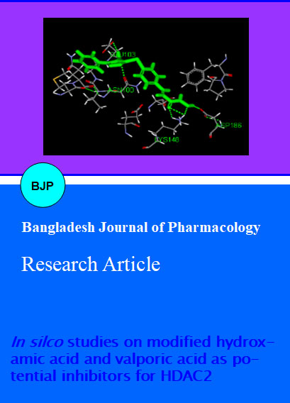
Figure 2: Docking of ligand p2 with 3 maxb at site 6
Figure 3: Docking of ligand V6 with 3 maxB at site 6
Figure 4: Docking of ligand V6 with 3 maxB at site 6
Table IV: Docking scores of compounds
| Name of the compound | Site 2 | Site 3 | Site 6 |
|---|---|---|---|
| P1 | 29.01(-2.746) | ** | 48.00(-2.832) |
| P2 | ** | ** | 64.70(-4.578) |
| P3 | 24.40(-2.261) | ** | 43.42(-2.645) |
| P4 | ** | ** | 45,73(-6.485) |
| P6 | ** | ** | 55.27(-5.648) |
| P9 | ** | ** | 51.00(-3.957) |
| V1 | 25.51(0.004) | 26.20(0.610) | 47.02(-0.237) |
| V3 | 28.41(-0.843) | 19.67(-5.88) | 43.50(-1.074) |
| V4 | 16.30(-2.807) | ** | 44.63(0.228) |
| V6 | 19.68(-1.573) | ** | 47.27(-4.001) |
| V9 | 21.75(-4.573) | ** | 46.03(-3.584) |
| V11 | 19.78(-3.191) | ** | 44.378(-2.07) |
| VAL1 | 18.13(1.991) | 19.71(0.638) | 35.48(0.493) |
| VAL2 | 16.92(-0.364) | ** | 36.68(-1.070) |
| VAL3 | ** | ** | 32.62(-1.684) |
| VAL4 | 0.07(7.71) | ** | 33.12(-2.785) |
| VAL6 | ** | ** | 26.83(-2.653) |
| VAL8 | 4.81(2.287) | ** | 42.36(1.316) |
| VAL9 | ** | ** | 36.03(-3.043) |
| VAL10 | ** | ** | 38.35(-2.870) |
Table V:Interaction of modified ligands with Hdac2 (3maxb chain) Homo sapiens at site 6
| Name of the compound | Hydrogen bond monitored |
|---|---|
| P1 | B:GLU154:HN-Molecule-1:N3 |
| P1 | B:ASP186:OD1- Molecule-1:H35 |
| P2 | B:GLU103:OE1- Molecule-1:H28 |
| P2 | B:SER153:OG- Molecule-1:H34 |
| P4 | B:ASN100:HD21-Molecule-1:O17 |
| P4 | B:GLU103:0E2- Molecule-1:H35 |
| P4 | B:GLU151:OE1- Molecule-1:H40 |
| P6 | B:ASN100:HD21-Molecule-1:O17 |
| P6 | B:GLU103:0E2- Molecule-1:H35 |
| P6 | B:GLU151:OE1- Molecule-1:H40 |
| P9 | B:ASN100:OD1- Molecule-1:H30 |
| P9 | B:GLU103:OE1- Molecule-1:H35 |
| P9 | B:SER153:OG- Molecule-1:H41 |
| V1 | B:GLY154:HN-Molecule-1:ON14 |
| V1 | B:GLU103:0E1- Molecule-1:H19 |
| V1 | B:ASO104:OD2- Molecule-1:H33 |
| V6 | B:MET 96: E21-Molecule-1:03 |
| V6 | B:GLU103:0E2- Molecule-1:H35 |
| V9 | B:LYS171:H23-Molecule-1:04 |
| V9 | B:GLU103:OE1- Molecule-1:H25 |
| V9 | B: VAL101 :O- Molecule-1:H26 |
| V9 | B:ANS153:OD1- Molecule-1:H39 |
Osiris property explorer used to find the in silico pharmacology features. The hydrophobicity of drugs could be inferred from Log P value. When its value is increasing, the drug will be more hydrophobic. When the drug is more hydrophobic, then the drug will be able to circulate longer in our body, because it would not be easy to secrete it. The Table VI shows, that the Log P values of the P2, P4-P8, V2-V8, VA1, VA3,VA4 and VA6 modified ligands are larger than the SAHA standard ligand. It shows that the modified ligands are more hydrophobic than SAHA. Normally, drugs, which interact with enzyme inside human body, have Log P value between 2 and 5 (Copeland, 2005). The drug likeness value of standard and modified ligand shows the fragment content of the drugs. If the drug likeness values are increasing, than it has the same fragment content with existing drugs. From Table VI, it is shown that the drug likeness values of most ligands are larger than the SAHA standard ligand. This result tells us, that the modified ligand has the most fragments content of drugs. The drug score values are the combination of drug likeness, Log P, solubility, molecular weight, and toxicity risk within one useful practical value. It could be used for evaluating the potential of the drug candidate (Lindemann and Johnstone, 2004). When the drug score is better, then the compound has a better chance to be a drug candidate. Table VI shows that only modified P3 and V3 ligands have better drug score than SAHA standard ligand.
Table VI: Drug likeness and scores of SAHA standard and modified ligands based on Osiris property explorer
| Compounds | cLogP | Solubility | Mol Wt | Drug likeness | Drug score |
|---|---|---|---|---|---|
| SAHA | 2.28 | -3.33 | 264 | -8.87 | 0.26 |
| P1 | 1.05 | -4.9 | 259 | -7.8 | 0.22 |
| P2 | 2.49 | -7.31 | 353 | -0.11 | 0.21 |
| P3 | 0.8 | -5.16 | 277 | -0.25 | 0.3 |
| P4 | 4.27 | -7.75 | 411 | -6.98 | 0.07 |
| P5 | 4.42 | -9.13 | 411 | -8.03 | 0.11 |
| P6 | 5.32 | -6.85 | 399 | -20.7 | 0.11 |
| P7 | 3.69 | -9.21 | 426 | -11.89 | 0.07 |
| P8 | 4.12 | -8.83 | 427 | -11.84 | 0.11 |
| P9 | 1.59 | -8.07 | 366 | -7.8 | 0.14 |
| P10 | 1.97 | -8.23 | 408 | -7.64 | 0.13 |
| P11 | 2.01 | -7.12 | 350 | -7.81 | 0.15 |
| P12 | 2.34 | -7.39 | 392 | -6.03 | 0.14 |
| V1 | 1.14 | -1.81 | 131 | -11.48 | 0.17 |
| V2 | 2.49 | -7.31 | 353 | -0.11 | 0.21 |
| V3 | 1.82 | -1.85 | 173 | -2 | 0.29 |
| V4 | 4.03 | -8.02 | 429 | 0.52 | 0.11 |
| V5 | 4.17 | -9.39 | 429 | -0.64 | 0.15 |
| V6 | 2.86 | -7.58 | 242 | -3.12 | 0.1 |
| V7 | 3.45 | -9.47 | 444 | -4.5 | 0.07 |
| V8 | 3.87 | -9.1 | 445 | -4.45 | 0.11 |
| V9 | 1.34 | -8.34 | 384 | -0.41 | 0.19 |
| V10 | 1.72 | -8.5 | 426 | -0.27 | 0.19 |
| V11 | 1.77 | -7.38 | 368 | -0.42 | 0.2 |
| V12 | 2.1 | -7.65 | 410 | 1.28 | 0.24 |
| VA1 | 3.15 | -2.24 | 158 | -6.27 | 0.36 |
| VA2 | 1.67 | -5.97 | 192 | -2.15 | 0.34 |
| VA3 | 3.36 | -8.06 | 268 | -2.76 | 0.25 |
| VA4 | 6.06 | -8.63 | 332 | -21.9 | 0.15 |
| VA5 | 2.63 | -8.13 | 283 | -6.57 | 0.09 |
| VA6 | 3.06 | -7.76 | 284 | -6.53 | 0.24 |
| VA7 | 0.53 | -7 | 223 | -2.45 | 0.18 |
| VA8 | 0.91 | -7.16 | 265 | -2.2 | 0.18 |
| VA9 | 0.95 | -6.05 | 207 | -2.41 | 0.33 |
| VA10 | 1.28 | -6.31 | 249 | -0.46 | 0.41 |
The toxicity of molecules is predicted using Osiris Property Explorer, and Lazar. All of them have different parameters for determining the toxicity of compounds. The prediction using Osiris Property Explorer was shown in colour codes. The result of toxicity analysis of SAHA standard ligand, first, and second modified ligands is shown in Table VII. Green colour shows the low toxicity tendency, yellow shows the mediocre tendency and red shows high tendency. Lazar is a software package with functionality of detecting mutagenic or carcinogenic properties based on the functional group similarity with mutagenic or carcinogenic ones. Lazar verified the mutagenicity of compounds by conducting assay test with Salmonella typhimurium. The carcinogenicity of compounds was verified by animal testing, with rat, and mouse (Table VIII). The best ligands for HDAC2 Homo sapiens could be determined based on drug scan and docking analysis (Alonso et al., 2006).
Table VII: Toxicity of SAHA standard and modified ligand based on Osiris property explorer
| Compounds | Mutagenic | Tumorigenic | Irritant | Reproductive effect |
|---|---|---|---|---|
| SAHA | Red | Green | Green | Green |
| P1 | Red | Green | Green | Green |
| P2 | Red | Green | Green | Green |
| P3 | Red | Green | Green | Green |
| P4 | Red | Red | Green | Green |
| P5 | Red | Green | Green | Green |
| P6 | Red | Green | Green | Green |
| P7 | Red | Red | Green | Green |
| P8 | Red | Green | Green | Green |
| P9 | Red | Green | Green | Green |
| P10 | Red | Green | Green | Green |
| P11 | Red | Green | Green | Green |
| P12 | Red | Green | Green | Green |
| V1 | Red | Green | Red | Green |
| V2 | Red | Green | Red | Green |
| V3 | Red | Green | Green | Green |
| V4 | Red | Red | Green | Green |
| V5 | Red | Green | Green | Green |
| V6 | Red | Yellow | Green | Yellow |
| V7 | Red | Red | Green | Green |
| V8 | Red | Green | Green | Green |
| V9 | Red | Green | Green | Green |
| V10 | Red | Green | Green | Green |
| V11 | Red | Green | Green | Green |
| V12 | Red | Green | Green | Green |
| VA1 | Green | Green | Green | Yellow |
| VA2 | Green | Green | Green | Green |
| VA3 | Green | Green | Green | Green |
| VA4 | Green | Green | Green | Green |
| VA5 | Red | Red | Green | Green |
| VA6 | Green | Green | Green | Green |
| VA7 | Red | Green | Green | Green |
| VA8 | Red | Green | Green | Green |
| VA9 | Green | Green | Green | Green |
| VA10 | Green | Green | Green | Green |
Table VIII: Toxicity analysis result by using lazar
| Name of the compound | Mutagenicity | Carcinogencity | |||
|---|---|---|---|---|---|
| DBS Mutagenicity | Salmonella typhimurium (Kazius/Bursi) | Mouse | Rat | Multi cell call | |
| P1 | No | No | No | No | No |
| P2 | No | No | No | No | No |
| P3 | No | No | No | No | No |
| P4 | No | Yes | No | No | No |
| P5 | No | Yes | No | No | No |
| P6 | No | No | No | No | No |
| P7 | No | No | No | Yes | No |
| P8 | Yes | No | No | No | No |
| P9 | No | No | No | No | No |
| P10 | No | No | No | No | No |
| P11 | No | Yes | No | No | No |
| P12 | No | Yes | No | No | No |
| V1 | No | No | No | No | No |
| V2 | No | No | Yes | No | No |
| V3 | No | No | Yes | Yes | No |
| V4 | Yes | No | Yes | No | No |
| V5 | No | No | Yes | No | No |
| V6 | No | No | No | No | No |
| V7 | Yes | Yes | Yes | Yes | Yes |
| V8 | Yes | No | No | No | Yes |
| V9 | No | No | No | No | Yes |
| V10 | Yes | Yes | No | No | Yes |
| V11 | Yes | Yes | Yes | - | Yes |
| V12 | Yes | Yes | Yes | - | No |
| VA1 | No | No | No | No | No |
| VA2 | No | No | Yes | No | No |
| VA3 | No | Yes | Yes | No | No |
| VA4 | No | No | No | No | No |
| VA5 | Yes | Yes | Yes | Yes | No |
| VA6 | No | Yes | No | No | No |
| VA7 | No | No | No | No | No |
| VA8 | No | No | No | No | No |
| VA9 | Yes | Yes | Yes | No | No |
| VA10 | Yes | Yes | Yes | No | No |
The docking result of SAHA standard, first, and second modified ligands toward HDAC2 shows that those ligands have same type of interaction toward HDAC2. The analysis of ΔG binding and Score show that modified ligand have smaller ΔG binding than SAHA standard ligand. It could be inferred modified ligand has better binding affinity than SAHA standard ligand. Every modified ligand has good pharmacological properties, and it could be inferred by its accordance with Lipinski's Rule, hydrophobicity based on log P value, and good drug likeness and drug score. However, the best ligands according to the binding energy and drug scan analysis are P2, P6 and V6 ligands, in this end; our SAR study has proven that P2, P6 and V6 inhibitors are the best inhibitor as alternatives of SAHA.
References
Alonso H, Bliznyuk AA, Gready JE. Combining docking and molecular dynamic simulation in drug design. Med Res Rev. 2006; 26: 531-68.
Bhakat S. SAR and pharmacophore based designing of some antimalarial and antiretroviral agents: An internet based drug design approach. Der Pharma Chemica. 2012; 4: 1247-63.
Bieliauskas AV, Pflum MKH. Isoform-selective histone deacetylase inhibitors. Chem Soc Rev. 2008; 37: 1402-13.
Copeland RA. Evaluation of enzyme inhibitors in drug discovery: A guide for medicinal chemists and pharmacologists. Methods Biochem Anal. 2005; 46: 1-265.
Lindemann RK, Johnstone RW. Histone deacetylase inhibitors: Promising candidates for chemotherapeutic drugs. Gene Ther Mol Biol. 2004; 8: 61-74.
Mai A, Massa S, Ragno R, Esposito M, Sbardella G, Nocca G, Scatena R, Jesacher F, Loidl P, Brosch G. Binding mode analysis of 3-(4-benzoyl-1-methyl-1H-2-pyrrolyl)-N-hydroxy-2-propenamide: A new synthetic histone deacetylase inhibitor inducing histone hyperacetylation, growth inhibition, and terminal cell differentiation. J Med Chem. 2002; 45: 1778-84.
Marks PA. Discovery and development of SAHA as an anticancer agent. Oncogene 2007; 26: 1351-56.
Monneret C. Histone deacetylase inhibitors. Eur J Med Chem. 2005; 40: 1-13.
Prince HM, Bishton M. Panobinostat (LBH589): A novel pan-deacetylase inhibitor with activity in T-cell lymphoma. Hematol Meet Rep. 2009; 3: 33-38.
Roffey J. Bioisosteres in medicinal chemistry. Maybridge Med Chem. 1997; 1: 23-44.
Walkinshaw DR, Yang XJ. Histone deacetylase inhibitors as novel anticancer therapeutics. Curr Oncol. 2008; 15: 237-43.
Venkatachalam CM, Jiang X, Oldfield T, Waldman M. LigandFit: A novel method for the shape-directed rapid docking of ligands to protein active sites. J Mol Graph Model. 2003; 21: 289-307.