Hepatoprotective and antioxidant effect of Carissa spinarum root extract against CCI4- and paracetamol-induced hepatic damage in rats
Abstract
Ethanolic extract of the roots of C. spinarum was evaluated for hepatoprotective and antioxidant activities in rats. Oral pre-treatment with ethanolic extract (100, 200 and 400 mg/kg) showed significant hepatoprotective activity against CCl4 and paracetamol-induced hepatotoxicity by decreasing the activities of bilirubin and lipid peroxidation, and significant increase in the levels of uric acid, glutathione, super oxide dismutase, catalase and protein in a dose-dependent manner, which was confirmed by the decrease in liver wet weight and histopathological examination. The extract possessed strong antioxidant activity. This suggests that the hepatoprotective activity of C. spinarum is possibly attributed to its free radical scavenging properties.
Introduction
Excessive production of reactive oxygen species (ROS) plays an important role in the pathogenesis and progression of various diseases involving different organs (Visioli et al., 2000). Liver disorders are mainly caused by toxic chemicals, excessive consumption of alcohol, infections and autoimmune disorders. Carbon tetrachloride and paracetamol being converted into reactive oxidative metabolites by hepatic microsomal enzymatic system, which causes hepatotoxicity (Brent and Rumack, 1993). In the present study, the CCU and paracetamol induced acute models have been used to assess hepatoprotective activity.
Carissa spinarum Linn. is a thorny, evergreen shrub. The roots of the plant have long been prescribed in Indian and Chinese system of medicine as purgative, for the treatment of rheumatism and hepatitis (Kirtikar and Basu, 2003). The extract of the plant possesses cardiotonic (Vohra and De, 1963) and antipyretic activity (Hegde and Joshi, 2010). The other related species like C. carandas was proved for its hepatoprotective activity (Hegde and Joshi, 2009).
However, no scientific data are available regarding the usefulness of C. spinarum as hepatoprotective agent.
Materials and Methods
Preparation of extract
The roots of C. spinarum were collected from Sirsi, Uttara Kannada District, Karnataka, India during May 2007. It was authenticated by Dr. Gopalakrishna Bhat, Department of Botany, Poorna Prajna College. Fresh roots were collected and dried by means of shade drying. The root powder (500 g) was soaked in 1.5 L of 95% ethyl alcohol and extracted for 4 days with occasional shaking. After 4 days the ethanol layer was decanted off and evaporated to dryness under reduced pressure on a rotary evaporator to give the ethanolic extract (13% w/w yield), which was stored at 4°C until use. Suspension of the extract was prepared in 1% Tween-80.
Experimental animals
Wistar albino rats of either sex, weighing about 150-180 g were used for experiments. Animals were maintained under standard conditions and were fed standard rat feed and water ad libitum. Approval for the experiment was obtained by the institutional animal ethical committee.
Acute toxicity study
Acute toxicity study of the extract was determined in rats according to OECD guidelines.
Phytochemical screening
Freshly prepared extract was subjected to phytochemical screening for the detection of major chemical constituents (Harborne, 1984).
CCl4-induced hepatotoxicity
Rats were randomly divided in to 6 groups of 6 animals each. Group I (control) was administered a single daily dose of normal saline (5 mL/kg body weight, orally). Group II (CCl4 control) was administered a single daily dose of normal saline (5 mL/kg body weight, orally) and CCl4/olive oil (1:1 v/ v, 0.7 mL/kg body weight, intraperitoneal) on alternate days for 7 days. Group III (standard) was administered a single daily dose of silymarin (25 mg/kg body weight, orally) and CCl4/ olive oil (1:1 v/v, 0.7 mL/kg, body weight, intraperitoneal) on alternate days for 7 days. Group IV, V and VI (test) were administered a single daily dose of the extract (100, 200 and 400 mg/kg body weight, orally, respectively) and CCU/olive oil (1:1 v/v, 0.7 mL/kg body weight, intraperitoneal) on alternate days for 7 days (Hegde and Joshi, 2009).
Paracetamol-induced hepatotoxicity
Randomly divided 6 groups of rats were treated similar to CCl4 induced hepatotoxicity for 7 days. On fifth day, after the administration of the respective drug treatments, all the animals of groups II, III, IV, V and VI were challenged with paracetamol 2 g/kg, orally, suspended in (40% w/v) sucrose solution.
Assessment of hepatoprotective activity
On the 7th day 2 hours after the administration of last dose, the animals were sacrificed by cervical decapitation; blood was withdrawn by intracardiac puncture. Serum was separated by centrifugation at 2,500 rpm for 10 min and stored at 4°C until use. The serum was used to estimate serum aminotransaminases (Retimen and Frankel, 1957), alkaline phosphatase (King, 1965), uric acid (Caraway, 1963), total protein (Lowry et al., 1951) and total bilirubin content (Malloy and Evelyn, 1937).
Histopathological studies
The liver was immediately excised and rinsed in ice cold normal saline, blotted with filter paper and weighed. Portions of the liver was fixed in 10% neutral formalin and further processed for pathological findings of hepatotoxicity.
Measurement of antioxidant activity
Liver was rinsed and 10% w/v of homogenate was prepared in 0.15 M Tris-HCl buffer (pH 7.4) and further processed for the estimation of lipid peroxidation in the form of malondialdehyde (MDA) (Okhawa et al., 1979). From part of the homogenate, after precipitating proteins with 20% trichloroacetic acid, the supernatant was used for reduced glutathione (GSH) estimation (Ellman, 1959). The rest of the homogenate was centrifuged at 2000 rpm for 10 min and supernatant was estimated for super oxide dismutase (SOD) (Kakkar et al., 1984) and catalase (CAT) activity (Aebi, 1974).
Statistical analysis
Data were analyzed using ANOVA followed by Tukey's multiple comparison post hoc test.
Results
There was no mortality amongst the graded dose groups of rats and was found to be safe up to 2 g/kg body weight of extract in rats. Phytochemical investigation of the extract led to the presence of saponins, cardiac glycosides, triterpenoids, flavonoids and tannins.
There was a significant elevation in the levels of serum marker enzymes like SGOT, SGPT and SALP content of CCl4/PCM intoxicated animals. In contrast, preÂtreatment with extract and silymarin exhibited significant (p<0.05) hepatoprotection by decreasing serum marker enzymes in a dose dependent manner. There was a significant increase in total bilirubin and significant reduction in uric acid and total protein content of CCl4 and PCM treated groups. Whereas, preÂtreatment with extract caused significant reduction in total bilirubin and increase in the activities of uric acid and total protein content dose-dependently (Table I).
Table I: Effect of ethanol extract of the roots of C. spinarum on serum enzyme and biochemical parameters in (A) CCI4 and (B) paracetamol-induced hepatic damage in rats
| Group | Dose (/kg body weight) | SGOT (U/L) | SGPT (U/L) | SALP (U/L) | Uric acid (mg/dL) | Total protein (mg/dL) | Total bilirubin (mg/dL) | |
|---|---|---|---|---|---|---|---|---|
| I (Vehicle control) | 5 mL | A | 69.1 ± 2.3 | 58.1 ± 1.9 | 71.4 ± 1.6 | 2.8 ± 0.9 | 7.0 ± 0.4 | 1.1 ± 0.1 |
| B | 76.2 ± 3.1 | 64.0 ± 1.9 | 69.5 ± 1.1 | 2.9 ± 0.5 | 6.8 ± 0.3 | 1.0 ± 0.2 | ||
| II (CCh/PCM control) | 0.7 mL | A | 181.5 ± 2.7a | 139.3 ± 3.1a | 121.9 ± 3.0a | 1.3 ± 0.2a | 5.1 ± 0.2a | 2.3 ± 0.3a |
| B | 186.3 ± 3.9a | 141.2 ± 3.2a | 129.0 ± 2.6a | 1.3 ± 0.3a | 5.1 ± 0.2a | 2.2 ± 0.2a | ||
| III (CCh/PCM + Silymarin) | 25 mg | A | 74.5 ± 1.9c | 67.3 ± 2.0c | 79.2 ± 1.0c | 2.5 ± 0.8c | 6.9 ± 0.4c | 1.2 ± 0.2c |
| B | 88.5 ± 1.2c | 73.0 ± 2.1c | 78.3 ± 1.7c | 2.5 ± 0.6c | 6.7 ± 0.1c | 1.1 ± 0.1c | ||
| IV (CCl4/PCM + extract) | 100 mg | A | 144.9 ± 3.2c | 128.1 ± 1.7b | 100.9 ± 2.1c | 1.9 ± 0.8§ | 5.2 ± 0.3§ | 2.1 ± 0.4§ |
| B | 151.6 ± 3.2c | 118.3 ± 2.2c | 110.5 ± 2.2c | 2.0 ± 0.5b | 5.7 ± 0.2b | 2.1 ± 0.2§ | ||
| V (CCl4/PCM + extract) | 200 mg | A | 106.9 ± 4.0c | 90.1 ± 1.3c | 94.3 ± 2.6c | 2.2 ± 0.5c | 6.0 ± 0.2c | 1.7 ± 0.16c |
| B | 121.6 ± 4.2c | 99.3 ± 2.0c | 101.1 ± 2.1c | 2.2 ± 0.6c | 6.1 ± 0.2c | 1.7 ± 0.2c | ||
| VI (CCb/PCM + extract) | 400 mg | A | 87.1 ± 2.2c | ± 1.5c | 89.4 ± 2.2c | 2.3 ± 0.8c | 6.5 ± 0.3c | 1.3 ± 0.2c |
| B | 99.2 ± 3.2c | ± 2.2c | 93.3 ± 2.1c | 2.4 ± 0.6c | 6.4 ± 0.3c | 1.3 ± 0.2c | ||
| Values are mean ± SE from 6 animals in each group. One-way ANOVA followed by Tukey's multiple comparison post hoc test. ap<0.001 when compared with vehicle treated control group, §p<0.05, bp<0.01, cp<0.001 when compared with CCl/PCM treated control group | ||||||||
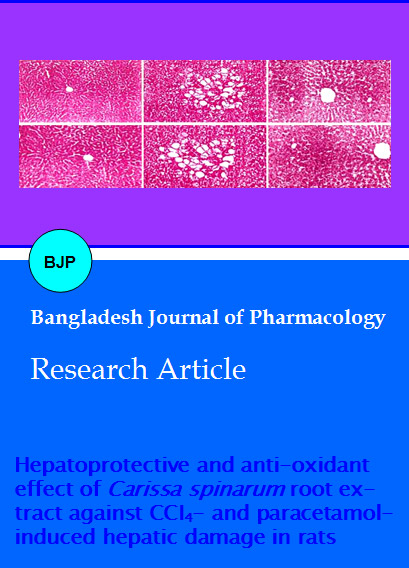
In histopathological study pre-treatment with extract exhibited significant liver protection against CCU/PCM induced liver damage, which is evident by the presence of more or less normal hepatocytes and reduced inflammatory infiltration and necrosis (Figure 1).
Figure 1: (A) Vehicle treated liver showing normal architecture of hepatic cells; (B) CCl4 treated showing inflammatory infiltration, fatty changes and necrosis; (C) Pretreated with silymarin prior to CCl4 showing normal architecture of hepatic cells with less fatty changes; (D) Pretreated with extract prior to CCl4 showing reduced inflammatory infiltration and necrosis; (E) PCM treated showing inflammatory infiltration, fatty changes and necrosis; (F) Pretreated with silymarin prior to PCM showing normal architecture of hepatic cells with less fatty changes; (G) Pretreated with extract prior to PCM showing reduced inflammatory infiltration and necrosis (H & E stain x 100)
There was a significant increase in MDA content and reduction in GSH, SOD and CAT activities of both CCl4/PCM intoxicated animals. Pre-treatment with extract significantly prevented the increase in MDA levels and brought them near to normal level, whereas GSH, SOD and CAT levels were significantly (p<0.05) raised. In both CCl4/PCM intoxicated groups, the weight of the liver was significantly increased, but it was normalized in extract treated groups (Table II).
Table II: Effect of ethanol extract of the roots of C. spinarum (extract) on lipid peroxidation (LPO), glutathione (GSH), superÂoxide dismutase (SOD), catalase (CAT) and liver weight in (A) CCU and (B) PCM induced hepatic damage in rats
| Group | Dose (/kg body weight) | LPO (nM MDA/ mg protein) | GSH (Pg/ mg protein) | SOD (U/mg protein) | CAT (U/mg protein) | Liver weight (Wt/100 g bw) | |
|---|---|---|---|---|---|---|---|
| Vehicle Control |
5 mL | A | 1.1 ± 0.1 | 5.1 ± 0.3 | 90.4 ± 2.7 | 336.9 ± 4.9 | 3.6 ± 0.1 |
| B | 1.0 ± 0.1 | 5.2 ± 0.2 | 89.6 ± 2.7 | 321.3 ± 4.7 | 3.6 ± 0.1 | ||
| CQ4/PCM Control |
0.7 mL | A | 6.8 ± 1.6a | 1.0 ± 0.1a | 49.8 ± 1.2a | 230.9 ± 3.0a | 6.2 ± 0.3a |
| B | 6.7 ± 0.7a | 1.0 ± 0.1a | 50.9 ± 1.4a | 234.3 ± 3.6a | 6.3 ± 0.3a | ||
| CQ4/PCM + Silymarin |
25 mg | A | 1.2 ± 0.4c | 5.0 ± 0.5c | 82.0 ± 2.0c | 320.4 ± 2.8c | 3.8 ± 0.2c |
| B | 1.2 ± 0.4c | 5.1 ± 0.5c | 81.1 ± 2.1c | 309.9 ± 3.9c | 3.8 ± 0.2c | ||
| CQ4/PCM + extract |
100 mg | A | 5.6 ± 0.9c | 1.9 ± 0.2c | 58.4 ± 1.9b | 265.6 ± 2.3b | 5.6 ± 0.3c |
| B | 5.8 ± 0.5c | 1.9 ± 0.2c | 56.0 ± 1.6§ | 254.1 ± 2.3b | 5.8 ± 0.3b | ||
| CQ4/PCM + extract |
200 mg | A | 3.9 ± 0.4c | 3.6 ± 0.3c | 63.9 ± 1.4c | 293.6 ± 3.8c | 5.0 ± 0.2c |
| B | 4.1 ± 0.7c | 3.5 ± 0.2c | 61.8 ± 1.6c | 292.0 ± 3.0c | 5.1 ± 0.1c | ||
| CQ4/PCM + extract |
400 mg | A | 1.9 ± 0.2c | 4.4 ± 0.2c | 72.2 ± 1.9c | 306.3 ± 2.6c | 4.1 ± 0.2c |
| B | 2.2 ± 0.2c | 4.5 ± 0.3c | 71.1 ± 1.3c | 301.9± 3.0c | 4.1 ± 0.2c | ||
| Values are mean ± SE from 6 animals in each group. One-way ANOVA followed by Tukey's multiple comparison post hoc test. ap<0.001 when compared with vehicle treated control group, §p<0.05, bp<0.01, cp<0.001 when compared with CCLj/PCM treated control group | |||||||
Discussion
CCl4 or PCM is biotransformed under the influence of microsomal cytochrome P-450 to reactive metabolites (Raucy et al., 1993). These free radicals bind to unsaturated lipid membrane and thereby provoking sharp increase in serum marker enzymes, depletion of GSH, increased lipid peroxidation and finally damage the hepatocytes (Kaplowitz et al., 1986).
In the present study, extract caused a significant inhibition in SGOT, SGPT and SALP activities towards the respective normal range with concurrent depletion of raised bilirubin and increase in total plasma protein content suggests the stability of biliary dysfunction during hepatic injuries (Mukherjee, 2002). This indicates that extract preserved the structural integrity of the hepatocellular membrane damaged by CCU/PCM which was confirmed by histopathological examination.
CCl4/PCM caused a significant increase in liver weight, which is due to blocking of secretion of hepatic triglycerides into the plasma (Aniya et al., 2005). The reduced level of uric acid is probably due to the increased utilization of uric acid against increased production of free radicals. Pre-treatment with extract significantly reduced the levels of MDA content. The decreased level of GSH has been associated with an enhanced level of lipid peroxidation in intoxicated groups of rats. Present study indicates that preÂtreatment with the extract caused a significant rise in hepatic SOD and catalase activities, thus protecting the liver from CCl4/PCM induced oxidative stress.
The presence of saponins, glycosides, triterpenoids, flavonoids and tannins in extract may attribute the hepatoprotective activity. Triterpenoids, flavonoids and saponins (Tran et al., 2001) are known to possess hepatoprotective activity in animals.
The hepatoprotective effect of the extract of C. spinarum may be due to its ability to block the bioactivation of toxicant and its potent antioxidant activity, and/or by scavenging the free radicals.
Conclusion
Acknowledgement
The authors are thankful to the authorities of A Shama Rao Foundation Mangalore, Karnataka, India and Nitte Education Trust Mangalore, Karnataka, India for the facilities.
References
Aebi H. Catalase. Methods of enzymatic analysis. Vol. 2, New York, Academic Press, 1974, pp 673-74.
Aniya Y, Koyama T, Miyagic C, Miyahira M, Inomata C, Kinoshita S, Ichiba T. Free radical scavenging and hepatoprotective actions of the medicinal herb, Crassocephalum crepietioides from the Okinowa Islands. Biol Pharm Bull. 2005; 28: 19-23.
Brent JA, Rumack BH. Role of free radicals in toxic hepatic injury. II. Are free radicals the cause of toxin-induced liver injury? J Toxicol Clin Toxicol. 1993; 31: 173-96.
Caraway WT. Standard methods of clinical chemistry. Vol. 4. New York, Academic Press, 1963, pp 239-47.
Ellman GL. Tissue sulphydryl groups. Arch Biochem Biophys. 1959; 82: 70-77.
Harborne JB. Phytochemical methods, A guide to modern techniques of plant analysis. 2nd ed. London, Chapman and Hall, 1984, p 84.
Hegde K, Joshi AB. Hepatoprotective effect of Carissa carandas Linn root extract against CCl4 and paracetamol induced hepatic oxidative stress. Indian J Exp Biol. 2009; 47: 660-67.
Hegde K, Joshi AB. Preliminary phytochemical screening and antipyretic activity of Carissa spinarum root extract. Der Pharmacia Lett. 2010; 2: 255-60.
Kakkar P, Das B, Viswanathan PN. A modified spectrophotometric assay of superoxide dismutase. Indian J Biochem Biophys. 1984; 21: 130-32.
Kaplowitz N, Aw TY, Simon FR, Stolz A. Drug-induced hepatotoxicity. Ann Intern Med. 1986; 104: 826-39.
King J. Practical clinical enzymology. London, Nostrand Company Ltd, 1965, pp 191-208.
Kirtikar KR, Basu BD. Indian medicinal plants. Vol. II. Allahabad, Lalit Mohan Basu, 2003, pp 1548-49.
Lowry OH, Rosebrough NJ, Farr AL, Randall RJ. Protein measurement with the folin-phenol reagent. J Biol Chem. 1951; 193: 265-75.
Malloy HT, Evelyn KA. The determination of bilirubin with the photometric colorimeter. J Biol Chem. 1937; 119: 481-90.
Mukherjee PK. Quality control of herbal drugs. 1st ed. New Delhi, Business Horizons Pharmaceutical Publication, 2002, p 531.
Okhawa H, Ohishi N, Yagi K. Assay for lipid peroxidation in animal tissue by thiobarbituric acid reaction. Anal Biochem. 1979; 95: 351-58.
Raucy JL, Kraner JC, Lasker JM. Bioactivation of halogenated hydrocarbons by Cytochrome P4502E1. Crit Rev Toxicol. 1993; 23: 1-20.
Retimen S, Frankel SA. Colorimetric method for determination of serum glutamic oxaloacetic and glutamic pyruvate transaminases. Am J Clin Pathol. 1957; 28: 56-63.
Tran QL, Adnyana IK, Tezuka Y, Nagaoka T, Tran QK, Kadota S. Triterpene saponins from Vietnamese ginseng (Panax vietnamensis) and their hepatocytoprotective activity. J Nat Prod. 2001; 64: 456-61.
Visioli F, Keaney JF, Halliwell B. Antioxidant and cardiovascular disease; panaceas or tonics for tired sheep? Cardivasc Res. 2000; 47: 409.
Vohra MM, De NN. Comparative cardiotonic activity of Carissa carandas and Carissa spinarum. Indian J Med Res. 1963; 51: 937-40.