Protective role of methanol extract of Phyllanthus polyphyllus against N-nitrosodiethylamine-induced liver tumor in rat
Abstract
The protective role of methanol extract of Phyllanthus polyphyllus was evaluated in N-nitrosodiethylamine-induced experimental liver tumor in male Wistar albino rats. Administration of extract (200 and 400 mg/kg) effectively suppressed liver tumor as revealed by decrease in elevated levels of Phase I enzymes like cytochrome P450, cytochrome b5, NADPH-cytochrome P450 reductase and glycoproteins like hexose, hexoseamine and sialic acid. The extract produced an increase in phase II enzymes like UDP-glucuronyl transferase and glutathione-S-transferase levels when compared to liver tumor bearing animals. This study suggests that methanol extract of P. polyphyllus may extend its protective role by modulating the levels of glycoproteins and phase I metabolizing enzymes and augmenting phase II metabolizing enzymes.
Introduction
Hepatocellular carcinoma is induced by toxic industrial chemicals, air and water pollutants, food additives and fungal toxins (Peers and Linsell, 1973). It is seldom detected at the early stage and once detected treatment has a poor prognosis in most cases (Jeena et al., 1999).
A number of natural and synthetic compounds have been identified as having potential cancer chemopreventive value. Plants and plant products have been shown to play an important role in the management of various liver disorders.
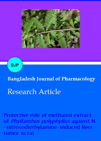
Phyllanthus polyphyllus Linn (Euphorbiaceae) is a deciduous shrub or small tree found mostly in hill areas of South India and Ceylon. Leaves are traditionally used for liver diseases by tribes of Kolli hills, Tamilnadu, India (Mathew, 1983). The phytochemical studies of the plant have revealed the presence of benzenoid, 4-0-methyl galic acid, together with three arylnapthalide lignans, namely phyllamyricin, justicidin B and diphyllin. Its extract shows dose-dependent inhibition
of inflammatory mediators such as LPS/INF-y stimulated by peritoneal exuded macrophages (Rao et al., 2006), monoacetylated triterpene arabinosides and terpenes found have cytotoxic activity against human cancer cell lines (Youkwan et al., 2005), antitumor activity against transplantable tumour, protective effect of human umbilical vein endothelial cells against glycated proteiniron chelate induced toxicity (Rajkapoor et al., 2007a, 2007b). The present study is aimed to evaluate the protective role of methanol extract of P. polyphyllus against N-nitrosodiethylamine-induced liver tumor in rats.
Materials and Methods
Chemicals
N-nitrosodiethylamine, bovine serum albumin, 2, 4, 6-trinitrobenzene sulfonate were obtained from Sigma Chemical Co., USA. All other chemicals used were of analytical grade obtained from Sisco Research Laboratories (India) and Glaxo Laboratories (India).
Plant materials
P. polyphyllus (euphorbiaceae) collected in the month of November 2009 from kolli hills, Tamilnadu, India and identified by Botanical survey of India, Coimbatore, Tamilnadu, India. A voucher specimen (PP-03) has been kept in our laboratory for future reference.
Preparation of extract
The leaves of P. polyphyllus were dried under shade and then powdered with a mechanical grinder. The powder was passed through sieve No 40 and treated with petroleum ether for dewaxing as well as to remove chlorophyll and it was later packed into soxhlet apparatus with methanol and subjected to hot continuous percolation. After the completion of extraction, it was filtered and the solvent was removed by distillation under reduced pressure. The extract was stored in desiccator.
Animals
Healthy male Wistar rats (6-8 weeks old) were used throughout the study. The animals were purchased from Sri Venkateswara enterprises, Bangalore, India and maintained in a controlled environmental condition of temperature (23 ± 2°C) and relative humidity (50-70%) on alternatively 12 hours light/dark cycles. All animals were fed standard pellet diet (Gold Mohor rat feed, Hindustan Lever Ltd., Mumbai) and water ad libitum.
Experimental protocol
The rats were divided into 4 groups, each group consisting of six animals. The rats of control group were treated with distilled water orally for 20 weeks. Liver tumor was induced in rest of the three groups by single intraperitoneal injection of N-nitrosodiethylamine at a dose of 200 mg/kg body weight in saline. Two weeks after the N-nitrosodiethylamine administration, the carcinogenic effect was promoted by 0.05% phenobarbitol (Yoshiji, et al., 1991), which was supplemented to the experimental animals through drinking water for up to 20 successive weeks. Third group of animals also treated with methanol extract (200 mg/kg body weight, dissolved in 0.3%carboxymethyl cellulose) simultaneously for 20 weeks from the first dose of N-nitrosodiethylamine and the fourth group were treated with methanol extract (400 mg/kg body weight, dissolved in 0.3% carboxymethyl cellulose) simultaneously for 20 weeks from the first dose of N-nitrosodiethylamine. At the end of experiments, animals were fasted overnight and were killed by cervical decapitation. Blood was collected and serum separated out. The liver were immediately removed and suspended in ice cold saline.
Measurement of phase I enzymes
Cytochrome P450 and cytochrome b5 was measured by the method of Omura and Sato (1964). The activity of NADPH-cytochrome P450 reductase was assayed by the method of Williams and Kamin (1962).
Measurement of phase II enzymes
The UDP-Glucuronyl transferase was estimated by the modified method of Hollman and Touster (1962). Glutathione-S-transferase was assayed by the method of Habig et al. (1974).
Measurement of glycoproteins
Hexose level was estimated by the method of Niebes (1972). Hexosamine content was estimated by the method of Wagner (1979). Sialic acid level was determined by the method of Warren (1957).
Statistical analysis
The values were expressed as mean ± SEM. Statistical analysis was performed by one-way analysis of variance (ANOVA) followed by Tukey multiple comparison tests. P values <0.05 were considered as significant.
Result and Discussion
There were significant (p<0.001) increase in the activities of cytochrome P450, cytochrome b5 and NADPH cytochrome C reductase in cancer bearing rats when compared with control rats (Table I). Methanol extract of P. polyphyllus treatment resulted in a significant (p<0.001) decrease in these enzymes on dose-dependent manner.
Table I: Effect of methanol extract of Phyllanthus polyphyllus on phase I enzymes in liver of rats
| Treatment | n | Cytochrome p450 (moles/ min/mg protein) | Cytochrome b5 (moles/ min/mg protein) | NADPH cytochrome C reductase (nmole of cytochrome C reduced/ min/mg protein) |
|---|---|---|---|---|
| Control | 6 | 0.7 ± 0.0 | 0.4 ± 0.0 | 110.4 ± 1.7 |
| Tumor bearing rat | 6 | 1.0 ± 0.0a | 0.8 ± 0.0a | 187.8 ± 2.4a |
| Extract (200 mg/kg) | 6 | 0.9 ± 0.0a,b | 0.6 ± 0.0a,b | 156.3 ± 1.6a,b |
| Extract (400 mg/kg) | 6 | 0.7 ± 0.0a,b | 0.5 ± 0.0a,b | 133.2 ± 2.4a,b |
| Data are mean ± SEM; ap<0.001 vs control; bp<0.001 vs tumor bearing animals; data were analyzed by one-way ANOVA followed by Tukey Kramer multiple comparison test | ||||
Table II represents the effect of MPP on the activities of UDP-GT, QR and GST enzymes in liver of control and experimental animals. Detoxification enzyme levels were significantly lowered in cancer bearing rats (p<0.001). Methanol extract of P. polyphyllus (200 and 400 mg/kg) treatment caused a significant (p<0.001) increases in the activities of these enzymes when compared with tumor bearing rats.
Table II: Effect of methanol extract of Phyllanthus polyphyllus on phase II enzymes in liver of rats
| Treatment | n | UDP-GT (Units/min/mg protein) | GST (pmoles o f CDNB conjugated / min/mg protein) | QR (nmol 2, 6- d ichlorophenol- indophenol induced min/ mg protein reduced) |
|---|---|---|---|---|
| Control | 6 | 1.1 ± 0.0 | 2.9 ± 0.1 | 202.2 ± 2.5 |
| Cancer bearing rat | 6 | 0.6 ± 0.0a | 1.8 ± 0.1a | 116.5 ± 1.6a |
| Extract (200 mg/kg) | 6 | 0.7 ± 0.0a,b | 2.1 ± 0.0a | 138.2 ± 1.9a,b |
| Extract (400 mg/ kg) | 6 | 1.0 ± 0.0a,b | 2.8 ± 0.0b | 187.6 ± 1.5a,b |
| Data are mean ± SEM; ap<0.001 vs control; bp<0.001 vs tumor bearing animals; data were analyzed by one-way ANOVA followed by Tukey Kramer multiple comparison test | ||||
The glycoproteins studied were hexose, hexosamine and sialic acid (Table III). These were found to rise (p<0.001) in the levels of all the three glycoproteins in cancer bearing animals, when compared with control. This rise was significantly decreased (p<0.001) by methanol extract treatment.
Table III: Effect of methanol extract of Phyllanthus polyphyllus on the levels of liver glycoproteins in rats
| Treatment | n | Hexose (mg/g of defatted tissue) | Hexoseamine (mg/g of defatted tissue) | Sialic acid (mg/g of defatted tissue) |
|---|---|---|---|---|
| Control | 6 | 1.4 ± 0.1 | 2.3 ± 0.1 | 1.9 ± 0.0 |
| Cancer bearing rat | 6 | 4.5 ± 0.3a | 3.9 ± 0.2a | 3.1 ± 0.1a |
| Extract (200 mg/kg) | 6 | 2.8 ± 0.2a,c | 2.9 ±0.1d | 2.6 ± 0.1a,c |
| Extract (400 mg/ kg) | 6 | 1.9 ± 0.3c | 2.6 ± 0.1c | 2.1 ± 0.0b,c |
| Data are mean ± SEM; ap<0.001; bp<0.05 vs control; Tukey Kramer multiple comparison test | ||||
In the present study, the activities of phase I enzymes like cytochrome P450, cytochrome b5 and NADPH cytochrome C reductase were increased considerably in the liver cancer animals compared to control animals. These activities were substantially altered in the methanol extract of P. polyphyllus (200 and 400 mg/kg).
Glutathione S-transferase is a critical detoxification enzyme that functions primarily in conjugating functional P450 metabolites with endogenous ligand (GSH), favoring their elimination from the organism (Hartman and Shankel, 1990). There is persuasive evidence to support the induction of GST and protection against a wide spectrum of cytotoxic, mutagenic and carcinogenic chemicals (Reed, 1990). The specific activity was the sum of all its isoforms, because we used CDNB as a nonspecific substrate for GST. The protective effects of many naturally occurring chemopreventive agents against carcinogenesis have been ascribed to decreased bioavailability of potential DNA-damaging entities and their destruction into excretable metabolites, facilitated through the induction of GST.
We observed a striking increase in UDP-GT, QR and GST activity. Hepatic microsomal UDP-GT, QR and GST are known to be important preneoplastic and neoplastic markers to evaluate the extent of free radical damage caused by exposure to various carcinogens (Thirunavukkarasu et al., 2002). The probable mechanism of this is that methanol extract arrests the formation of free radicals and the oxidative threat to the rats generated by exposure to N-nitrosodiethylamine. The results of the present study suggest that methanol extract could play an important role against N-nitrosodiethylamine-induced and PB-promoted hepatic carcinogenesis by protecting the glutathione metabolizing enzymes, phase I and phase II xenobiotic enzymes, and could be beneficial for cancer chemoprevention.
Glycoproteins are important groups of compounds involved in the cellular function. They play a significant role in contributing to the surface properties of the cells and also important role in tumorigenesis and as mediators of immunological specificity. Carbohydrates moieties of glycoproteins have also been implicated in the transport of metabolites across cell membranes and also observed a direct relationship between glycol-proteins and tumorigenesis (Thirunavukkarasu and Sakthisekaran, 2003). The biochemical markers, hexose, hexosamine and sialic acid, have been measured in the liver tissues. Many chemical changes in the host's lung and liver are detectable before the onset of secondary physiological and nutritional changes that may be associated with the condition of tumor-bearing host (Herzfeld and Greengard, 1997).
This study shows that methanol extract administration can inhibit glycoprotein synthesis in tumor cells. Our results were in accordance with the findings that 5-flurouracil an antitumor agent can reduce serum sialic acid in mice inoculated with transplantable hepatoma (Lu et al., 2000). The possible mechanism of MPP, especially in relation to antioxidant property is inhibition of free radical formation and reduced cancer incidence. It was found in the present study that MPP was more effective during the initiation of treatment. This may be due to the inhibitory action of methanol extract on the initiation of N-nitrosodiethylamine activation/detoxification process.
Conclusion
This study demonstrates that oral supplementation with methanol extract of P. polyphyllus can modulate phase I and II enzyme activities and protecting the glycoprotein levels in tissues.
Ethical Issue
The research has followed the national ethical standards for the care and use of laboratory animals and it was approved by the Institutional Animal Ethics Committee constituted for the purpose.
References
Habig WH, Pabst MJ, Jakoby WB. Glutathion S-transferase, the first enzymatic step in mercapturic acid formation. J Biol Chem. 1974; 249: 7130-39.
Hartman PE, Shankel DW. Antimutagens and anticarcinogens: A survey of putative interceptor molecules. Environ Mol Mutagen. 1990; 15: 145-82.
Herzfeld A, Greengard O. The effect of lymphoma and other neoplasms on hepatic and plasma enzymes of the host rat. Cancer Res. 1997; 37: 231-38.
Hollmann S, Touster O. Alterations in tissue levels of uridine diphosphate glucose dehydrogenase, uridine diphosphate glucuronic acid pyrophosphatase and glucuronyl transferase induced by substances influencing the production of ascorbic acid. Biochim Biophys Acta. 1962; 62: 338-52.
Jeena KJ, Joy KK, Kuttan R. Effect of Emblica officinalis, Phyllanthus amarus and Picrorrhiza kurroa on N-nitrosodiethylamine-induced hepatocarcinogenesis. Cancer Lett. 1999; 136: 11-16.
Lu CQ, Lu J, Wangb L, Cui Y. Changes in ganglioside contents, plasma sialic acid and cAMP levels in experimental hepatoma in mice. Mol Cell Biochem. 2000; 207: 29-33.
Mathew KM. The flora of the Tamilnadu Carnatic. Vol. 3. The Rapinet Herbarium, St. Joseph's College, Trichirapalli, 1983, p 1468.
Niebes P. Determination of enzymes and degradation products of glycosaminoglycan metabolism in the serum of healthy and varicose subjects. Clin Chim Acta. 1972; 42: 399-408.
Omura T, Sato R. The carbon monoxide-binding pigment of liver microsomes. Evidence for its hemoprotein nature. J Biol Chem. 1964; 239: 2370-78.
Peers PG, Linsell CA. Dietary aflatoxins and liver cancer: A population based study in Kenya. Br J Cancer. 1973; 27: 473Â84.
Rajkapoor B, Venugopal R, Sakthisekaran D, Zhang HQ, Nishigaki I. Protective effect of Phyllanthus polyphyllus on human umbilical vein endothelial cells against glycated protein-iron chelate induced toxicity. J Med Sci. 2007; 7: 961Â68.
Rajkapoor B, Sankari M, Sumithra M, Anbu J, Harikrishnan N, Gobinath M, Suba V, Balaji R. Antitumor and cytotoxic effects of Phyllanthus polyphyllus on Ehrlich ascites carcinoma and human cancer cell lines. Biosci Biotechnol Biochem. 2007; 71: 2177-83.
Rao YK, Fang SH, Tzeng YM. Anti-inflammatory activities of constituents isolated from Phylanthus polyphyllus. J Ethnopharmacol. 2006; 103: 181-86.
Reed DJ. Glutathione: Toxicological implications. Ann Rev Pharmacol Toxicol. 1990; 30: 603-31.
Thirunavukkarasu C, Prince Vijeya Singh J, Thangavel M, Selvendiran K, Sakthisekaran D. Dietary influence of selenium on the incidence of N-nitrosodiethylamine-induced hepatoma with reference to drug and glutathione metabolizing enzymes. Cell Biochem Funct. 2002; 20: 347-56.
Thirunavukkarasu C, Sakthisekaran D. Influence of sodium selenite on glycoprotein contents in normal and N- nitrosodiethylamine initiated and phenobarbital promoted rat liver tumors. Pharmacol Res. 2003; 48: 167-71.
Williams CH Jr, Kamin H. Microsomal triphosphopyridine nucleotide-Cytochrome c reductase of liver. J Biol Chem. 1962; 237: 587-95.
Wagner WD. A more sensitive assay discriminating galactosamine and glucosamine in mixtures. Anal Biochem. 1979; 94: 394-96.
Warren L. The thiobarbituric acid assay of sialic acids. J Biol Chem. 1959; 234: 1971-75.
Yoshiji H, Nakae D, Kinugasa T, Matsuzaki M, Denda A, Suiji T. Inhibitory effect of the iron deficiency on the induction of putative preneoplastic foci rat liver initiated with diethylnitrosamine and promoted by phenobarbital. Br J Cancer. 1991; 64: 939-82.
Youkwan J, Srisomphot P, Sutthivaiyakit S. Bioactive constituents of the leaves of Phyllanthus polyphyllus. J Nat Prod. 2005; 68: 1006-09.