Cardioprotective activity of ethanolic extract of Callistemon lanceolatus leaves on doxorubicin-induced cardiomyopathy in rats
Abstract
The cardioprotective effect of ethanol extract of Callistemon lanceolatus leaves (100 and 200 mg/kg) against doxorubicin-induced cardiomyopathy was studied. Rats were pretreated with the extract for 7 days and simultaneous treatment with doxorubicin along with the extracts for the next 14 days. After 24 hours of last dose of doxorubicin, ECG, invasive blood pressure, serum markers (creatine kinase-MB, lactate dehydrogenase, serum transaminases), tissue antioxidant markers (catalase, superoxide dismutase) and extent of lipid peroxidation (malondialdehyde) were studied. The elevated ST segment, decreased blood pressure, increased level of serum enzymes and decreased level of tissue antioxidant markers were observed in doxorubicin treatment (p<0.01). While 200 mg/kg extract significantly reduced the elevated levels of the serum enzymes and restores the ECG and blood pressure to normal, also significantly increased the tissue antioxidant levels, while decreased the malondialdehyde level (p<0.01) when compared with the control. The histopathological study confirmed the cardioprotection.
Introduction
Doxorubicin-induced myocardial injury has been believed to be mediated through different mechanisms. A common denominator to most of the proposed mechanisms is the formation of an iron-anthracycline complex that generates free radicals, which in turn, causes severe damage to the plasma membrane, and interferes with the cytoskeletal structure (Billingham et al., 1977). Oxygen free radical formation by doxorubicin (Chopra et al., 1995; Van Acker et al., 1996) enhances the susceptibility of cardiac tissue to lipid peroxidation leading to a progressive dose-related irreversible loss of myofibrils, dilation of the sarcoplasmic reticulum, cytoplasmic vacuolization, swelling of mitochondria, increased number of lysosomes, myocyte necrosis (Minotti et al., 2004), inhibition of nucleic acid as well as protein synthesis (Monti et al., 1995), release of vasoactive amines (Bristo et al., 1980), change in adrenergic function (Singal et al., 1998), decreased activity of Na+K+ATPase (Geetha and Devi, 1992), alteration in sarcoplasmic calcium transport and imbalance of myocardial electrolytes in response to the doxorubicin (Siveski-Iliskovic et al., 1994).
In vivo, doxorubicin is converted to semiquinone by mitochondrial, lysosomal and cystolic enzymes. Semiquinone is a charged moiety that readily donates an electron to an oxygen molecule, resulting in generation of an oxygen free radical or superoxide ion/hydroxyl radicals. Although superoxide ion can cause tissue damage, these are not very reactive. Superoxide can be converted further to hydrogen hydroxide catalyzed by the action of superoxide dismutase (Jain, 2000).
Callistemon lanceolatus (Family: Myrtaceae) commonly known as bottle brush because of its crimson red colored spikes, is frequently cultivated throughout India in gardens as ornamental plants (Sharma et al., 2006; Anonymous, 2006). It is indigenous to Australia. The Lambadi tribals of north telangana districts of Andhra Pradesh use this plant for the treatment of pain, gastrointestinal disorders and infectious diseases (Sudhakar et al., 2004). Aqueous extracts of the leaves and flowers have antifungal and antibacterial activity. The plant extracts inhibit urd bean leaf crinkle virus in vitro. The extract also shows cholinesterase activity (Chowdhary and Saha, 1985; Gupta and Gupta, 1997). The ethanolic extract of C. lanceolatus showed strong elastase inhibition and DPPH radical scavenging activities (Kim et al., 2009). The essential oils from leaves possess antimicrobial, fungitoxic, antinociceptive and anti–inflammatory activities. Several triterpenoids, flavonoides, fatty acids, tannins and phenolic compounds have been isolated from its leaves (Sudhakar et al., 2004; Jeong et al., 2009). The leaves was also reported for its hepatoprotective effect (Jain et al., 2007), antidiabetic, hypolipidemic and antioxidant activity (Kumar et al., 2011). The fruit of C. lanceolatus was reported for its calcium channel blocking activity (Ali et al., 2011). The present work was undertaken to study the cardioprotective activity of ethanol extract of C. lanceolatus leaves on doxorubicin-induced cardiac toxicity.
Materials and Methods
Plant material
The leaves of C. lanceolatus were collected in the month of August 2010 from local area of Sangli, Maharashtra and were authenticated by Dr. U.S. Yadav, Department of Botany, Willingdon College, Sangli. The herbarium has been submitted for further reference (V. No. WC/2010/125). The plant material was shade dried at room temperature.
Preparation of extract
The dried leaves of C. lanceolatus were grinded and made into a coarse powder. The coarse powder was then subjected to continuous hot extraction in soxhlet apparatus with ethanol as a solvent and extracted till the solvent becomes colorless. The extract was evaporated under reduced pressure using rotary evaporator (Equitron Roteva) at a low temperature of 45ºC until the extract turns syrupy and then the syrupy extract is transferred to an evaporating dish for drying at room temperature. The yield was found to be 19.6% w/w.
Preliminary phytochemical investigation
The extract was subjected to chemical tests qualitatively for the identification of different phytoconstituents like glycosides, saponins, carbohydrates, sterols, alkaloids, flavonoids, tannins, proteins, triterpenoids (Khandelwal, 2004).
Animals
Albino Wistar rats of 150-250 g of either sex were used for the study. The inbred species of rats were obtained from animal house of Appasaheb Birnale College of Pharmacy, Sangli for experimental purpose. The animals were maintained under controlled conditions of temperature (23 ± 2ºC) before the study. The animals were randomized into experimental, normal and control groups, housed individually in sanitized polypropylene cages containing sterile paddy husk as bedding. They had free access to standard pellets as basal diet and water ad libitum. Animals were habituated to laboratory conditions for 48 hours prior to experimental protocol to minimize if any of non-specific stress.
Toxicity studies
Acute toxicity study was carried using rats by up and down/staircase method as per OECD guidelines. The ethanol extract of C. lanceolatus was orally administered to different groups of rats at the doses of 50, 300, 1,000, 2,000 and 3,000 mg/kg body weight respectively. The animals were observed for 48 hours to study the general behavior and for any sign of discomfort to the animals. The mortality was found at dose 3,000 mg/kg. Hence the dose of 2,000 mg/kg was selected and the 1/10th dose ie. 200 mg/kg and 1/20th dose i.e., 100 mg/kg was used to study the cardioprotective activity.
Chemicals
Sodium carboxymethyl cellulose, thiobarbituric acid, heparin, ketamine (Neon Labs, India), xlyazine (Indian Immunologicals, India), doxorubicin (Cipla, India), sodium hydroxide, pyridine, n-butanol, pyrogallol, anesthetic ether etc. the enzyme kits used were creatine kinase-MB (CK-MB; Pathozyme, India), lactate dehydrogenase (Teco Diagnostics, India), SGOT (Span Diagnostics Ltd., India), SGPT (Span Diagnostics Ltd., India), total protein (Pathozyme, India) etc, all the chemicals used was of analytical grade.
Doxorubicin-induced cardiac stress
The rats were divided into four groups of six animals each. Group 1: Rats treated with 1% sodium carboxymethyl cellulose (~2 mL/Kg/day, per oral); Group 2: Rats treated with doxorubicin with total cumulative dose of 15 mg/kg i.p for 2 weeks in six divided dosage; Group 3: Rats pretreated with ethanol extract of C. lanceolatus 100 mg/Kg per oral along with doxorubicin treatment; Group 4: Rats pretreated with ethanol extract of C. lanceolatus 200 mg/Kg per oral along with doxorubicin treatment. Group 2, 3 and 4 received the treatment of doxorubicin at alternate days for a period of 2 weeks (the days selected for doxorubicin injection was on the 8th, 10th, 14th, 16th, 18th, and 21st day after the 7 days pretreatment with the extract.
On the 22nd day parameters studied were general appearance, heart weight and heart/body weight ratio. Electrocardiography (ECG) was recorded at the end of the treatment after the last dosing of doxorubicin. All rats were fasted overnight but had free access to water after the last dose administration. Biopac MP-35 (Santa Barbara, USA) was used to record and monitor ECG tracings. Rats from each group were anesthetized with ketamine-xylazine anesthesia, a needle electrodes were inserted under the skin for the limb lead at position II. For each ECG tracing P wave, QRS complex, QT interval, RR interval and cardiac cycle were measured.
Blood pressure
Blood pressure was determined by invasive method (carotid artery cannulation) by using BIOPAC MP-35. Carotid artery was cannulated by using PE-50 tube which was attached to the pressure transducer loaded previously with the heparinized saline. Various parameters such as systolic, diastolic, mean blood pressure and heart rate were determined.
Biochemical parameters
Soon after the blood pressure had been recorded, the blood was collected from the retroorbital route and subjected to centrifugation to isolate serum out of it. The serum was further used for estimation of CK-MB by immunoinhibition method, lactate dehydrogenase by kinetic method, SGOT/aspartate aminotransferase (AST) by 2,4-DNPH method and SGPT/alanine aminotransferase by 2,4-DNPH method. Heart tissue homogenate was prepared in 0.05 M phosphate buffer pH 7.4 and homogenated in tissue homogenizer at 2,000 rpm for 10 min. Antioxidant enzymes estimated were catalase (CAT) (Aebi, 1974), superoxide dismutase (SOD) (Marklund, 1985) and extent of lipid peroxidation malondialdehyde (Okhawa et al., 1979).
Histopathological study
At the end of study, the heart was isolated, washed with ice cold saline. The tissue was fixed in 10% buffered neutral formalin solution. After fixation tissues were embedded in paraffin-wax and sections were cut and stained with hematoxylin and eosin. The slides were observed under light microscope.
Statistical analysis
Statistical analysis was performed using one-way ANOVA followed by Dunnett t test (GraphPad Instat, USA).
Results
Phytochemical investigation revealed the presence of steroids, tannins, flavonoids, phenolic compounds and alkaloids (data not shown).
In doxorubicin-treated group, the animal fur became scruffy and developed a yellowish to reddish tinge. These rats had red exudates around the eyes and nose, soft watery feces, presence of peritoneal fluid, necrosis at the site of injection. Animals were sicker, lethargic and enlargement of abdomen and liver when compared with the normal. These observations were markedly reduced in the group treated with ethanol extract of C. lanceolatus leaves (200 mg/kg), when compared with the doxorubicin control group.
In doxorubicin-treated rat, decrease in body weight, heart weight and heart/body weight ratio were seen at the end of the study. Extract (200 mg/kg) demonstrated significant increase in body weight gain, increase in weight of heart (p<0.01) and increase in heart/body weight ratio (p<0.01), when compared with doxorubicin control group (Table I).
Table I: Body weight, heart weight and heart/body weight ratio
| Groups | Initial body weight (g) | Final body weight (g) | Heart weight (g) | Heart/body weight ratio (x10-3) |
|---|---|---|---|---|
| Normal | 221.7 (1.2) | 227.2 (1.6) | 1.0 (0.0) | 4.5 (0.0) |
| Control DOX treated | 219.6 (1.5) | 199 (1.7) | 0.8 (0.0)a | 3.8 (0.0)a |
| DOX + extract 100 mg/Kg | 226.8 (1.8) | 210.5 (1.4) | 0.9 (0.0)b | 4.2 (0.1)c |
| DOX + extract 200 mg/Kg | 225 (1.3) | 215.4 (1.4) | 0.9 (0.0)c | 4.3 (0.0)c |
| DOX means doxorubicin; Data are mean (SEM); n = 6; ap<0.01, when compared with normal; bp<0.05, when compared with control group; cp<0.01, when compared with control group using one-way ANOVA coupled with "Dunnett's t test" | ||||
The rats treated with doxorubicin showed a decrease in the systolic, diastolic, mean blood pressure and heart rate. Pretreatment with the extract (100 mg and 200 mg/kg) showed a significant increase in the systolic blood pressure (p<0.01), diastolic blood pressure (p<0.01), mean blood pressure (p<0.01) and the heart rate (p<0.01) respectively, when compared with the control group (Table II).
Table II: Effect of ethanol extract of Callistemon lanceolatus leaves on blood pressure of rats by doxorubicin-induced cardiac toxicity
| Groups | Blood pressure (mm Hg) | Blood pressure (mm Hg) | Blood pressure (mm Hg) | Heart rate | |
|---|---|---|---|---|---|
| Groups | Systolic | Diastolic | Mean | Heart rate | |
| Normal | 123.89 (0.79) | 119.2 (0.27) | 112.11 (0.4) | 272.31 (1.25) | |
| Control DOX treated | 74.16 (0.22)a | 69.35 (0.27)a | 72.07 (0.28)a | 210.55 (1.79)a | |
| DOX + extract 100 mg/Kg | 118.47 (0.57)b | 110.93 (0.49)b | 116.03 (0.40)b | 253.42 (1.56)b | |
| DOX + extract 200 mg/Kg | 113.69 (0.38)b | 105.96 (0.58)b | 110.61 (0.38)b | 270.29 (1.48)b | |
| DOX means doxorubicin; Data as mean (SEM); n=6; heart rate is expressed as rate per minute; ap<0.01, when compared with normal group; bp<0.01, when compared with control group using one way ANOVA coupled with "Dunnett t test" | |||||
Figure 1 shows the ECG pattern of normal, control and extract-treated rats (100 and 200 mg/kg). Normal group showed a normal ECG pattern, whereas rats treated with doxorubicin alone showed significant elevation in ST segment, prolongation in P wave, QRS complex and R-R interval. In addition, there was decreased in cardiac cycles and prolongation of QT interval as compared to normal rats. Treatment with extract 200 mg/Kg for 21 days alternatively with doxorubicin-treated rats exhibited near to normal ECG pattern with a decreased elevation in ST segment.
Figure 1: Electrocardiogram changes (Upper tracing is normal; Second tracing is control DOX treated; Third tracing is DOX and extract 100 mg/kg; Lower tracing is DOX and extract 200 mg/kg)
Furthermore, treatment also resulted in decrease in P wave (extract-200 mg/kg, p<0.01), QRS complex (extract-200 mg/kg, p<0.01), QT interval (extract- 100 and 200 mg/Kg, p<0.01) and R-R interval (extract-100 and 200 mg/Kg, p<0.01), whereas cardiac cycle was increased (extract- 200 mg/kg, p<0.01) when compared with the control (Table III).
Table III: Effect of ethanolic extract of Callistemon lanceolatus leaves on ECG changes of rats by doxorubicin-induced cardiac toxicity
| Groups | P wave | QRS complex | QT interval | RR interval | Cardiac cycle |
|---|---|---|---|---|---|
| Normal | 0.023 (0.001) | 0.033 (0.002) | 0.068 (0.001) | 0.199 (0.002) | 0.144 (0.006) |
| Control DOX treated | 0.0425 (0.001)a | 0.0433 (0.003)a | 0.1092 (0.002)a | 0.33 (0.010)a | 0.105 (0.002)a |
| DOX + extract 100 mg/kg | 0.037 (0.018)b | 0.035 (0.002)b | 0.094 (0.003)c | 0.238 (0.004)c | 0.124 (0.005)b |
| DOX + extract 200 mg/kg | 0.033 (0.002)c | 0.031 (0.002)c | 0.078 (0.004)c | 0.219 (0.002)c | 0.136 (0.003)c |
| The ECG parameters are expressed in seconds (sec.). Data are mean ± SEM; n = 6; ap<0.01, when compared with normal group; bp<0.05, when compared with control group; cp<0.01, when compared with control group using one way ANOVA coupled with "Dunnett's t test" | |||||
Table IV shows the effect of ethanol extract on the level of CK–MB, lactate dehydrogenase, SGOT and SGPT enzymes. Treatment with DOX causes an elevation in level of these enzymes which are considered as the selective biomarkers of myocardial damage (except SGPT) when compared with the normal. Our study showed decrease in the elevated levels of these enzymes. Pretreatment with extract 200 mg/kg showed significant decrease in CK-MB (p<0.01), lactate dehydrogenase (p<0.01) and SGPT (p<0.01), while extract 100 and 200 mg/kg showed a significant decrease in the level of SGOT (p<0.01).
Table IV: Effect of ethanolic extract of Callistemon lanceolatus leaves on CK-MB, LDH, SGOT and SGPT levels in rats by doxorubicin-induced cardiac toxicity
| Groups | CK-MB (U/L) | LDH (IU/L) | SGOT (IU/L) | SGPT (IU/L) |
|---|---|---|---|---|
| Normal | 44.9 (0.9) | 73.9 (0.4) | 41.2 (0.2) | 20.6 (0.1) |
| Control DOX treated | 164.6 (0.4)a | 155.5 (0.8)a | 186.1 (0.8)a | 43.4 (0.3)a |
| DOX + extract 100 mg/Kg | 161.9 (0.9)b | 153 (0.7)b | 163.6 (0.4)b | 41.4 (0.8)b |
| DOX + extract 200 mg/Kg | 132.4 (0.6)c | 136.3 (0.6)c | 124.3 (0.2)c | 33.6 (0.2)c |
| Data are mean ± SEM; n = 6; ap<0.01, when compared with normal group; bp<0.01, when compared with control group; cp<0.01, when compared with control group using one way ANOVA coupled with "Dunnett's t test" | ||||
Doxorubicin caused a decrease in the level of endogenous antioxidant reserves viz. SOD and CAT and shows an increased in the lipid peroxidation of the heart when compared with the normal. Pretreatment with extract 100 and 200 mg/kg showed a significant increased in the SOD (p<0.01) and CAT (p<0.01). While significantly decreased the level of malondialdehyde (p<0.01), when compared with the control (Table V).
Table V: Antioxidant enzymes and lipid peroxidation of heart tissue homogenate of rats
| Groups | CAT (U/mg protein) | SOD (U/ mg protein) | MDA (umole/ mg protein) |
|---|---|---|---|
| Normal | 32.9 (0.5) | 2.3 (0.1) | 0.9 (0.0) |
| Control DOX treated | 15.8 (0.3)a | 0.6 (0.1)a | 2.9 (0.0)a |
| DOX + extract 100 mg/Kg | 19.2 (0.3)b | 1.3 (0.1)b | 2.1 (0.0)b |
| DOX + extract 200 mg/Kg | 24.2 (0.2)b | 1.9 (0.1)b | 1.6 (0.0)b |
| Data are mean ± SEM; n = 6; ap<0.01, when compared with normal group; bp<0.01, when compared with control group using one-way ANOVA coupled with "Dunnett's t test" | |||
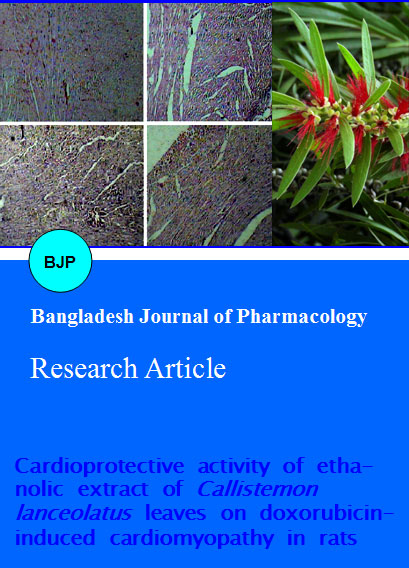
Histopathological examination of myocardial tissue obtained from normal animals exhibited clear integrity of myocardial membrane. Normal rats showed normal cardiac fibers without any damage. The heart sections obtained from doxorubicin-treated animals showed abundant areas of necrosis and aggregation of acute inflammatory cells and damaged vascular spaces. Animals pretreated with extract 200 mg/kg showed improvement in the cell integrity evidenced by absence of necrosis, marked decrease in infiltration of inflammatory cells and maintenance of normal integrity of the cardiac muscles (Figure 2).
Figure 2: Histopathological images of heart pretreated with ethanol extract of Callistemon lanceolatus leaves by doxorubicin induced cardiac toxicity. (A) Normal, (B) Control, (C) extract: 100 mg/kg, (D) extract: 200 mg/kg
Discussion
The ethanol extract of C. lanceolatus has shown significant cardioprotection by doxorubicin-induced cardiac toxicity. Repeated administration of doxorubicin beyond a certain dose has been shown to cause cardiopathic changes in patients (Steinhertz et al., 1991) as well as in a variety to animal species (Van Acker et al., 1996). In this study, we utilized a rat model of doxorubicin-induced cardiotoxicity and it has been shown that these animals have scruffy fur as well as significantly decreased body weight, heart weight, heart/body weight ratio (You et al., 2006). The decrease in body weight in this study is in accordance with other studies (Herman et al., 2000; Hoekman et al., 1999) and it may be attributed to reduced food intake and inhibition of protein synthesis due to doxorubicin. The ethanol extract demonstrated increase in the body weight gain along with the gain in weight of heart and the heart/body weight ratio.
Doxorubicin treatment causes prolongation of P wave, QRS complex, QT interval and RR interval while reduces cardiac cycle (Rossi et al., 1994; Danesi et al., 1986). Results of this study showed that, in comparison to the group treated with doxorubicin alone, rats pretreated with extract have nearly normal ECG data and significantly prevented ST segment prolongation throughout the study period.
Doxorubicin causes an decrease in the systolic, diastolic, mean blood pressure and heart rate (Kalender et al., 2010). This is probable due to effect of doxorubicin on the myofibrils, causes its disruption (Weinberg and Singal, 1987). Hence the systolic, diastolic, mean blood pressure and heart rate decrease, while the extract restores the systolic, diastolic, mean blood pressure and heart rate near to normal.
Adriamycin administration produced significant elevation of CK–MB (Wakade et al., 2008), lactate dehydrogenase (Garba and Ubom, 2005), SGOT (Dewar et al., 1958) and SGPT (Saad et al., 2001). Treatment with extract demonstrated a significant decrease in the elevated levels of these enzymes.
Oxidative stress and mitochondrial dysfunction are associated with disease and toxic process. It results from over production of reactive oxygen species, often leading to peroxidation of membrane phospholipids and production of reactive aldehydes (Hanasaki et al., 1994; Ferrari et al, 1996; Korshunov et al., 1997 and Morin et al., 2001).
The antioxidant enzymes viz. SOD and CAT of heart upon doxorubicin treatment has shown a significant decrease while the extent of lipid peroxidation; malondialdehyde increases, when compared with the normal group. Pretreatment with ethanol extract reflected an significant increase in the SOD and CAT while causes a decrease in the malondialdehyde level in a dose-dependant manner. This proves the prevention of oxidative damage by C. lanceolatus.
Histopathological examination of myocardial tissue obtained from normal animal exhibited clear integrity of myocardial membrane. Normal rats showed cardiac fibers without any infarction. The heart sections obtained from doxorubicin-treated animals showed disruption of several subcellular elements including loss of myofibrils, swelling of mitochondria, vacuolization of the cytoplasm, formation of lysosomal bodies and dilation of the sarcotubur and dilation of the sarcotubular system (Olson and Young, 1974; Young and Myers, 1977). Treatment with extract 200 mg/kg restores the architecture of the heart near to normal when compared with the control group.
The preliminary phytochemical investigation also revealed the presence of steroids, terpenoids, saponins, fatty acids, flavonoids, phenolic compounds, alkaloids and carbohydrates. This phytochemical might be responsible to blunt the oxidative stress produced by the doxorubicin which is a big factor for causing the alterations in the normal physiology of heart and other body tissues.
Conclusion
C. lanceolatus shows a potent cardioprotective effect in rats by doxorubicin-induced cardiotoxicity.
Ethical Issue
All the studies conducted were approved by the Institutional Animal Ethical Committee (IAEC) of Appasaheb Birnale College of Pharmacy, Sangli, Maharashtra (Registration No. 843/AC/04 /CPCSEA), India.
Acknowledgements
The authors extends sincere thanks to the Principal D. D. Choughule for providing excellent laboratory facilities and last but not the least Ms. S. C. Shaikh for her help during the work.
References
Aebi H. Catalase. In: Methods of enzymatic analysis. Bergmeyer HU (ed). Verlag, Chemic Academic Press Inc, 1974, pp 673-85.
Ali N, Syed WAS, Ahmad B. Calcium channel blocking activity of fruits of Callistemon citrinus. J Chem Soc Pak. 2011; 33: 245-48.
Anonymous. The wealth of India raw material. New Delhi, Council of Scientific & Industrial Research, 1992, pp 63-65.
Billingham ME, Bristow MR, Glatstein E, Spina A, Tacconi MT, Veroni E. Adriamycin cardiotoxicity: Endomyocardial evidence of enhancement by irradiation. Am J Surg Pathol. 1977; 1: 17-23.
Bristo MR, Sageman WS, Scott RH. Acute and chronic cardiovascular effects of doxorubicin in dog. J Cardiovasc Pharmacol. 1980; 2: 487-515.
Chopra S, Pillai KK, Husain SZ, Girl DK. Propolis Protects against doxorubicin-induced myocardiopathy in rats. Exp Mol Pathol. 1995; 62: 190-98.
Chowdhary AK, Saha NK. Inhibition of urd bean leaf crinkle virus by different plant extracts. Indian Phytopathol. 1985; 38: 566-68.
Danesi R, Del Tacca M, Soldani G. Measurement of the ST segment as the most reliable electrocardiogram parameter for the assessment of adriamycin-induced cardiotoxicity in the rat. J Pharmacol Methods. 1986; 6: 251-59.
Dewar HA, Rowell NR, Smith AJ. Serum glutamic oxalacetic transaminase in acute myocardial infarction. Br Med J. 1958; 8: 1121-25.
Ferrari R. The role of mitochondria in ischemic heart disease. J Cardiovasc Pharmacol. 1996; 28: S1-S10.
Garba IH, Ubom GA. Total serum lactate dehydrogenase activity in acute Plasmodium falciparum malaria infection. Singapore Med J. 2005; 46: 632-34.
Geetha A, Devi CS. Effect of doxorubicin on heart mitochondrial enzymes in rats: A protective role for alphatocopherol. Indian J Exp Biol. 1992; 30: 615-18.
Gupta A, Gupta R. A survey of plants for anticholinesterase activity. Phytochemistry 1997; 46: 827-31.
Hanasaki Y, Ogawa S, Fukui S. The correlation between active oxygen scavenging and antioxidative effects of flavonoids. Free Radical Biol Med. 1994; 16: 845-50.
Herman EH, Zhang J, Chadwick DP, Ferrans V. Comparison of the protective effects of amifostine and dexrazoxane against the toxicity of doxorubicin in spontaneously hypertensive rats. Cancer Chemother Pharmacol. 2000; 45: 329-34.
Hoekman K, Van der WJF, Vermorken JB. Clinical and preclinical modulation of chemotherapy-induced toxicity in patients with cancer. Drugs 1999; 57: 133-56.
Jain AK, Dubey SK, Sikarwar MS, Jain SK. Hepatoprotective activity of methanolic extract of leaves of Callistemon lanceolatus. Internat J Plant Sci. 2007; 2: 185-86.
Jain D. Cardiotoxicity of doxorubicin and other anthracycline derivatives. J Nuclear Cardiolog. 2000; 7: 53-62.
Jeong W, Su HS, Kim N, Yang YT, Su SY, Lee C, Hwang BY, Lee D. Bioactive triterpenoids from Callistemon lanceolatus. Arch Pharm Res. 2009; 32: 845-49.
Kalender O, Eylem T, Nurcan D. Protective effect of carnosine on adriamycin-induced oxidative heart damage in rats. Anadolu Kardiyol Derg. 2010; 11: 3–10.
Khandelwal KR. Practical pharmacognosy, techniques and experiments. 12th ed. Pune, Nirali Prakashan, 2004, pp 152-56.
Kim JH, Byun JC, Bandi AKR, Hyun CG, Lee NH. Compounds with elastase inhibition and free radical scavenging activities from Callistemon lanceolatus. J Med Plants Res. 2009; 3: 914-20.
Korshunov SS, Skulachev VP, Starkov AA. High protonic potential actuates a mechanism of production of reactive oxygen species in mitochondria. FEBS Lett. 1997; 416: 15-18.
Kumar S, Kumar V, Prakash O. Antidiabetic, hypolipidemic and antioxidant activities of Callistemon lanceolatus leaves extract. J Herbs Spices Med Plants. 2011; 17: 144-53.
Marklund S. Pyrogallol autooxidation. In: Handbook of methods for oxygen radical research. Greenwald RA (ed). Boca Raton, CRC Press, 1985, pp 243-47.
Minotti G, Menna P, Salvatorelli E, Cairo G, Gianni L. Anthracyclines: Molecular advances and pharmacologic developments in antitumor activity and cardiotoxicity. Pharmacol Res. 2004; 56: 185-229.
Monti EE, Prosperi R, Supino Bottiroli G. Free radical-dependant DNA lesions are involved in the delayed cardiotoxicity-induced by adriamycin in the rat. Anticancer Res. 1995; 15: 193-97.
Morin D, Barthelemy S, Zini R, Labidalle S, Tillement JP. Curcumin induces the mitochondrial permeability transition pore mediated by membrane protein thiol oxidation. FEBS Lett. 2001; 495: 131-36.
Okhawa H, Ohishi N, Yaki K. Assay for lipid peroxidation in animal tissues by thiobarbituric acid reaction. Anal Biochem. 1979; 95: 351-58.
Olson HM, Young DM. Electrolyte and morphologic alterations of myocardium in adriamycin treated rabbits. Am J Pathol. 1974; 77: 439-54.
Rossi F, Filippelli W, Russo S, Filippelli A, Berrino L. Cardio-toxicity of doxorubicin: Effects of drugs inhibiting the release of vasoactive substances. Pharmacol Toxicol. 1994; 75: 99-107.
Saad SY, Najjar TA, AL-Rikabi AC. The preventive role of deferoxamine against acute doxorubicin-induced cardiac, renal and hepatic toxicity in rats. Pharmacol Res. 2001; 43: 211-18.
Sharma RK, Kotoky R, Bhattacharyya PR. Volatile oil from the leaves of Callistemon lanceolatus D. C. grown in north-eastern India. Flavour Fragr J. 2006; 21: 239-40.
Singal PK, Iliskovic N. Doxorubicin-induced cardiomyopathy. N Engl J Med. 1998; 339: 900-05.
Siveski-Iliskovic N, Kaul N, Singal PK. Probucol promotes endogeneous antioxidants and provides protection against adriamycin-induced cardiacmyopathy in rats. Circulation 1994; 89: 2829-35.
Steinhertz LJ, Steinhertz PG, Tan CTC, Heller G, Murphy L. Cardiac toxicity 4 to 20 years after completing anthracycline therapy. J Am Med Assoc. 1991; 266: 1672-77.
Sudhakar M, Rao CV, Rao AL, Raju DB. Antinociceptive and anti-inflammatory effects of the standardized oil of Indian Callistemon lanceolatus leaves in experimental animals. Acta Pharmaceutica Turcica. 2004; 46: 131-39.
Van Acker SABE, Kramer K, Voest EE, Grimbergen JA, Zhang J, Van der Vijg WJF, Bast A. Doxorubicin–induced cardiotoxicity monitored by ECG in freely moving mice. Cancer Chemother Pharmacol. 1996; 38: 95-101.
Wakade AS, Sha AS, Kulkarni MP, Juvekar AR. Protective effect of Piper longum L on oxidative stress induced injury and cellular abnormality in adriamycin-induced cardiotoxicity in rats. Indian J Exp Biol. 2008; 46: 528-33.
Weinberg LE, Singal PK. Refractory heart failure and age-related differences in adriamycin-induced myocardial changes in rats. Can J Physiol Pharmacol. 1987; 65: 1957-65.
You JS, Huang HF, Chang YL, Lee YS. Sheng-Mai-San reduces adriamycin–induced cardiomyopathy in Rats. Am J Chinese Med. 2006; 34: 295-305.
Young RC, Myers CE. Adriamycin: The role of lipid peroxidation in cardiac toxicity and tumor response. Science 1977; 19: 165-67.