Phytochemical screening and antibacterial activity of leaf and callus extracts of Centella asiatica
Abstract
The present study shows the phytochemical analysis and antibacterial activity of leaf and callus of Centella asiatica. Leaf explants of C. asiatica were cultured on MS medium supplemented with different concentration of plant growth regulators for callus initiation. The maximum percentage of callusing was achieved in medium supplemented with 6-benzylaminopurine 4.0 mg/L and 2,4-dichlorophenoxyacetic acid 2.0 mg/L. In the preliminary phytochemical screening, alkaloids, glycosides, terpenoids, steroids, flavonoids, tannins, saponins and reducing sugars were present in most of the tested extracts of leaf and in vitro grown callus of C. asiatica. Methanol, acetone, chloroform and water extracts of leaf and callus were evaluated for in vitro antibacterial activity against Bacillus cereus, Escherichia coli, Staphylococcus aureus and Pseudomonas aeruginosa by agar plate well diffusion method. All the extracts from leaf and callus of C. asiatica showed significant antibacterial activity against the tested organisms. However, methanol extracts of leaf and callus showed maximum inhibitory effect.
Introduction
Centella asiatica L. Urban (syn. Hydrocotyle asiatica L.) belongs to the family Mackinlayaceae is native to most of the countries of Asia. In Ayurvedic system of medicine, C. asiatica is used as brain tonic, and to treat chronic diseases and mental disorders. The plant contains several valuable compounds viz., centella-saponin, asiaticoside, madecassoside and sceffoleoside (James and Dubery, 2009; Matsuda et al., 2001), pectin (Wang et al., 2005), castilliferol 1 and castillicetin 2 (Subban et al., 2008). The fatty oil isolated from the plant consists of glycerides of oleic, linolic, centoic, linolenic, lignoceric, palmitic, and steric acids; the leaves contain triterpene madasiatic acid as well as 3-glycosyl quercetin, 3-glycosyl kaempferol and 7-glycosyl kaempferol (Martin, 2004). A bitter principle vellarine, pectic acid and resin present in the leaf and root; asiaticoside and oxyasiaticoside shown to be active in the treatment of leprosy and tuberculosis (Chopra et al., 1980).
C. asiatica possesses a wide range of pharmacological effects, being used for wound healing, mental disorders, antibacterial, antioxidant and anticancer purposes. The plant is highly effective in ulcer-preventive (Cho, 1981), anti-depressive sedative and ability to improve the venomous insufficiency (Zheng and Qin, 2007). The plant is found to improve the power concentration, general ability and behavior of mentally retarded in children (Appa Rao et al., 1973) and to treat rheumatic disorders (Howes and Houghton, 2003). Asiaticoside is one of the prime triterpene saponin found in leaves in large amount is utilized commercially as a wound healing agent due to its potent anti-inflammatory effect (Pointel et al., 1987; Shukla et al., 1999) and showed the potential use as anti-gastric ulcers drugs (Cheng et al., 2004).
The development of drug resistance in human pathogens against commonly used antibiotics has necessitated a search for new antimicrobial substances from other sources including plants. Many of the plant species has been evaluated for antimicrobial properties but majority of them have not been systematically evaluated and a lot of attention is being derived to evaluate plant extracts as antibacterial agents against resistant plant pathogens. It is important to develop an efficient protocol for callus proliferation in order to start in vitro selection for the development of maximum amount of callus using various explants and media having different composition of growth hormones. Efforts to produce large quantities of active secondary compounds by plant tissue culture techniques have been developed for the rapid, large scale production of cells and their secondary compounds (Lee et al., 2011). Through this approach we can isolate active components through callus without exploiting the plants from natural resources. Therefore, the present study has been carried out to evaluate the preliminary screening of bioactive compounds and antibacterial activity of leaf and in vitro developed callus from the leaves of C. asiatica.
Materials and Methods
Plant materials
asiatica plants were collected in the month of November 2008 from medicinal plant garden of Irula Tribal Women's Welfare Society, Thandarai, Chenglepet. The mother plants were maintained in green house of Sathyabama University, Chennai. The leaf explants were cut into 1.0-1.5 cm and washed under running tap water for 15 min to remove the surface contaminants and soil particles and immersed in detergent (Tween 20) for 5 min and rinsed with distilled water for four times. Then the explants were soaked in 0.1% (w/v) mercuric chloride solution for 5 min and thoroughly rinsed with distilled water for four times.
Callus induction
MS media (Murashige and Skoog, 1962) supplemented with 2,4-dichlorophenoxyacetic acid and 6-benzylaminopurine at the concentrations of 0.5, 1.0, 2.0, 3.0 and 4.0 mg/L were used for callus initiation and proliferation. MS medium without any plant growth regulators (2,4-dichlorophenoxyacetic acid and 6-benzylaminopurine) are used as control. The leaves of C. asiatica were cut into required sizes and inoculated onto MS medium fortified with various concentrations of plant growth regulators to initiate the callus phase. The cultures were incubated under 24 hours dark cycle at 25 ± 2ºC initially and transferred to light (16 hours light and 8 hours dark) after one week of dark incubation. Details regarding quantity of callus color, type and number of days to callus formation were observed and results were recorded.
Ultrasonic extraction
Shade dried leaves and in vitro grown callus (100 g) were coarsely powdered and subjected to successive solvent extraction by continuous ultrasonic. The ultrasonic assisted extraction was performed in an ultrasonic bath (Sartorius, Labsonic® M) and its working frequency was 33 KHz. 100 g of C. asiatica plant material was extracted with 90% methanol, acetone, chloroform and water (100 mL) in 250 mL volumetric flasks and kept in sonication for 15, 30 and 60 min at room temperature. After extraction, the contents were stirred and evaporated to dryness and dissolved in respective solvents followed by centrifugation at 14,000 rpm for 10 min. The procedure of ultrasonic extraction was repeated three times in the same manner.
Phytochemical study
For preliminary phytochemical analysis, extract was prepared by weighing the dried and powdered material of leaf and callus. The powdered leaf and callus were defatted by methanol and subjected to successive continuous extraction in soxhlet apparatus with different solvents with increase in polarity, viz., acetone, chloroform and finally with water. The extracts were filtered in each step, concentrated and the solvent was removed by vacuum distillation. The extracts were dried in the vacuum desiccator and the residues were weighed. The presence or absence of phytoconstituents such as alkaloids, glycosides, terpenoids, steroids, flavonoids, tannins, saponins and reducing sugars in leaf and in vitro grown callus extracts were assessed by standard phytochemical methods (Siddiqui and Ali, 1997; Evans, 2002).
Test organisms
The plant extracts were assayed against following test organisms; gram negative bacteria Pseudomonas aeruginosa (MTCC-2295) and Escherichia coli (MTCC-890); gram positive bacteria Staphylococcus aureus (MTCC-7443) and Bacillus cereus (MTCC-1305). All the stock cultures were obtained from Micro lab, University of Madras, India.
Culture media and preparation of inoculums
The components of Muller Hinton Agar (MHA) were dissolved in distilled water and the volume was brought to 500 mL. The media was autoclaved for 15 min at 15 psi pressure at 120ºC. The MHA plates were prepared by pouring 15 mL of media in sterile petriplates and the plates were allowed to solidify. 0.1% of inoculum suspension was swabbed uniformly in the petriplates and the inoculum was allowed to dry.
Antibacterial activity
In vitro antibacterial activity was done by agar well diffusion method (Parekh and Chanda, 2007) to determine the inhibitory activity of the tested extracts. Solution of different extracts in varying concentrations ranging 10 to 100 µg/L was prepared in DMSO. The well on cups of 8 mm size was made with sterile cork borer in agar plates containing bacterial inoculum. Penicillin (antibacterial) was used as standard. Different concentrations (10, 25, 50 and 100 µg/mL) of plant extracts were added using sterilized dropping pipettes into the wells and allowed to diffuse at room temperature for 2 hours. The plates were incubated at 37°C for 18-24 hours. Respective solvent controls for leaf and callus extracts were also maintained and the diameter of zone of inhibition were recorded in mm and compared with standard values. Triplicates were maintained and the experiment was repeated thrice.
Result and Discussion
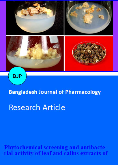
Callus development was observed from the leaf explants of C. asiatica within a week of inoculation and the highest frequency of callus was observed after 3 weeks on MS medium supplemented with different concentrations of 2,4-dichlorophenoxyacetic acid and 6-benzylaminopurine (Figure 1). Patra et al. (1998) induced callus on semisolid and modified MS medium with 2.0 mg/L kinetin and 4.0 mg/L alpha-naphthaleneacetic acid from stem and leaf explants of C. asiatica, whereas Martin (2004) developed callus on MS medium with 2,4-dichlorophenoxyacetic acid and alpha-naphthaleneacetic acid (1.0 mg/L) in combination with 0.5 mg/L kinetin. In the present study, morphology and growth of callus was affected by varying concentration of 2,4-dichlorophenoxyacetic acid and 6-benzylaminopurine (with friable and more friable, yellowish and yellowish-white) and no callus was observed in MS medium without plant growth regulators. Loc and Tam (2010) reported that MS medium supplemented with 1.0 mg/L 6-benzylaminopurine and alpha-naphthaleneacetic acid resulted in the most favorable induction of compact, friable and yellow colored callus after 21 days of culture from the petiole of C. asiatica. They also reported that other combinations of plant growth regulators (6-benzylaminopurine, alpha-naphthaleneacetic acid and 2,4-dichlorophenoxyacetic acid) resulted in poor callus induction i.e., callus were either white or green in color, soft or succulent or not induced.
Figure 1: Stages of callus proliferation from leaf explants of Centella asiatica. (A) Callus induction after two weeks of culture on 6-benzylaminopurine 4.0 mg/L; (B) Callus induction after two weeks of culture on 2,4-dichlorophenoxyacetic acid 2.0 mg/L; (C) More friable and yellowish callus formation on 6-benzylaminopurine 4.0 mg/L after 4 weeks of culture; (D) Dried callus harvested (6-benzylaminopurine 4.0 mg/L) for the study of preliminary phytochemical screening and antibacterial activity
Higher concentration of 6-benzylaminopurine (4.0 mg/L) and 2,4-dichlorophenoxyacetic acid (2.0 mg/L) showed 95% callusing response with yellowish and more friable callus. The media fortified with 2,4-dichlorophenoxyacetic acid at 2.0 mg/L proved to be optimum for callus initiation and proliferation and 6-benzylaminopurine with 4.0 mg/L was the most responsive for mass induction of callus and proliferation. Whereas Rao et al. (1999) reported that alpha-naphthaleneacetic acid (2.0 mg/L) in combination with kinetin (0.2 mg/L) is best for callus induction from leaf explants of C. asiatica. Likewise, Gupta et al. (2010) stated that the leaf explants of Stevia rebaudiana cultured on MS medium supplemented with 0.8 mg/L alpha-naphthaleneacetic acid in combination with 1.0 mg/L 2,4-dichlorophenoxyacetic acid could be a suitable medium and noble approach to produce maximum amount of callus within short time period. It is suggested that, development of callus through in vitro method offers the possibility of obtaining desirable medicinal compounds as well as ensuring sustainable conservation and rational utilization of biodiversity (Coste et al., 2011).
Table I: Preliminary phytochemical analysis of C. asiatica leaf and callus extracts
| Phytocontituents | Leaf | Callus | ||||||
|---|---|---|---|---|---|---|---|---|
| Methanol | Acetone | Chloroform | Aqueous | Methanol | Acetone | Chloroform | Aqueous | |
| Alkaloids | + | + | + | - | + | + | + | + |
| Glycosides | + | + | + | + | + | - | + | + |
| Terpenoids | + | - | + | + | + | + | + | + |
| Steroids | + | + | + | - | - | + | + | - |
| Flavonoids | + | - | + | + | + | - | + | + |
| Tannins | + | + | - | + | - | + | - | + |
| Saponins | - | - | - | - | - | - | - | - |
| Reducing sugars | + | - | + | + | + | + | - | |
| (+) presence; (-) absence | ||||||||
Phytochemical screening of the leaf and callus extracts of C. asiatica revealed the presence of alkaloids, glycolsides, terpenoids, steroids, flavonoids, tannins and reducing sugars (Table I). These compounds have significant application against human pathogens, including those that cause enteric infections and are reported to have curative properties against several pathogens and therefore could suggest their use in the treatment of various diseases (Hassan et al., 2004). In general, the total phenolic compounds found in the leaf, root and petiole are the major contributions to the antioxidant activities of the plant (Zainol et al., 2003). Saponin is not detected in C. asiatica in the present study, whereas alkaloids are present in all the tested extracts. Asiatic acid and asiaticoside found in C. asiatica showed great promise in prevention and treatment of cancer either as a plant alone or in combination with other forms of chemotherapy such as vincristine from Catheranthus roseus (Bridgman, 2003). Triterpenoids are reported to have useful for antibacterial activity and can be applied against various bacterial pathogens like S. aureus, Shigella flexneri, Pasteurella multocida, E. coli, Salmonella and etc. (Utami et al., 2011).
As phytochemicals often play an important role in plant defense against prey, microorganism, stress as well as interspecies protections, these plant components have been used as drugs for millennia and hence, screening of phytochemicals serves as the initial step in predicting the types of potential active compounds from plants (Chew et al., 2011). With regard to yield from the powdered materials of leaf and callus extracts (100 g), maximum amount of yield from leaf was obtained in methanol extract (26.3 g), it was followed by acetone (25 g), aqueous (20.1 g) and chloroform (17.9 g) extracts; likewise methanol extract of 100 g of callus yielded 25 g and it was followed by acetone (22.6 g), aqueous (18.1 g) and chloroform (15.8 g) extracts.
The antibacterial activity of the tested extracts of C. asiatica showed significant reduction in bacterial growth in terms of zone of inhibition. All the leaf and callus extracts showed dose dependent activity i.e., while increase in the concentration of extract, the zone of inhibition is also increased (Table II). In the present study, maximum growth of inhibition (30 mm) was observed in methanol extract of leaves at 100 µg/mL against E. coli, which was followed by B. cereus (29 mm), P. aeruginosa and S. aureus (28 mm). Similarly, methanol extract of in vitro grown callus at the concentration of 100 µg/mL showed maximum growth of inhibition (29 mm) against P. aeruginosa, E. coli and S. aureus which was followed by B. cereus (28 mm). Methanol extract of leaf and callus at the concentration of 10, 25 and 50 µg/mL also showed significant activity (24-27 mm of zone of inhibition) against all the tested organisms. Whereas Zaidan et al. (2005) reported that, methanol extract of the leaves of C. asiatica showed moderate activity against S. aureus and penicillin resistant S. aureus.
Table II: Antibacterial activity of leaf and callus extracts of C. asiatica against human pathogenic microorganisms
| Plants extracts | Concentration (µg/mL) |
Zone of growth inhibition (mm) |
|||||||
|---|---|---|---|---|---|---|---|---|---|
| Staphylococcus aureus | Bacillus cereus | Pseudomonas aeruginosa | Escherichia coli | ||||||
| Leaf | Callus | Leaf | Callus | Leaf | Callus | Leaf | Callus | ||
| Methanol | 10 | 24 | 25 | 26 | 25 | 23 | 23 | 25 | 24 |
| 25 | 26 | 26 | 26 | 27 | 26 | 25 | 26 | 25 | |
| 50 | 27 | 27 | 27 | 27 | 27 | 27 | 28 | 27 | |
| 100 | 28 | 29 | 29 | 28 | 28 | 29 | 30 | 29 | |
| Acetone | 10 | 7 | 6 | 6 | 5 | 10 | 7 | 7 | 7 |
| 25 | 7 | 7 | 7 | 6 | 12 | 8 | 8 | 8 | |
| 50 | 8 | 9 | 9 | 8 | 12 | 9 | 9 | 9 | |
| 100 | 9 | 10 | 11 | 9 | 14 | 10 | 16 | 11 | |
| Chloroform | 10 | 9 | 8 | 9 | 6 | 10 | 12 | 12 | 14 |
| 25 | 13 | 10 | 10 | 8 | 12 | 13 | 13 | 15 | |
| 50 | 11 | 11 | 11 | 9 | 13 | 13 | 15 | 16 | |
| 100 | 21 | 12 | 13 | 11 | 16 | 14 | 19 | 16 | |
| Aqueous | 10 | 6 | 5 | 5 | 6 | 5 | 5 | 5 | 4 |
| 25 | 6 | 6 | 6 | 7 | 6 | 5 | 7 | 5 | |
| 50 | 7 | 7 | 6 | 8 | 8 | 6 | 8 | 6 | |
| 100 | 8 | 8 | 7 | 7 | 9 | 8 | 10 | 7 | |
| Penicillin | 10 | 4 | 3 | No activity | No activity | 3 | 2 | No activity | No activity |
The aqueous and acetone extracts of leaf and callus were found to be less effective and chloroform extract of leaf and callus showed moderate zone of inhibition against all the tested organisms in the present study. Jagtap et al. (2009) reported that, the aqueous extract of C. asiatica did not showed any antibacterial effects at lower concentrations, but it was effective at the concentrations above 125 µg/mL against S. aureus and E. coli. The extracts of C. asiatica are effective to kill the bacteria that can survive in extreme conditions like high or low temperature especially B. cereus (Utami et al., 2011). In Malaysian traditional medicine, C. asiatica has been used as an antibacterial agent and recommended as an alternative for skin diseases and nervous system disorders (Zaidan et al., 2005).
The results of the present study showed that, leaf and leaf derived callus extracts of C. asiatica especially methanol extract possess bioactive compounds with antibacterial activity against many pathogens. It is suggested that the methanol extract of leaf and callus revealed a significant scope to develop a novel broad spectrum of antimicrobial drug formulation and can be used to carry out further pharmacological evaluation to be used as antibacterial agents/drugs.
Acknowledgements
The authors are thankful to Dr. Valli Nachiyar, Department of Biotechnology, Sathyabama University for providing necessary facilities. Prof. Thangam Menon, Department of Microbiology, University of Madras, India is acknowledged for providing the test organisms.
References
Appa Rao MVR, Srinivas K, Koteshwar Rao T. The effect of Mandookaparni (Centella asiatica) on the general mental ability (Medhya) of mentally retarded children. J Res Indian Med. 1973; 8: 9-16.
Bridgman KE. Herbal medicines. Faculty of Pharmacy, University of Sydney, 2003.
Cheng CL, Guo JS, Luk J, Koo MW. The healing effects of Centella extract and asiaticoside on acetic acid induced gastric ulcers in rats. Life Sci. 2004; 74: 2237-49.
Chew YL, Chan EWL, Tan PL, Lim YY, Stanslas J, Goh JK. Assessment of phytochemical content, polyphenolic composition, antioxidant and antibacterial activities of Leguminosae medicinal plants in Peninsular Malaysia. BMC Complem Altern Med. 2011; 11: 12.
Cho KH. Clinical experiences of madecassol (Centella asiatica) in the treatment of peptic ulcer. Korean J Gastroenterol. 1981; 13: 49-56.
Chopra RN, Nayar SL, Chopra IC. Glossary of Indian Medicinal Plants (Including the Supplement). New Delhi, Council of Scientific and Industrial Research, 1980.
Coste A, Vlase L, Halmagyi A, Deliu C, Coldea G. Effects of plant growth regulators and elicitors on production of secondary metabolites in shoot cultures of Hypericum hirsutum and Hypericum maculatum. Plant Cell Tissue Organ Cult. 2011; 106: 279-88.
Evans WC. Trease and Evans Pharmacognosy. 5th ed. London, Cambridge University Press, 2002, pp 336-93.
Gupta P, Sharma S, Saxena S. Callusing in Stevia rebaudiana (Natural Sweetener) for steviol glycoside production. Int J Agri Biol Sci. 2010; 1: 30-34.
Hassan MM, Oyenwale AO, Abduallahi MS, Okonkwo EM. Preliminary phytochemical and antibacterial investigation of crude extract of the root bark of Datarium microcarpum. J Chem Soc Nigeria. 2004; 29: 26-29.
Howes MR, Houghton PJ. Plants used in Chinese and Indian traditional medicine for improvement of memory and cognitive function. Pharm Biochem Behav. 2003; 75: 513-27.
Jagtap NS, Khadabadi SS, Ghorpade DS, Banarase NB, Naphade SS. Antimicrobial and antifungal activity of Centella asiatica (L.) Urban, Umbeliferae. Res J Pharm Tech. 2009; 2: 329-30.
James JT, Dubery IA. Pentacyclic triterpenoids from the medicinal herb, Centella asiatica (L.) Urban. Molecules 2009; 14: 3922-41.
Lee Y, Lee DE, Lee HS, Kim KS, Lee WS, Kim SH, Kim MW. Influence of auxins, cytokinins, and nitrogen on production of rutin from callus and adventitious roots of the white mulberry tree (Morus alba L.). Plant Cell Tissue Organ Cult. 2011; 105: 9-19.
Loc NH, Tam NT. An Asiaticoside production from Centella (Centella asiatica L. Urban) cell culture. Biotech Bioproc Eng. 2010; 15: 1065-70.
Martin KP. Plant regeneration through somatic embryogenesis in medicinally important Centella asiatica L. In vitro Cell Dev Biol Plant. 2004; 40: 586-91.
Matsuda H, Morikawa T, Ueda H, Yoshikawa M. Medicinal foodstuffs, XXVII. Saponin constituents Gotu Kola (2): Structures of new ursane- and oleanane-type triterpene oligoglycosides, centellsaponin B, C, and D, from Centella asiatica cultivated in Sri Lanka. Chem Pharm Bull. 2001; 49: 1368-71.
Murashige T, Skoog F. A revised medium for rapid growth and bioassays for tobacco tissue cultures. Physiol Plant 1962; 15: 473-97.
Parekh J, Chanda S. Antibacterial and phytochemical studies on twelve species of Indian medicinal plants. Afri J Microbiol Res. 2007; 1: 175-81.
Patra A, Rai B, Rout GR, Das P. Successful plant regeneration from callus cultures of Centella asiatica (L.) Urban. Plant Growth Regul. 1998; 24: 13-16.
Pointel JP, Boccalon H, Cloarec M, Ledebehat C, Joubert M. Titrated extract of Centella asiatica (TECA) in the treatment of venous insufficiency of the lower limbs. Angiol. 1987; 38: 46-50.
Rao KP, Rao SS, Sadanandam M. Tissue culture studies of Centella asiatica. Indian J Pharm Sci. 1999; 61: 392-94.
Shukla A, Rasik AM, Jain GK, Shankar R, Kulshrestha DK, Dhawan BN. In vitro and in vivo wound healing activity of asiaticoside isolated from Centella asiatica. J Ethnopharmacol. 1999; 65: 1–11.
Siddiqui AA, Ali M. Practical Pharmaceutical chemistry. 1st ed. New Delhi, CBS Publisher and Distributors, 1997; pp 126-31.
Subban R, Veerakumar A, Manimaran R, Hashim KM, Balachandran I. Two new flavonoids from Centella asiatica (Linn.). J Nat Med. 2008; 62: 369-73.
Utami CV, Hatane SE, Gorjian M. The Application of three herbs; Chrysanthemum indicum, Centella asiatica and Andrographis paniculata to reduce bacteria in cow milk. Proceedings of The First International Conference on Interdisciplinary Research and Development, 31 May-1 June 2011, Thailand, pp 51.1-51.6.
Wang XS, Liu L, Fang JN. Immunological activities and structure of pectin from Centella asiatica. Carbohyd Polym. 2005; 60: 95–101.
Zaidan MRS, Rain AN, Badrul AR, Adlin A, Norazah A, Zakiah I. In vitro screening of five local medicinal plants for antibacterial activity using disc diffusion method. Trop Biomed. 2005; 22: 165-70.
Zainol MK, Abd-Hamid A, Yusuf S, Muse R. Antioxidative activity and total phenolic compounds of leaf, root and petiole of four accessions of Centella asiatica (L.) Urban. Food Chem. 2003; 81: 575–81
Zheng CJ, Qin LP. Chemical components of Centella asiatica and their bioactivities. J Chin Integ Med. 2007; 5: 348–51.