Nephroprotective effect of Berberis baluchistanica against gentamicin-induced nephrotoxicity in rabbit
Abstract
The present study was conducted to explore the protective effect of Berberis baluchistanica against gentamicin-induced renal toxicity in rabbit. Phytochemical investigations lead to the isolation of berberine and palmatine. The crude hydromethanolic extract at various doses (100, 200 and 300 mg/kg body weight) elicited strong nephroprotective effects by restoring various biomarkers which were deranged by gentamicin such as creatinine, urea, serum uric acid levels (p<0.001) in plasma and urine output creatinine clearance, urinary protein and γ-glutamyl transferase level (p<0.001) in urine in dose-dependent manner. The mediators involved in oxidative stress such as malondialdehyde, reduced glutathione, glutathione peroxidase, and catalase levels were significantly (p<0.05-0.001) modulated in kidney tissue homogenate. Correspondingly, there was a significant (p<0.05) recovery in kidney weight and %loss in the body weight compare to gentamicin-treated group. In conclusion, B. baluchistanica exhibits nephroprotective effect.
Introduction
Gentamicin is an effective aminoglycoside antibiotic used against infections caused by Gram positive and Gram negative aerobic bacteria. But nephrotoxicity and oxidative damage limit its long-term clinical use (Alarifi et al., 2012).
The oxidative stress are diminished upon supplementation with certain dietary antioxidants such as vitamin E, C, these vitamins was seems to ameliorate the suppression in enzymatic activity of GSH-Px and catalase in the renal tissues induced by gentamicin (Kavutcu et al., 1996). The other non-nutrient antioxidant such as flavonoids, medicines derived from plant extracts are being increasingly utilized as adjunct treatment options for a wide variety of clinical disease (Rahimi et al., 2005; Al-Azzawie and Alhamdani, 2006). More interest has developed to the protective effects of medicinal plants against chemically induced toxicities (Frei and Higdon, 2003). For example, Azima tetracantha (Konda et al., 2016) and Capparis spinosa (Tlili et al., 2017), Cestrum nocturnum (Saleem et al., 2017), Morus alba (Ullah et al., 2016), Nigella sativa (Begum et al., 2006), Sechium edule (Mumtaz et al., 2012), Terminalia muelleri (Fahmy et al., 2016) show nephroprotective effect.
Berberis baluchistanica (Family Berberidaceae), which in local language “Brahvi†known as Zarch, found in Kalat district, Balochistan province, Pakistan. It is a wild plant and is used as medicine by the local people, roots decoction is used for cure of internal injuries. Beside this, it is also used for the removal of kidney related disorders as it contains berberine which is known to be useful in the treatment of number of diseases (Baloch et al., 2013). Strong antibacterial effects of the roots extracts against a variety of pathogenic bacteria have been reported (Kakar et al., 2012). The phytochemical investigations showed that the genus, berberis is rich in berberine and berberine like compounds, while from B. baluchistanica several alkaloids such as pakistanine, pakistanamine and (+)-baluchistine have been isolated (Shamma et al., 1973; Miana et al., 1979).
Keeping in mind, the strong in vitro antioxidant potential and traditional use of B. baluchistanica in the treatment of kidney disorders, the present study was designed to evaluate the nephroprotective like effect of B. baluchistanica against gentamicin-induced nephrotoxicity.
Materials and Methods
Chemicals
Gentamicin sulfate (80 mg/2 mL; Abbott Laboratories Pakistan Ltd.), silliver® (Silymarin, 200 mg; Searl Pakistan Ltd.), reduced glutathione, glutathione reductase, thiobarbituric acid, fosfotungustic acid, hydrogen peroxide, NADPH and other reagents were used.
Plant material
The roots of the plant (35 kg dry wt.) of B. baluchistanica were collected from, District Kalat, Balochistan Province, Pakistan, during the month of April 2012. The plant was identified by Prof. Muhmmad Ibrar of the Department of Botany, University of Peshawar. A voucher No. Bot/20105 has been placed in the herbarium of the Department of Botany, University of Peshawar as a reference.
Preparation of plant extracts
The freshly collected shade dried roots was powdered and extracted by maceration in 90% methanol for 10 days (3 × 50 L). The combined methanol extract was filtered with a muslin cloth, evaporated and concentrated under vacuum using rotary evaporator (Buchi – R210, Switzerland) at a temperature of 40°C and a viscous extract (2.2 kg) was obtained. The crude methanolic extract was investigated for the nephroprotective activity.
Phytochemical investigation
The B. baluchistanica extract was screened for alkaloids, flavonoids, saponins, phenols, and diterpines (Baloch et al., 2013) The crude extract is further screened for the presence of triterpine, tannins and saponins.
Isolation and structure elucidation of compounds
The viscous extract (2.2 kg) was mixed with 3L distilled water and extracted three times with 6L n-hexane, chloroform, ethyl-acetate, and n-butanol to get n-hexane (59 g), ethyl acetate (39 g), chloroform (88 g), butanol (65 g) and water (89 g) soluble fractions respectively. The ethyl acetate soluble subfraction was chromatographed over silica gel and eluted with mixtures of n-hexane-chloroform, and chloroform–methanol in increasing order of polarity to obtain 8 major fractions EtA1- EtA8 . Fraction EA3 obtained from n-hexane–chloroform (2.0:8.0) was rechromatographed and eluted with chloroform–methanol (4.5:5.5 and 2.0:8.0) to afford compound (1) (12 mg), and compound (2) (14 mg), respectively. The purity of compounds were analyzed using TLC followed by spraying with Dragendorff’s reagent. Structures were elucidated using various techniques such as 1H-NMR, 13C-NMR, 2D-NMR, EI-MS, FAB-MS, UV and IR.
Animals and experimental layout
The healthy male rabbits (Oryctolagus cuniculus) (1,200-1,500 g) were purchased from the Veterinary Research Institute, Peshawar and maintained under optimal conditions at the University of Peshawar. The rabbits were fed on chow pellets along with fresh green vegetables and grasses and free access to fresh water ad libitum. Before feeding experimental diet, animals were acclimatized for two weeks.
Animals grouping and dosing
The rabbits were divided into groups, each having six animals. Three doses (100, 200 and 300 mg/kg p.o) were tested for the crude extract. During the experiment, gentamicin 100 mg/kg i.p. while silymarin at 50 mg/kg i.p. were administered (Shahbazi et al., 2012).
Group 1: Normal saline (control); Group 2: gentamicin (100 mg/kg/day, i.p); Group 3: silymarin (50 mg/kg/day, i.p); Groups 4: methanol extract (100 mg/kg/day, p.o) plus gentamicin (100 mg/kg/day, i.p); Groups 5: methanol extract (200 mg/kg/day, p.o) plus gentamicin (100 mg/kg/day, i.p); Groups 6: methanol extract (300 mg/kg/day, p.o) plus gentamicin (100 mg/kg/day, i.p). The experiment was continue for 15 days
Samples collection and processing
After dosage completion, 24 hours later on day 16, rabbits were anesthetized by intraperitoneal injection of ketamine. The blood samples were taken by cardiac puncture, transferred to EDTA and non-EDTA containing tubes. The serum was separated by centrifugation (K240R, Centurion Scientific, UK) for 15 min at 3,000 rpm and 37°C, stored at 4°C till determination of biochemical parameters. The serum sample was assayed for urea, creatinine, and uric acid through standard procedures (Cobas c 311 analyzer kit, UK).
Collection and analysis of urine
At the end of the experimental period, 24 hours urine samples were collected in standard metabolic cages from each animal in all the groups. After measuring volume, these urine samples were used for analyze for urinary protein, creatinine clearance and gamma-glutamyl transferase levels. These parameters were estimated through COBAS chemistry automation using Roche Diagnostic kits.
Measurement of body and organs weight
The body weight was measured on a daily basis throughout the treatment period before administration of drug or saline and at the end of experiment in all the groups. Similarly, at the end of 15-day treatment, each rabbit was euthanized and theabdominal cavity was dissected out and liver was removed and weighted.
Preparation of liver homogenate
The right kidney was immediately removed, washed thoroughly in the cold saline solution to remove the blood. The 10% homogenate was prepared in phosphate buffer (0.05M, pH 7) using a homogenizer at 25°C. The homogenate was centrifuged to remove the cell debris and the supernatant was used for the determination of in vivo antioxidant assay.
In vivo antioxidant activities
The levels of lipid peroxidation is based on the reaction of malondialdehyde, a product of lipid peroxidation with thiobarbituric acid to form thiobarbituric acid reactive substances, which has absorption at 514 nm and shows pink color (Placer et al., 1966; Rao and Mohan, 2017).
Catalase activity was determined according to the method of Aebi (1984). In brief, kidney tissue homogenate supernatant approximately 10.5 mL was added to 3 mL of phosphate-buffered saline and the absorbance was read at 240 nm using a UV spectrophotometer. The principle of the assay is based on the H2O2 decomposition rate at 240 nm. Results were obtained kg/ protein.
Glutathione peroxidase (GSH-Px) was measured by the method of Goldberg and Spooner described as oxidized glutathione is instantly converted to its reduced form with a simultaneous oxidation of NADPH to NADP+ (Burk et al., 1980). In the presence of glutathione reductase and NADPH, The decrease in absorbance at 340 nm is measured. GSH-Px activity was expressed as U/mg protein. Tissue reduced glutathione activity was determined according to the previously published method (Kaur et al., 2006). The kidney tissue homogenate 0.8 mL was mixed with 0.5 mL 20% solution of trichloroacetic acid, the mixture was kept on ice bath for 10 min, then subjected to centrifugation at 3,000 rpm for 15 min, 0.5 mL of the supernatant was mixed 2 mL of 0.6 mM dithionitrobenzene (prepared in 0.2M buffer, pH 8) with 0.8 mL of 0.2M sodium phosphate buffer (pH 8) when yellow color obtained absorbance was measured at after 20 min at 412 nm The reduced glutathione content was expressed as U/mg protein.
Histopathalogical examination
For histopathalogical examination, the left kidney was fixed in 10% buffered formalin for 48 hours and was dehydrated in graded ethanol solution and embedded in paraffin blocks. The sections of the kidney were oriented perpendicular to the plane of section in the block and 6 micrometer thick transverse sections were cut and mounted on the glass slides and stained with Hematoxylin and Eosin (Tomita et al., 1993; Miyamoto et al., 2002). The microscopic observation (Labomed Lx400 with digital camera iVu 3100, USA) was done to keep the count of histopathological changes, which were graded as (–) none; (+) mild; (++) moderate; (+++) severe (Shahid and Subhan, 2014; Shahid et al., 2016).
Statistical analysis
Data are presented as means ± (SEM). To compare means, all groups were subjected to one-way analysis of variance (ANOVA) followed by Tukey' smultiple comparison post hoc test using GraphPad Prism5 (GraphPad Software, USA), to determine the difference in all parameters. Results were considered statistically significant at p<0.05.
Results
Preliminary phytochemical analysis
Phytochemical investigation of the methanol extract revealed the presence of alkaloids, flavonoids and tanins (Table I).
Table I: Phytochemical screening of B. baluchistanica
| Test | Observation | Result |
|---|---|---|
| 1 drop of extract on TLC plate + Dragendorff’s reagent | Appearance of orange /red color |
Alkaloids present |
| 150 mg extract + 1.5 mL chloroform → warmed for 0.5 hour → 1 mL sulphuric acid added | No color change observed | Tri-terpenoids absent |
| 100 mg extact → boil + 2.5 mL distilled water → shake→ foam formation | No foam appear | Saponin absent |
| 250 mg extract + 2.5 mL dilute ammonia solution → 0.5 mL sulphuric acid added |
Appearance of yellowish color | Flavonoids present |
| Aqueous aliquot of extract + ferric chloride reagent | Appearance of greenish black color | Tannins present |
| 150 mg extract + sulphuric acid → boil/cool→ add chloroform → separate chloroform layer + dilute ammonia solution |
No color change | Anthraquinone absent |
| 1 g extract + 10 mL of distilled water + add few drops of copper acetate→ | Emerald green color | Diterpenes present |
| 500 mg of MBHE + 0.5 mL Fehling A solution + 0.5 mL Fehling solutions + heat |
Not any change observed | Reducing sugar absent |
Table II: Effect of B. baluchistanica extract on gentamicin-induced nephrotoxic rabbit
| Groups | Normal saline(10 mL/kg) | Gentamicin (80 mg/kg) | Silymarin (50 mg/kg) | Methanol extract (100 mg/kg) plus gentamicin | Methanol extract (200 mg/kg) plus gentamicin | Methanol extract (300 mg/kg) plus gentamicin |
|---|---|---|---|---|---|---|
| Kidney weight (g/100 g bw) | 0.8 ± 0.1 | 1.1 ± 0.1 | 0.9 ± 0.1c | 1.0 ± 0.1b | 0.8 ± 0.1b | 0.8 ± 0.1c |
| %Loss in body weight | 0.2 ± 0.9 | 10.8 ± 1.1a | 2.7 ± 0.9c | 3.6 ± 0.8c | 4.7 ± 0.9c | 5.6 ± 1.5c |
| Blood urea (mg/dL) | 12.0 ± 2.6 | 24.3 ± 2.3a | 11.5 ± 3.4b | 14.5 ± 1.5c | 12.3 ± 1.3b | 10.3 ± 2.0c |
| Blood uric acid (mg/dL) | 2.5 ± 0.0 | 4.2 ± 0.2a | 1.2 ± 0.2b | 2.9 ± 0.2b | 1.6 ± 0.3a | 1.1 ± 0.2c |
| Serum creatinine (mg/dL) | 0.5 ± 0.1 | 3.5 ± 0.3a | 0.4 ± 0.3b | 0.5 ± 0.1c | 0.4 ± 0.0b | 0.2 ± .1c |
| Urine output (mL/hour) | 200 ± 9.2 | 168 ± 12.0a | 200.6 ± 19.6b | 199 ± 26.0b | 200 ± 26.0b | 208 ± 21.8c |
| Creatinine clearance (mL/min) | 4.7 ± 2.8 | 0.4 ± 1.3a | 1.7 ± 0.8 | 3.3 ± 1.0c | 4.0 ± 0.9b | 4.7 ± 1.0c |
| Urinary protein (mg/dL) | 1.8 ± 0.2 | 3.86 ± 0.3a | 1.74 ± 0.1b | 1.7 ± 0.3c | 1.8 ± 0.2b | 1.7 ± 0.3c |
| Urinary γ-glutamyl transferase (U/L) | 48.8 ± 5.1 | 104.7 ± 8.1a | 50.6 ± 4.2b | 72.7 ± 3.2b | 60.6 ± 2.2c | 56.4 ± 1.1c |
| MDA (nmolg/protein) | 4.8 ± 0.6 | 12.3 ± 1.9a | 4.9 ± 0.7 | 10.5 ± 1.0b | 6.1 ± 0.3c | 5.2 ± 0.3c |
| Catalase (kg/protein) | 133.0 ± 11.4 | 70.3 ± 1.9a | 137.0 ± 11.5c | 90.4 ± 2.2b | 103.5 ± 7.3b | 121.5 ± 8.3c |
| GSH (nmol g/tissue) | 2.0 ± 0.0 | 1.8 ± 0.0 | 2.2 ± 0.4a | 1.8 ± 0.0a | 2.0 ± 0.3b | 2.3 ± 0.2b |
| GSH-Px (IU/g protein) | 33.0 ± 1.2 | 18.7 ± 3.0a | 33.9 ± 1.5b | 24.3 ± 2.0b | 26.4 ± 1.6b | 27.3 ± 2.4b |
| Histopathological findings | ||||||
| Interstitial inflammation | ++ | - | - | - | ||
| Tubular dilatation | +++ | + | + | + | ||
| Necrosis of epithelium | ++ | - | - | - | ||
| Glomerular congestion | ++ | - | - | - | ||
| Tubular cast | ++ | + | - | - | ||
| Data are expressed as mean ± SEM followed by Tukey’s post hoc test;. ap<0.05 significant difference compared to the normal control; bp<0.001, cp significant difference compare to gentamicin-treated group (n = 6); (–) none; (+) mild; (++) moderate; (+++) severe | ||||||
Isolated compounds
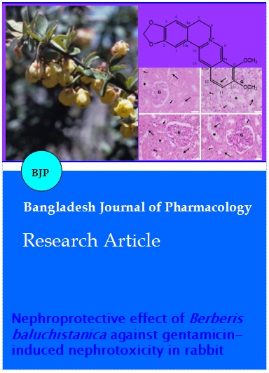
Compound 1 (berberine; Figure 1) was isolated from the ethyl acetate fraction through normal phase column chromatography using normal phase silica gel as stationary phase. Mobile phase used was CHCl3– MeOH (4.5:5.5). The EI-MS showed a molecular ion peak at 336.1295. High resolution EI-MS confirmed the molecular formula to be C20H18NO4.
Figure 1: Chemical structure of berberine and palmatine
The broad band 13C-NMR showed 20 signals, out of which six were methines and three methylenes confirmed by dept 90° and dept 135° respectively.
H-1 resonated at δ 7.656 showing HMBC correlation with C-2 (δ 149.918) and C-3 (δ 152.174). H-4 resonated at δ 6.957 (s) showing HMBC correlation with C-3 (δ 152.174), C-2 (δ 149.918) and C-14a (δ121.855). The methylene protons H-5 resonated at δ 3.261 as triplet and H-6 at δ 4.930 as triplet. H-5 showed HMBC correlation with C-4a (δ131.884), C-4 (δ109.397), C-6 (δ57.203) and C-14a (δ121.855). The correlation of H-5 and H-6 was also confirmed by COSY spectrum. H-6 methyle protons showed HMBC correlation with C-4a (δ 131.884), C-5 (δ 28.20), C-8 (δ 146.403) and C-14 (δ 139.661). H-8 proton resonated at δ 9.764 as singlet, showed HMBC correlation with C-8a (δ 123.321), C-9 (δ 145.770) and C-14 (δ 139.661). H-11 resonated as doublet at δ 8.1211, showed HMBC interaction with C-9 (δ 149.770), C-10 (δ 152.022) and C-12 (δ 124.521). H-12 methine protons resonated at δ 8.005 as doublet shoed HMBC correlation with C-9 (δ 145.770), C-10 (δ 152.022), C-12a (δ 135.162) and C-13 (δ 121.482). the COSY spectrum showed the correlation of H-11 and H-12.
H-13 showed resonance signals as singlet at δ8.70. the HMBC interrelation of H-13with C-8a (δ 123.321) and C-14 (δ 139.661) was observed. The methylene protons of O-CH2-O group resonated as singlet at δ 6.105. The HMBC spectrum showed correlation of this methylene with C-2 (δ 149.918) and C-3 (δ 152.174).
Two methyls resonated as singlets at δ 4.200 and δ 4.104 in H-NMR spectrum. These singlets were assigned to two methoxy groups attached with C-9 (δ 145.770) and C-10 (δ 152.022) confirmed by HMBC spectrum.
Compound 2 (palmatine; Figure 1) was isolated from the ethyl acetate fraction using repeated column chromatography The EI-MS shows the molecular ion peak at m/z: 352 [M+] (calculated for C21H22NO4+). The HREI-MS was in agreement with molecular formula for C21H22NO4+) at m/z (rel. int. %): 352 (11, M+), 351 (35), 337 (100), 294 (46), 278 (34), 219 (7), 190 (8), 138 (5), 94(5), 43 (10).
1HNMR of compound 2 showed six aromatic protons at δ 7.83, (1H, s, H-1), 7.23 (1H, s, H-4), 9.75 (1H, s, H-8), 8.40 (1H, d, J = 9.0 Hz, H-11), 8.05 (1H, d, J = 9.0 Hz, H-12) and 9.12 (1H, s, H-13), three methylenes at δ 6.09 (2H, s, H-14), 3.24 (2H, t, J = 10.0 Hz, H-5) and 4.23 (2H, s, J = 10.0 Hz, H-6) and two methoxy peaks at δ 4.06 (3H, s, 9-OMe) and 4.09 (3H, s, 10-OMe).
The 13C-NMR spectrum of compound 2 showed twenty one carbon atoms which included six methine, two methylene, four methoxy and nine quaternary carbon atoms on the basis of DEPT-q spectral data.
The methine proton at δ 9.12 (H-8) showed correlations with δ 138.75 (C-13a), 56.43 (C-6), 123.33 (C-8a), 135.10 (C-12a) and 145.40 (C-9), the methine proton at δ 8.40 (H-11) correlated to δ 148.68 (C-10), 124.30 (C-12), 135.10 (C-12a) and 145.40 (C-9), the methine proton appearing at δ 8.04 (H-12) showed correlations with δ 125.53 (C-11), 148.68 (C-10), 135.10 (C-12a), 123.33 (C-8a) and 118.81 (C-13), the methine proton at δ 9.12 (H-13) correlated to δ 135.10 (C-12a), 123.33 (C-8a), 138.75 (C-13a) and 123.40 (C-12).
The methylene protons appearing at δ 3.23 (H-5) correlated to δ 56.43 (C-6), 127.45 (C-4a), 119.80 (C-13b) and 113.25 (C-4), the methylene protons at δ 4.84 (H-6) showed correlations with δ 138.75 (C-13a), 143.35 (C-8), 127.45 (C-4a) and 24.84 (C-5) and four methoxy peaks at δ 4.09 (9-OMe), 4.06 (10-OMe), 3.24 (2-OMe) and 4.23 (3-OMe) correlated to δ 145.40 (C-9) , 148.68 (C-10), 152.52 (C-2) and 152.23 (C-3) respectively.
The data of the compounds were clearly matched with reported data from the literature of berberine and palmatine (Küpeli et al., 2002).
Effect on physical parameters
The treatment with the extract significantly (p<0.05) improved the weight changes induced by gentamicin. The overall effect was dose-dependent at 100, 200 and 300 mg/kg p.o. Similarly, the methanol extract caused significant (p<0.05) recovery in the kidney weight and %loss in the body weight compare to gentamicin-treated group.
Effect on biochemical markers
The treatment of roots extract of the plant significantly (p<0.05) ameliorated the toxicological changes induced by gentamicin in blood urea, serum uric acid and creatinine levels. The overall effect was dose-dependent at 100, 200 and 300 mg/kg p.o. Similarly, the crude extract of the plant caused significant (p<0.05) nephro-protective effect on the urine biomarker including creatinine clearance, gamma-glutamyl transferase, urinary output and urinary protein levels.
Effect on oxidative stress biomarkers
Animals treated with gentamicin showed significant increase in kidney malondialdehyde, while animals treated along with crude methanolic extract (all doses) significantly controlled the increase in kidney malondialdehyde level. Similarly catalase, reduced glutathione and GSH-PX activity were decreased in gentamicin-treated group while groups treated with crude methanol extract significantly recovered the levels of above biomarkers.
Effects of gentamicin and B. baluchistanica on kidney histopathology
Histological findings of this study showed photomicrograph of a section of kidney from a rabbit treated with normal saline/vehicle showing normal appearance of glomerulus in a Bowman’s capsule interspersed among renal tubules lined by epithelial cells with a brush border extending into the lumen (Figure 2A). Similarly, Figure 2B demonstrated the section of kidney from a rabbit treated with gentamicin (100 mg/kg) representing mild atrophy of glomerulus, inflammation of interstitial spaces, dilatation of renal tubules, containing cellular cast in their lumen. Correspondingly improved architecture is observed in crude methanolic extract-treated groups (100 mg/kg) plus gentamicin, (Figure 2C), crude methanolic extract (200 mg/kg) plus gentamicin, (Figure 2D) and crude methanolic extract (300 mg/kg) plus gentamicin (Figure 2E) in dose-dependent manner with marked histological amelioration compared with the gentamicin-treated group.
Figure 2: Histopathological evaluation of B. baluchistanica on GM-induced nephrotoxicity (H and E staining; x40, scale bar =100 μm) (n = 6 rabbits per group). (A): Photomicrograph of a section of kidneys from a rabbit treated with normal saline/vehicle showing normal appearance of glomerulus (G) in a Bowman’s capsule interspersed among renal tubules (large arrows) lined by epithelial cells (small arrows) with a brush border extending into the lumen (asterisk). (B): Photomicrograph of a section of kidney from a rabbit treated with Gentamicin (100 mg/kg) showing mild atrophy of glomerulus (G), inflammation of interstitial spaces, dilatation of renal tubules (large arrows) containing cellular cast (small arrows) in their lumen (asterisk). Normal histological appearance of glomerulus (G), renal tubules (large arrows) lined by epithelial cells (small arrows) with a brush border extending into the lumen (asterisk) are observed in groups of rabbits treated with (C): mBBE (100 mg/kg) plus GM, (D): mBBE (200 mg/kg) plus GM, and (E): mBBE (300 mg/kg) plus GM.
Discussion
The current study revealed significant nephroprotective like effect of crude methanol extract B. baluchistanica roots in gentamicin-induced toxicity by ameliorating biochemical markers and physical parameters strongly augment by histopathalogical changes in experimental rabbits.
The exact mechanism by which gentamicin induced nephrotoxicity is unknown, however, several researchers reported that aminoglycoside antibiotics are a class of drug capable of causing the formation of reactive oxidative species (ROS) which can be directly involved in gentamicin-induced damage. Malondialdehyde, end product of lipid peroxidation in tissues, results in a decrease in polyunsaturated fatty acid content, which serves as substrate for free radicals (Eisenberg et al., 1987). It is also reported that iron is important in models of tissue injury. Gentamicin acts as an iron chelator and that have been shown to cause release of iron from renal cortical mitochondria and iron-gentamicin complex is a potent catalyst of free-radical formation and enhance the generation of ROS (Priuska and Schacht, 1995; Baliga et al., 1999). GGT levels may be a prominent and sensitive marker of oxidative stress (Lee et al., 2004). Studies have shown that GGT, like other antioxidant enzymes, is inducible by oxidative stress, which increases GGT activity (Zhang et al., 2006). The impairment in kidney functions was accompanied by either increase in serum creatinine and urea levels or kidney tissue malondialdehyde levels that indicated lipid peroxidation (Cuzzocrea et al., 2002). The reduced glutathione is one of the essential compounds for maintaining cell integrity participation in the cell metabolism. However, xenobiotics or peroxide-dependent alterations in tissue reduced glutathione levels and antioxidant enzyme activities are currently controversial problem. In another study (AteÅŸÅŸahin et al. 2003), it was found that gentamicin caused depletion in kidney reduced glutathione levels and unaffected antioxidant enzyme activities such as GSH-Px and catalase. Controversial results are reported by several investigations (Nakajima et al., 1994; Maldonado et al., 2003; Parlakpinar et al., 2005). Gentamicin-induced nephrotoxicity was associated with low activity of GSH-Px, catalase, and levels of reduced glutathione in the renal cortex. These decreases in renal antioxidant enzymatic protection could aggravate the oxidative damage. The increased production of ROS in gentamicin-induced nephrotoxicity may cause inactivation of anti-oxidant enzymes such as GSH-Px, catalase. Similarly, in this study, the increases in malondialdehyde levels, decreases in GSH-Px and catalase activities, but no alteration in reduced glutathione levels in rabbits treated with gentamicin alone appear to support above investigations that gentamicin-induced nephrotoxicity is associated with lipid peroxidation in renal tissue as reflected by an increase in malondialdehyde. These observations are correlated well with the renal histological findings, which explained extensive and marked tubular necrosis. The nephro-protection by medicinal plants may be by inhibiting the production of ROS and lipid peroxidation (Fahmi et al., 2016; Konda et al., 2016; Tilili et al., 2017).
B. baluchistanica is a wild plant used as medicine by local people. They used the roots decoction for the cure of internal injuries and ophthalmic problems. Beside of this, it is also used for the removal of kidney stones, as it contains berberine and berberine like compounds, which is known to be useful for a number of diseases (Li et al., 2014).
A marked restoration in lipid peroxidation paralleled with GSH-Px and catalase activity in methanol extract- treated group were observed. These findings were in conformity with above parameters, as similar results observed in this study. Phytochemical strength of genus Berberis is berberine, an isoquinolone alkaloid found in many plants of genus berberis and its derivatives (Imanshahidi and Hosseinzadeh, 2008; Guamán Ortiz et al., 2015; Khan, 2016). These compounds possessed strong in vitro and in vivo antioxidant potentials (Somova et al., 2003; Lyamzaev et al., 2011) which support nephroprotective like effect of B. baluchistanica. Interestingly, it is reported that berberine at the doses of 3 mg/kg caused significant neprohroprotective like effect (Kumar et al., 2015). In another study, berberine ameliorates proteinuria in type 2 diabetic (Liu et al., 2008; Ashraf et al., 2013). It is also reported that berberine reduce oxidative stress in streptozotocin diabetic rats (Moghaddam et al., 2014). In this phytochemical investigation, it had been isolated a number of compounds including berberine and palmatine. Therefore, the current findings on methanol extract can be attributed to these isolated compounds.
Conclusion
B. baluchistanica root extract possesses marked protective effect against gentamicin-induced nephrotoxicity by ameliorating various biomarkers of nephrotoxicity and oxidative stress.
Ethical Issue
The study protocols were approved by the Ethical Committee of Department of Pharmacy, University of Peshawar (01/EC- 17/pharm).
References
Aebi H. Catalase in vitro. Method Enzymol. 1984; 105: 121-26.
Al-Azzawie HF, Alhamdani MSS. Hypoglycemic and antioxidant effect of oleuropein in alloxan-diabetic rabbits. Life Sci. 2006; 78: 1371-77.
Alarifi S, Al-Doaiss A, Alkahtani S, Al-Farraj S, Al-Eissa MS, Al-Dahmash B, Al-Yahya H, Mubarak M. Blood chemical changes and renal histological alterations induced by gentamicin in rats. Saudi J Biol Sci. 2012; 19: 103-10.
Ashraf H, Heidari R, Nejati V, Ilkhanipoor M. Aqueous extract of Berberis integerrima root improves renal dysfunction in streptozotocin induced diabetic rats. Avicenna J Phytomed. 2013; 3: 82.
Ateşşahin A, Karahan I, Yilmaz S, Çeribaşi A, Princci I. The effect of manganese chloride on gentamicin-induced nephrotoxicity in rats. Pharmacol Res. 2003; 48: 637-42.
Baliga R, Ueda N, Walker PD, Shah SV. Oxidant mechanisms in toxic acute renal failure. Drug Metab Rev.1999; 31: 971-97,
Baloch N, Nabi S, Yasser M, Kahraman A In vitro antileishmanial, cytotoxic, antioxidant activities and phytochemical analysis of Berberis baluchistanica roots extracts and its fractions. J Phytopharmacol. 2013; 4: 282-87.
Begum N, Dewan ZF, Nahar N, Mamun M. Effect of n-hexane extract of Nigella sativa on gentamicin-induced nephrotoxicity in rats. Bangladesh J Pharmacol. 2006; 1: 16-20.
Burk RF, Lawrence RA, Lane JM. Liver necrosis and lipid peroxidation in the rat as the result of paraquat and diquat administration: Effect of selenium deficiency. J Clin Invest. 1980; 65: 1024.
Cuzzocrea S, Mazzon E, Dugo L, Serraino I, Di Paola R, Britti D, De Sarro A, Pierpaoli S, Caputi AP, Masini E. A role for superoxide in gentamicin-mediated nephropathy in rats. Eur J Pharmacol. 2002; 450: 67-76.
Eisenberg JM, Koffer H, Glick HA, Connell ML, Loss LE, Talbot GH, Shusterman NH, Strom BL. What is the cost of nephrotoxicity associated with aminoglycosides? Ann Intern Med. 1987; 107: 900-09.
Fahmy NM, Al-Sayed E, Abdel-Daim MM, Karonen M, Singab AN. Protective effect of Terminalia muelleri against carbon tetrachloride-induced hepato- and nephrotoxicity in mice and characterization of its bioactive constituents. Pharm Biol. 2016; 54: 303-13.
Frei B, Higdon JV. Antioxidant activity of tea polyphenols in vivo: Evidence from animal studies. J Nut. 2003; 133: 3275-84.
Guamán Ortiz LM, Croce AL, Aredia F, Sapienza S, Fiorillo G, Syeda TM, Buzzetti F, Lombardi P, Scovassi AI. Effect of new berberine derivatives on colon cancer cells. Acta Biochimica et Biophysica Sinica. 2015; 47: 824-33.
Imanshahidi M, Hosseinzadeh H. Pharmacological and therapeutic effects of Berberis vulgaris and its active constituent, berberine. Phytother Res. 2008; 22: 999-1012.
Kakar SA, Tareen RB, Kakar MA, Jabeen H, Kakar S-u-R, Al-Kahraman Y, Shafee M. Screening of antibacterial activity of four medicinal plants of Balochistan- Pakistan. Pakistan J Bot. 2012; 44: 245-50.
Kaur G, Alam MS, Jabbar Z, Javed K, Athar M. Evaluation of antioxidant activity of Cassia siamea flowers. J Ethnopharmacol. 2006; 108: 340-48.
Kavutcu M, Canbolat O, Öztürk S, Olcay E, Ulutepe S, Ekinci C, Gökhun IH, Durak I. Reduced enzymatic antioxidant defense mechanism in kidney tissues from gentamicin-treated guinea pigs: Effects of vitamins E and C. Nephron 1996; 72: 269-74.
Khan H. Berberine: As a therapeutic target for treating obese diabetes. Sci Forschen J Dia Res Ther. 2016; 2: 1-2.
Konda VR, Arunachalam R, Eerike M, Rao R, Radhakrishnan AK, Raghuraman LP, Meti V, Devi S. Nephroprotective effect of ethanolic extract of Azima tetracantha root in glycerol induced acute renal failure in Wistar albino rats. J Trad Complement Med. 2016; 6: 347-54.
Kumar A, Chopra K, Mukherjee M, Pottabathini R, Dhull DK. Current knowledge and pharmacological profile of berberine: An update. Eur J Pharmacol. 2015; 761: 288-97.
Küpeli E, Koşar M, Yeşilada E, Başer KH. A comparative study on the anti-inflammatory, antinociceptive and antipyretic effects of isoquinoline alkaloids from the roots of Turkish Berberis species. Life Sci. 2002; 72: 645-57.
Lee D-H, Blomhoff R, Jacobs DR. Review is serum gamma- glutamyltransferase a marker of oxidative stress? Free Rad Res. 2004; 38: 535-39.
Li Z, Geng YN, Jiang JD, Kong WJ. Antioxidant and anti-inflammatory activities of berberine in the treatment of diabetes mellitus. Evid Based Complement Altern Med. 2014; 2014.
Liu WH, Hei ZQ, Nie H, Tang FT, Huang HQ, Li XJ, Deng YH, Chen SR, Guo FF, Huang WG. Berberine ameliorates renal injury in streptozotocin-induced diabetic rats by suppression of both oxidative stress and aldose reductase. Chin Med J. 2008; 121: 706.
Lyamzaev KG, Pustovidko AV, Simonyan RA, Rokitskaya TI, Domnina LV, Ivanova OY, Severina II, Sumbatyan NV, Korshunova GA, Tashlitsky VN Novel mitochondria-targeted antioxidants: Plastoquinone conjugated with cationic plant alkaloids berberine and palmatine. Pharm Res. 2011; 28: 2883-95.
Maldonado PD, Barrera D, Medina-Campos ON, Hernández-Pando R, Ibarra-Rubio MaE, Pedraza-Chaverrı J. Aged garlic extract attenuates gentamicin induced renal damage and oxidative stress in rats. Life Sci. 2003; 73: 2543-56.
Miana G, Foy J, Minard R, Shamma M. Baluchistanine, a new bisbenzylisoquinoline alkaloid. Cell Mol Life Sci. 1979; 35: 1137-38.
Miyamoto A, Nakayama K, Imaki H, Hirose S, Jiang Y, Abe M, Tsukiyama T, Nagahama H, Ohno S, Hatakeyama S. Increased proliferation of B cells and auto-immunity in mice lacking protein kinase Cδ. Nature 2002; 416: 865-69.
Moghaddam HK, Baluchnejadmojarad T, Roghani M, Khaksari M, Norouzi P, Ahooie M, Mahboobi F. Berberine ameliorate oxidative stress and astrogliosis in the hippocampus of STZ-induced diabetic rats. Mol Neurobiol. 2014; 49: 820.
Mumtaz SMF, Paul S, Bag AK. Effect of Sechium edule on chemical induced kidney damage in experimental animals. Bangladesh J Pharmacol. 2012; 8: 28-35.
Nakajima T, Hishida A, Kato A. Mechanisms for protective effects of free radical scavengers on gentamicin-mediated nephropathy in rats. Am J Physiol Renal Physiol. 1994; 266: 425-31.
Parlakpinar H, Tasdemir S, Polat A, Bay-Karabulut A, Vardi N, Ucar M, Acet A. Protective role of caffeic acid phenethyl ester (cape) on gentamicin-induced acute renal toxicity in rats. Toxicology 2005; 207: 169-77.
Placer ZA, Cushman LL, Johnson BC. Estimation of product of lipid peroxidation (malonyl dialdehyde) in biochemical systems. Anal Biochem. 1966; 16: 359-64.
Priuska EM, Schacht J. Formation of free radicals by gentamicin and iron and evidence for an iron/gentamicin complex. Biochem Pharmacol. 1995; 50: 1749-52.
Rahimi R, Nikfar S, Larijani B, Abdollahi M. A review on the role of antioxidants in the management of diabetes and its complications. Biomed Pharmacother. 2005; 59: 365-73.
Rao PS, G. K. Mohan GK. In vitro alpha-amylase inhibition and in vivo antioxidant potential of Momordica dioica seeds in streptozotocin-induced oxidative stress in diabetic rats. Saudi J Biol Sci. 2017; 24: 1262-67.
Saleem U, Ali N, Ahmad B. Protective and curative effects of Cestrum nocturnum on rabbit kidney. Bangladesh J Pharmacol. 2017; 12: 284-91.
Shahbazi F, Dashti-Khavidaki S, Khalili H, Lessan-Pezeshki M. Potential renoprotective effects of silymarin against nephrotoxic drugs: A review of literature. J Pharm Pharm Sci. 2012; 15: 112-23.
Shahid M, Subhan F. Protective effect of Bacopa monniera methanol extract against carbon tetrachloride induced hepatotoxicity and nephrotoxicity. Pharmacol Online. 2014; 2: 18-28.
Shahid M, Subhan F, Ullah I, Ali G, Alam J, Shah R. Beneficial effects of Bacopa monnieri extract on opioid induced toxicity. Heliyon 2016; 2: 1-30.
Shamma M, Moniot J, Yao S, Miana G, Ikram M. Pakistanine and pakistanamine, two new dimeric isoquinoline alkaloids. J Am Chem Soc.1973; 95: 5742-47.
Somova L, Nadar A, Rammanan P, Shode F. Cardiovascular, antihyperlipidemic and antioxidant effects of oleanolic and ursolic acids in experimental hypertension. Phytomedicine 2003; 10: 115-21.
Tlili N, Feriani A, Saadoui E, Nasri N, Khaldi A. Capparis spinosa leaves extract: Source of bioantioxidants with nephroprotective and hepatoprotective effects. Biomed Pharmacother. 2017; 87: 171-79.
Tomita Y, Aozasa K, Myoui A, Kuratsu S, Matsumoto K, Uchida A, Ono K. Histologic grading in soft tissue sarcomas: An analysis of 194 cases including agnor count and mast cell count. Int J Cancer.1993; 54: 194-99.
Ullah N, Khan MA, Khan S, Ahmad H, Asif AH, Khan T. Nephroprotective potential of Morus alba, a prospective experimental study on animal models. Pharmaceutical Biol. 2016; 54: 530-35.
Zhang H, Liu H, Iles KE, Liu R-M, Postlethwait EM, Laperche Y, Forman HJ. 4-Hydroxynonenal induces rat γ-glutamyl transpeptidase through mitogen-activated protein kinase–mediated electrophile response element/nuclear factor erythroid 2–related factor 2 signaling. Am J Respir Cell Mol Biol. 2006; 34: 174-81.
Apply citation style format of Bangladesh Journal of Pharmacology
Copyright (c) 2018 Samreen Pervez, Muhammad Saeed, Haroon Khan, Muhammad Shahid, Irfan Ullah

This work is licensed under a Creative Commons Attribution-NonCommercial 4.0 International License.
