Antimycotic potential of a diterpenoid taxoquinone against Candida species isolated from Metasequoia glyptostroboides
Abstract
The objective of this study was to confirm the antimycotic potential of a diterpenoid compound taxoquinone, isolated from Metasequoia glyptostroboides against pathogenic isolates of Candida species. The taxoquinone (100 μg/disc) displayed potential anticandidal effect against Candia albicans KBN06P00076, KBN06P00074, C. glabrata KBN06P00066, KBN06P00068, C. tropicalis KBN06P00682, KBN06P00058, C. parapsilosis KBN06P00060, KBN06P00055, and C. guilliermondii KBN06P00492 and KBN06P00867 as a diameter of zones of inhibition, found in the range of 10 ± 0.6 to 13 ± 1.1 mm. The minimum inhibitory and minimum fungicidal concentrations of taxoquinone against the tested clinical isolates were found in the range of 125 to 500 and 125 to 1,000 ug/mL, respectively. On the viable counts of the tested fungal isolates, the taxoquinone evoked a remarkable anticandidal effect. Elaborative study of SEM revealed potential detrimental effect of taxoquinone on the morphology of C. parapsilosis KBN06P00060 at MIC concentration. These findings confirmed therapeutic potential of taxoquinone.
Introduction
Candida species have been shown to be the causative agents of many infections in an increasing range of anatomical sites and clinical settings (Langenbeck, 1939). In normal healthy individuals, the yeast Candida is classified as a commensal organism that can colonize both internal and external surfaces (Truss, 1981). Although Candida species have been implicated in the early stages of AIDS, infections due to other Candida species are becoming more widespread (Hood and Denning, 1996). The management of serious and life-threatening candidiasis remains severely hampered by the lack of reliable antifungal drugs that allow both fungemia and tissue invasion by Candida species.
Although mycotic infections have been treated with polyene antifungals, such as amphotericin B, and with azoles, high relapse rate, resistance and reported toxic side effects have led to their limited use as the first line treatment (Gallies et al., 1990; Lyman and Walsh, 1992; Baily et al., 1994). Because of the rising incidences of failures in the treatment of mycoses, there is a need for the development of new therapeutic agents that support the anticandidal activity of antimycotics (Wakabayashi et al., 1996).
In order to overcome these problems, plant-based bio-active constituents have been served as potential biomedicinal tools to control serious fungal infection caused by Candida species (Inouye et al., 2006; Picman et al., 1990; Rao et al., 2010; Yin et al., 2008). Previously we isolated an abietane type diterpenoid taxoquinone from Metasequoia glyptostroboides, and first time reports its antimycotic potential against clinical isolates of Candida species.
Materials and Methods
Microorganisms
The test clinical isolates of Candida species such as C. albicans KBN06P00076, C. albicans KBN06P00074, C. glabrata KBN06P00066, C. glabrata KBN06P00068, C. tropicalis KBN06P00682, C. tropicalis KBN06P00058, C. parapsilosis KBN06P00060, C. Parapsilosis KBN06P00055, C. guilliermondii KBN06P00492 and C. guilliermondii KBN06P00867 were provided by the National Biobank of Korea, Chonbuk National University Hospital, supported by the Ministry of Health, Welfare and Family Affairs, Republic of Korea. All materials derived from the National Biobank of Korea were obtained with informed consent under institutional review board-approved protocols.
Plant material
The cones of M. glyptostroboides were collected locally from Pohang city, Republic of Korea, in November and December, 2008, and identified by the morphological features and the database present in the library at the Department of Biotechnology, Daegu University, Korea. A voucher specimen was deposited in the herbarium of College of Engineering, Department of Biotechnology, Daegu University, Korea.
Extraction, isolation and purification of taxoquinone
Dried cones of M. glyptostroboides (2 kg) were milled into powder and then extracted with ethyl acetate at room temperature for 12 days. The extract was evaporated under reduced pressure using a rotary evaporator (EYELA N1000, Japan). The dried ethyl acetate extract (7 g) was subjected to column chromatography over silica gel (mesh 230-400 mesh, Merck, Darmstadt, Germany) and was eluted with hexane-ethyl acetate-methanol solvent system to give 20 fractions. Of the fractions obtained, fraction-12 was further purified by preparative TLC over silica gel GF254 using hexane-ethyl acetate (1:2) as a mobile phase to give one compound (152 mg) which on the basis of spectral data analysis was characterized as taxoquinone as shown in Figure 1.
Anticandidal activity assay
Anticandidal activity was assayed by the standard agar diffusion method (Chandrasekaran and Venkatesalu, 2004). Briefly, a 100 uL of standardized inoculum (approximately 107 CFU/mL) of fungal suspension was loaded uniformly on petri plates with 20 mL of potato-dextrose agar (PDA) medium, and allowed to dry for 5 min. A sterile Whatman No. 1 filter paper disc with 6 mm diameter was impregnated with 100 ug/disc of test compound taxoquinone dissolved in 5%dimethyl sulfoxide (DMSO). Negative controls were prepared using the same solvent employed to dissolve the sample. Standard reference antibiotic, amphotericin B (10 ug/disc, Sigma-Aldrich Co., USA) was used as the positive control against the tested clinical isolates of Candida species. After incubating the plates at 28°C for 2-3 days, anticandidal activity was evaluated by comparing the diameter of the zones of inhibition against the tested Candida species. For the anticandidal activity assay, the experiment was replicated at least three times.
Minimum inhibitory (MIC) and minimum fungicidal (MFC) concentrations
The MIC of taxoquinone was tested by the 2-fold serial dilution method (Murray et al., 1995). The taxoquinone dissolved in 5% DMSO was incorporated into PDB medium to obtain a concentration of 1,000 ug/mL, and then serially diluted to achieve 500, 250, 125, 62.5, 31.3, 15.6 and 7.8 ug/mL. A 10 μL standardized suspension of each tested Candida species (107 CFU/mL approximately) was transferred to each tube. The control tubes with only candidal suspension were incubated at 28°C for 2-3 days. The MIC expressed in μg/mL was defined as the lowest concentration of taxoquinone not showing any growth of test Candida species by macroscopic evaluation. Further, the concentrations showing complete inhibition of visual growth of fungal isolates were identified, and 50 uL of each diluted culture broth transferred on to the agar plates were incubated for specified time and at temperature as mentioned above. The complete absence of growth on the agar surface in the lowest concentration of sample was defined as MFC.
Cell viability assay
Active cultures for viable count assay were prepared in PDB medium (Bajpai et al., 2009). For viable counts, each of the tubes containing resuspended candidal suspension (approximately 107 CFU/mL) was inoculated with 125 ug/mL concentration of the test compound taxoquinone in 10 mL PDB broth, and incubated at 28°C. For viable cell counts, samples were taken out at 0, 20, 40, 60, 80, 100 and 140 min time intervals, and the viable cells were counted as followed: After incubition, 1 mL of the resuspended culture was diluted into 9 mL buffer peptone water, there by diluting it 10-fold. A 0.1 mL sample of each treatment was diluted further and spread on the surface of PDA plates. Newly formed colonies were counted after 2-3 days of incubation at 28°C. The controls were composed of inoculums without test compound for each Candida isolate with same experimental conditions. Each assay in this experiment was replicated three times.
Scanning electron microscopy
Based on the susceptibility and MIC concentration, one of the clinical isolates of Candida species was selected for further elaborative study of scanning electron microscopy (SEM), to determine the anticandidal efficacy of bioactive compound taxoquinone on the morphology of Candida parapsilosis KBN06P00060. Controls were prepared without test compound. To observe the morphological changes, the method of SEM was modified from Kockro method (Kockro et al., 2000). The fungal samples were washed gently with 0.1 M phosphate buffer solution (pH 7.2), and fixed with 2.5% (w/v) glutaraldehyde. The specimen was dehydrated using sequential exposure per ethanol concentrations ranging from 50 to 100%, and then ethanol was replaced by tertiary butyl alcohol. After dehydration, the specimen was dried with CO2. Finally, the specimen was sputter-coated with gold in an ion coater for 2 min, followed by examinations by an SEM (S-4300; Hitachi, Japan).
Statistical analysis
Each experiment was run in triplicate and the average values were calculated. The statistical analysis was carried out employing one way ANOVA (p<0.05) with a SPSS statistical package (version 11.0).
Results
The ethyl acetate cone extract of M. glyptostroboides after column chromatography over silica gel yielded a pure compound which was obtained as orange needles with a specific melting point (mp 212-214°C). The 1H NMR spectrum (CDCl3, 250 MHz) of the compound showed a hydroxyl methine signal at d 4.77 (1H, ddd, J = 2.2, 7.4, 9.8 Hz), an oxygenated proton at d 3.80 (1H, d, J = 2.2 Hz), an aliphatic methine at d 3.14 (1H, sept, J = 7.1 Hz), and proton signals for methylene (d 2.65-1.01) and five terminal methyl groups (d 1.33, 1.20, 1.19, 0.92, and 0.90). Further analysis of the COSY data established the connectivity through H-7a (d 4.77, ddd, J = 2.2, 7.4, 9.8 Hz), 7b-OH (d 3.80, d, J = 2.2 Hz), and H-6b (d 2.18, dd, J = 7.4, 12.5 Hz), and through H-1b (d 2.65, br d, J = 13.0 Hz), H-2b (d 1.71, m), and H-1a, 2a, 3a, 3b (1.65-1.01, m). In addition, two methyl signals at d 1.20 (3H, d, J = 7.1 Hz) and 1.19 (3H, d, J = 7.1 Hz) coupling with a methine signal at d 3.14 (1H, sept, J = 7.1 Hz, H-15) in the 1H NMR data as well as 20 carbon signals including two carbonyl groups at d 189.6 and 183.7 in the 13C NMR data strongly suggested that this compound should be an abietane diterpenoid. On the basis of the interpretation of HMQC and HMBC data, this compound was proposed to be taxoquinone. By comparison of the multiplicity of H-7 and the chemical shifts both in the 1H and 13C NMR data, the structure of this compound (Figure 1) was determined to be taxoquinone (Bajpai et al., 2010).
Figure 1: Chemical structure of an abietane type diterpenoid, taxoquinone isolated from M. glyptostroboides
The anticandidal effect of taxoquinone against the employed isolates of Candida species was monitored by the diameter of inhibition zones. The taxoquinone at 100 ug/disc exhibited potent inhibitory effect (Figure 2). C. albicans KBN06P00076, KBN06P00074, C. tropicalis KBN06P00683, C. parapsilosis KBN06P00060 and KBN-06P00055 were found to be the most inhibited fungal isolates of Candida species by the taxoquinone, with their respective diameter of zones of inhibition of 11 ± 0.8, 13 ± 1.1, 11 ± 0.9, 13 ± 1.1 and 12 ± 1.0 mm. However, the diameters of zones of inhibition of taxoquinone against C. glabrata KBN06P00066, KBN06P00068, C. tropicalis KBN06P00058, C. guilliermondii KBN06-P00492 and KBN06P00867 were found to be 10 ± 0.7, 10 ± 0.8, 10 ± 0.8, 10 ± 0.8 and 10 ± 0.6 mm, respectively (Figure 2). As a negative control, only solvent had no anticandidal effect. As shown in Figure 2, it was confirmed in this assay that taxoquinone significantly inhibited the growth of some of the tested clinical isolates of Candida species (diameter of zones of inhibition: 10 ± 0.6 - 13 ± 1.1 mm) than that of standard antibiotic amphotericin B (diameter of zones of inhibition: 12 ± 0.7 - 14 ± 0.9 mm).
Figure 2: Antimycotic effect of taxoquinone (100 ug/disc) against clinical isolates of Candida species
Ca-1: Candia albicans KBN06P00076, Ca-2: C. albicans KBN06P00074, Cg-1: Candia glabrata KBN06P00066, Cg-2: C. glabrata KBN06P00068, Ct-1: Candida tropicalis KBN06P00682, Ct-2: C. tropicalis KBN06P00058, Cp-1: Candida parapsilosis KBN06P00060, Cp-2: C. parapsilosis KBN06P00055, Cgl-1: Candida guilliermondii KBN06P00492 and Cgl-2: C. guilliermondii KBN06P00867
The taxoquinone displayed potential anticandidal effect as MIC and MFC values against the tested clinical isolates of Candida species. The MIC and MFC values of taxoquinone against the tested isolates of Candida species were placed in the range of 125 to 500 and 125 to 1,000 ug/mL, respectively (Figure 3). When standard amphotericin B was tested for the MIC values against the clinical isolates of Candida species, the MIC values were found in the range of 62.5 to 250 μg/mL. C. guilliermondii KBN06P00492 and KBN06P00867 were found less sensitive fungal isolates to amphotericin B as compared to the other isolates of Candida species. C. albicans KBN06P00074, C. tropicalis KBN06P00682, C. Parapsilosis KBN06P00060 and KBN06P00055 were found to be the most susceptible Candida species to taxoquinone (MIC: 125 ~ 250 ug/mL) followed by C. albicans KBN06P00076, C. glabrata KBN06P00066, KBN-06P00068, C. tropicalis KBN06P00058, C. guilliermondii KBN06P00492 and KBN06P00867 (MIC: 500 ug/mL for each fungal isolate).
Figure 3: Minimum inhibitory (MIC) and minimum fungicidal (MFC) concentrations of taxoquinone against clinical isolates of Candida species
Ca-1: Candia albicans KBN06P00076, Ca-2: C. albicans KBN06P00074, Cg-1: Candida glabrata KBN06P00066, Cg-2: C. glabrata KBN06P00068, Ct-1: Candida tropicalis KBN06P00682, Ct-2: C. tropicalis KBN06P00058, Cp-1: Candida parapsilosis KBN06P00060, Cp-2: C. parapsilosis KBN06P00055, Cgl-1: Candida guilliermondii KBN06P00492 and Cgl-2: C. guilliermondii KBN06P00867
A cell viable count assay was carried out to evaluate the anticandidal effect of taxoquinone on the selected clinical isolates of Candida species. The taxoquinone had negative effect on the growth of tested fungal isolates of Candida species at the used concentrations. At 100 min exposure, near about 70-90% inhibition of the viable cells was observed in all the tested isolates of Candida species (Figure 4). However, taxoquinone completely inhibited the CFU numbers against C. albicans KBN06-P00074, C. glabrata KBN06P00682, C. parapsilosis KBN06-P00060 and KBN06P00055 at 140 min exposure time, and no CFUs were observed.
Figure 4: Effect of taxoquinone on the viable counts of the tested clinical isolates of Candida species
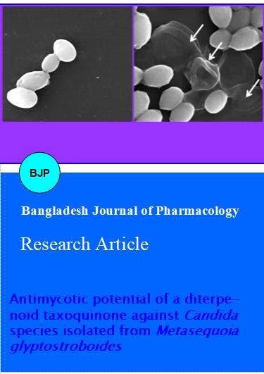
The effect of the bioactive diterpenoid taxoquinone on the morphology of C. parapsilosis KBN06P00060 was visualized by the elaborative study of SEM. As shown in Figure 5, the taxoquinone was found potentially capable to alter the cell morphology of C. parapsilosis KBN06P00060 as compared to the control group, served without any treatment (Figure 5). In contrast to the regular and smooth surface of control cells of C. parapsilosis KBN06P00060 (Figure 5A), the inoculated cells with the taxoquinone at the MIC concentration (125 µg/mL) revealed severe detrimental effect on the morphology of the tested isolate showing swelling, abnormal cell morphology or lysed cell formation (Figure 5B).
Figure 5: Scanning microscopy study showing antimycotic effect of a diterpenoid compound taxoquinone on the morphology of C. parapsilosis KBN06P00060. A): Control, showing regular and smooth surface; B): Treatment, showing swelling, abnormal cell formation and/or lysed cell formation
Discussion
This research demonstrated potential anticandidal effect of taxoquinone, inhibiting the growth of various clinical isolates of Candida species. The data of this study suggest that taxoquinone may act as an effective agent when applied to a clinical Candida infection situation. Only solvent, as a negative control had no anti-candidal effect against any of the tested fungal isolates of Candida species. This study also describes the complex effect of taxoquinone as MIC and MFC values as well as its inhibitory effect on cell viable counts of the tested clinical isolates of Candida species. A diterpenoid taxoquinone exhibited a wide range of anticandidal activity against the tested isolates of Candida species. Several terpenoid compounds have been shown to exert inhibitory effect against the fungal pathogens of Candida species (Inouye et al., 2006; Picman et al., 1990; Rao et al., 2010). Previous studies suggest that various terpenoid compounds of plant origin can be available for trials to control severe fungal infections caused by Candida species. These compounds may be applied in the same way against the pathogenic isolates of Candida species as alternative medicinal products where these pathogens have developed resistance against the specific fungicides (Sandven, 2000; Yin et al., 2008).
Furthermore, the anticandidal efficacy of the taxoquinone was also confirmed by the study of SEM, which revealed potential detrimental effect of the bioactive terpenoid compound taxoquinone on the morphology of one of the tested isolates C. parapsilosis KBN-06P00060. These altered morphological features might be occurred due to the detrimental effect on membrane integrity, thereby resulting in the lysis of Candida cell wall followed by the loss of intracellular dense material of treated cells, similar findings were also confirmed by others (Ghannoum, 1988; Wang et al., 2005). Although the putative mechanism of action is not clear but it might be assumed from the above results that the anticandial effect of the taxoquinone could be attributed due to its effect on cell wall synthesis, enzymes inhibition, leakage of cellular material and ergosterol biosynthesis. As reported previously, treatment of Candida species with various antifungal agents accumulates phosphatidylserine with a lower proportion of phosphatidylcholine. Synthesis of phosphatidylcholine starts from phosphatidylserine, therefore, accumulation of phosphatidylserine was suggested to interference the enzymes, catalyzing the biosynthetic pathway. Changes in membrane fluidity usually occur due to alterations in lipid composition of cell membrane (Sikkema et al., 1995; Weete, 1980), which are suspected to be a compensatory mechanism to counter the lipid disordering effects of the treatment agent. Hence, relevance of taxoquinone as antimycotic treatment to the accumulation of phosphatidylserine is clearly required in further investigation.
Candida species are getting serious worldwide due to several pathogenic disorders in human beings, although control measures are available, but limitedly effective (Yin et al., 2008). Hence, biologically active compounds of plant origin may be considered as an effective and alternative biological approach to develop new and novel types of antifungal agents for preventive treatment of serious fungal infections caused by pathogenic Candida species.
Information on the anticandidal effects of plant-based terpenoid compounds is scant, and these results show, for the first time that an abietane type diterpenoid taxoquinone isolated from M. glyptostroboides possessed substantial antimycotic effect against different clinical isolates of Candida species. Candida species are getting serious worldwide due to pathogenic disorders in human beings, although control measures are available but limitedly effective (Cowen et al., 2002). Hence, plant-based natural bioactive molecules may be considered as effective alternatives to develop new and novel types of antifungal agents for preventive treatment of serious fungal infections in animals and human beings caused by various pathogenic and clinical isolates of Candida species (Demirci et al., 2009).
Antimycotic effects of plant-based bioactive molecules against pathogenic and clinical isolate of Candida species may offer many new applications of clinical trials for medicine industry. The availability of terpenoid compounds may contribute to providing sustainable anticandidal tools, and are highly desired with significant biomedicinal potential. These findings reinforce the suggestions that bioactive diterpenoid taxoquinone from M. glyptostroboides could be included as an affective addition to medicine industry as a potential anticandidal agent for its possible applications of clinical efficacy. Thus, as a consequence plant-based bioactive constituents could be used as possible alternatives or complementary therapeutic agents for the treatment of serious fungal infections in antimycotic therapy. Further studies are warranted to confirm the in vivo efficacy of taxoquinone.
References
Baily GG, Perry FM, Denning DW, Mandal BK. Fluconazole-resistant candidiasis in an HIV cohort. AIDS. 1994; 8: 787-92.
Bajpai VK, Al-Reza SM, Choi UK, Lee JH, Kang SC. Chemical composition, antibacterial and antioxidant activities of leaf essential oil and extracts of Metasequoia glyptostroboides Miki ex Hu. Food Chem Toxicol. 2009; 47: 1876-83.
Bajpai VK, Na MK, Kang SC. The role of bioactive substances in controlling foodborne pathogens derived from Metasequoia glyptostroboides Miki ex Hu. Food Chem Toxicol. 2010; 48: 1945-49.
Chandrasekaran M, Venkatesalu V. Antibacterial and antifungal activity of Syzygium jambolanum seeds. J Ethnopharmacol. 2004; 91: 105-08.
Cowen LE, Anderson JB, Kohn LM. Evaluation of drug resistance in Candida albicans. Annu Rev Microbiol. 2002; 56: 139-65.
Demirci B, Toyota M, Demirci F, Dadandi MY, Can-Baser KH. Anticandidal pimaradiene diterpene from Phlomis essential oils. C R Chim. 2009; 12: 612-21.
Gallis HA, Drew RH, Pickard WW. Amphotericin B: 30 years of clinical experience. Rev Infect Dis. 1990; 12: 308-29.
Ghannoum MA. Studies on the anticandidal mode of action of Allium sativum (Garlic). J Gen Microbiol. 1988; 134: 2917-24.
Hood S, Denning DW. Treatment of fungal infection in AIDS. J Antimicrob Chemother. 1996; 37: 71-85.
Inouye S, UchidaK, Takizawa T, Yamaguchi H, Abe S. Evolution of the effect of terpenoid quinines on Trichophyton mentagrophytes by solution and vapor contact. J Infect Chemother. 2006; 12: 100-04.
Kockro RA, Hampl JA, Jansen B, Peters G, Scheihing M, Giacomelli R, et al. Use of scanning electron microscopy to investigate the prophylactic efficacy of rifampin-impregnated CSF shunt catheters. J Med Microbiol. 2000; 49: 441-50.
Langenbeck B. Auffingung von Pilzen aus der Schleimhaut der Speisero¨hre einer Typhus-Leiche. Neue Not Geb Natur-u-Heilk (Froriep) 1939; 12: 147.
Lyman CA, Walsh TJ, Systemically administered antifungal agents. A review of their clinical pharmacology and therapeutic applications. Drugs 1992; 44: 9-35.
Murray PR, Baron EJ, Pfaller MA, Tenover FC, Yolke RH. Manual of clinical microbiology (6th ed), Washington, DC: ASM Press, 1995.
Picman AK, Schneider EF, Gershenzon J. Antifungal activity of sunflower terpenoids. Biochem Syst Ecol. 1990; 18: 325-28.
Rao A, Zhang Y, Muend S, Rao R. Mechanism of antifungal activity of terpenoid phenols resembles calcium stress and inhibition of the TOR pathway. Antimicrob Agent Chemother. 2010; 54: 5062-69.
Sandven P. Epidemiology of candidemia. Revis Iberoamericana de Micol. 2000; 17: 73-81.
Sikkema J, de Bont JAM, Poolman B. Mechanisms of membrane toxicity of hydrocarbons. Microbiol Rev. 1995; 59: 201-22.
Truss CO. The role of Candida albicans in human illness. J Orthomol Psychiatry. 1981; 10: 228-38.
Wakabayashi H, Abe S, Okutomi T, Tansho S, Kawase K, Yamaguchi H. Cooperative anti-candida effects of lactoferrin or its peptides in combination with azole antifungal agents. Microbiol Immunol. 1996; 40: 821-25.
Wang SL, Yen YH, Tzeng GC, Hsieh C. Production of antifungal materials by bioconversion of shellfish chitin wastes fermented by Pseudomonas fluorescens K-188. Enzyme Microb Technol. 2005; 36: 49-56.
Weete JD. Lipid Biochemistry of Fungi and Other Organisms. New York, Plenum Press, 1980.
Yin S, Su ZS, Yue JM. Antimicrobial diterpenes from Trigonostemon chinensis. J Nat Prod. 2008; 71: 1414-17.