In vitro antibacterial activity and phytochemical analysis of hexane extract of Vicia sativa
Abstract
Vicia sativa is traditionally used medicinal plant in skin infections, asthma, bronchitis, urinary diseases and also used as antiseptic, antipoison, aphrodisiac, anti rheumatic and antipyretic. In the present study n-hexane extract of V. sativa was evaluated for the antibacterial activity against pathogenic bacteria Staphylococcus aureus, Bacillus atrophaeus, Escherichia coli and S. epidermidis by disc diffusion method. Minimum inhibitory concentration of the n-hexane extract against all bacteria was determined by broth dilution method. Preliminary phytochemical analysis and HPLC analysis showed the presence of a number of bioactive constituents which exhibits antibacterial activity. So the current study showed that V. sativa possesses the significant antibacterial activity.
Introduction
Plants and their active constituents have a diverse history as clinical source of chemotherapeutic agents (Cushnie and Lamb, 2005). Many thousands of plant species have been evaluated for the antimicrobial activity but very less was found to be active (Meng et al., 1998) and non toxic to human beings (Izzo, 2004). There are many researches in the literature which shows the antimicrobial activity of the crude extracts obtained from the plants (El-seedi et al., 2002; Rojas et al., 2003). From the past two decades, antibacterial activity have been reported from various plant parts like leaves, root, stem, flower, fruits, seeds of some of the medicinal plants (Levan et al., 1979; Erturis and Demirbag, 2003; Sudharameshwari and Radhika, 2007). Since from 1940s, many bacteria are now becoming resistant due to the emergence of the problem of resistance. The speed of antibiotic resistance development is increasing as the frequency of antibiotic use increases. Thus the effectiveness of antibiotics becomes reduced (Livermore, 2004). Antibiotic resistance is now a days the greatest problem to the proper treatment of infections globally. Resistance adversely affects both clinical and financial therapeutic outcomes, with effects results in the failure of an individual patient to respond to therapy and the need for expensive alternative drugs increases, longer duration of hospitalization, and the need for changes in empirical therapy.
Materials and Methods
Collection of the plant material
The plant of Vicia sativa was collected from the botanical garden of University of Agriculture Faisalabad in April 2013. The plant was thoroughly washed, dried and identified by Dr. Mubashir Niaz, Department of Botany, G.C University Faisalabad, Pakistan. Voucher specimen was kept in the herbarium as future reference.
Extraction
Plant was shade dried and grinded into fine powder. Powdered plant material was soaked in n-hexane for 3 to 5 days with occasional shaking. Then plant material was filtered and the filtrate was evaporated in rotary evaporator until the solid mass extract was obtained which was stored at 4°C.
Test microorganisms
Antibacterial activity of n-hexane extract of V. sativa was evaluated against pathogenic bacteria Staphylococcus aureus, Bacillus atrophaeus, Escherichia coli and S. epidermidis. These strains were obtained from the Department of Microbiology, Saffron Pharmaceuticals Pvt Ltd.
Antibacterial assay of plant extracts
Antibacterial activity was determined by standard agar disc diffusion method (Heatley, 1944). Sterile filter paper discs were loaded with different concentrations of extract (300, 200 and 100 mg/mL) and placed on the surface of inoculated agar plates of bacteria S. aureus, E. coli, S. epidermidis and B. atrophaeus by using sterile forceps. A standard disc of tetracycline was also placed along with extract discs to check the comparison of inhibition of bacterial growth. These plates for antibacterial assay were incubated in incubator at 37°C for 24 hours. After 24 hours, the plates were observed for zones of inhibitions, the diameters of zone of inhibition were determined by digital varnier caliper in mm.
Determination of minimum inhibitory concentration
Minimum inhibitory concentration of extract against S. aureus, E. coli, S. epidermidis and B. atrophaeus was determined by broth dilution method using 96 microwel plate (Mothana et al., 2009).
Preliminary phytochemical analysis
The phytochemical screening of the plant extracts were performed by standard procedures as given below:
Test for alkaloids
0.2 g of the extract was added in 2N HCl (5 mL) then heated on the boiling water bath. The mixture was filtered after cooling and the filtrate was divided in the two equal halves. Few drops of Mayer's reagent were added in one portion and the Dragendeoff's reagent was added in other portion. Turbidity of the formed precipitate in the both reagents indicates the presence of alkaloids (Mojab et al., 2003; Sharma et al., 2010)
Test for tannins
0.2 g extracts was dissolved in 10 mL of distilled water and warm on water bath. The mixture was filtered and 5% solution of ferric chloride was added to filtrate. The formation of dark green solution indicates the presence of tannins (Mojab et al., 2003).
Test for saponins
0.2 g of extract was mixed in test tube with 5 mL of distilled water and warm on water bath until it begins to boil. Formation of foam that persists for 10 min indicates the presence of saponins (Mojab et al., 2003; Sofowora, 1993).
Test for terpenoids
0.2 g of extract was shaken separately with 2 mL of chloroform (CHCl3), and then 3 mL of concentrated H2S04 was added carefully to form a layer. The formation of reddish brown color at inert face of the solution indicates the presence of terpenoids (Sofowora, 1993; Sharma et al., 2010).
Test for flavonoids
0.2 g of extract was taken separately and dissolved in 5 mL diluted NaOH then 1M of 5 mL HCl was added. A yellow color solution that changes into colorless solution indicates the presence of flavonoids (Sofowora, 1993).
Test for anthraquinones
0.5 g of extract was boiled with 10% of HCl in water bath for few min and then filtered. The filtrate was cooled and then equal amount of CHCl3 was added in the filtrate. Few drops of 10% NH3 was added in the mixture and heated. The formation of rose-pink coloration indicates the presence of authraqui nones (Mojab et al., 2003; Sharma et al., 2010).
Test for glycosides
1.2 g of extract was hydrolyzed with 10 mL of 1% HCl and then neutralized by 10% of NaOH solution. Few drops of Fehling's solution A and B were poured in it. Formation of red color precipitates shows the presence of glycosides (Mojab et al., 2003; Sharma et al., 2010).
HPLC of the plant extracts
Flavonoids and phenolics present in n-hexane extract of V. sativa was determined by some modification in procedure as determined by (Sultana et al., 2008). Sample of plant extract (n-hexane) was prepared by adding small amount of extract in 5 mL distilled water, mix it well then 12 mL methanol was added in it, shake it well then stay for 5 min. 6 mL distilled water was added and again stay for 5 min. 10 mL of 15 M HCl was added to it and place this in digital drying oven for 2 hours. Filter this with syringe filter and analyze it by HPLC. Kaempferol and phenolics were separated using a shim-pack CLC-ODS (C-18) column, 25 cm x 4.6 mm, 5 µm. mobile phase used was acetonitrile and acetic acid at a flow rate of 1 mL/min. Detector used was gradient HPLC detector, range was bipolar, 1250 mV, 10 samples per sec. Threshold was set at 0.005 Mv. Length of column used was 100 mm. Width of peak was 0.2 min. Ethanol was used for calibration. Samples were analyzed by UV-visible detector at 280 nm for phenolics at 248 nm for kaempferol at room temperature.
Results
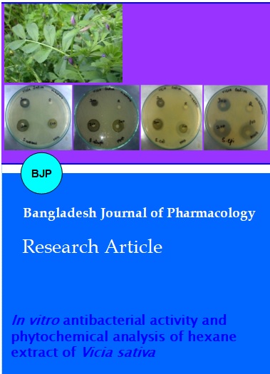
The results of antibacterial activity of n-hexane extracts of V. sativa are tabulated in Table I, and zones of inhibitions are shown in Figure 1. n-Hexane extract of V. sativa showed maximum antibacterial activity at 100 mg/mL concentration against E. coli (40.7 mm ± 0.8) while tetracycline positive control showed zone of (23.3 ± 0.5) and MIC was 1.6 mg/mL. V. sativa showed maximum antibacterial activity against other bacteria at 100 mg/mL concentration; B. atrophaeus (32.2 mm ± 0.2), S. epidermidis (31.3 mm ± 0.6), S. aureus (25.8 mm ± 0.2) and their MIC were 3.1 mg/mL, 6.3 mg/mL, 6.3 mg/mL respectively.
Figure 1: Zones of inhibitions of Vicia sativa (n-hexane extract) against (A) Staphylococcus aureus, (B) Bacillus atrophaeus, (C) Escherichia coli and (D) Staphylococcus epidermidis
Table I: Antibacterial activity of Vicia sativa (n-hexane extract) against Staphylococcus aureus, Bacillus atrophaeus, Escherichia coli and Staphylococcus epidermidis (mean ± SE)
| Pathogen | Conc. of n-hexane extract (mg/mL) | Mean zone of inhibition (mm) | Positive control tetracycline(mm) |
Negative control |
p value | MIC (mg/mL) |
|---|---|---|---|---|---|---|
| Staphylococcus aureus | 300 | 21.9 ±0.1 |
24.7 ± 0.2 |
0 | 0.000 | 6.3 |
| 200 | 25.6 ±0.2 | 0.014 | ||||
| 100 | 25.8 ±0.2 | 0.002 | ||||
| Bacillus atrophaeus | 300 | 15.2 ± 0.1 | 18.7 ± 0.2 | 0 | 0.000 | 3.1 |
| 200 | 26.7 ± 0.1 | 0.000 | ||||
| 100 | 32.2 ± 0.2 | 0.000 | ||||
| Escherichia coli | 300 | 23.6 ±0.1 | 23.3 ± 0.5 | 0 | 0.010 | 1.6 |
| 200 | 31.2 ± 0.1 | 0.000 | ||||
| 100 | 40.4 ± 0.8 | 0.000 | ||||
| Staphylococcus epidermidis | 300 | 20.7 ± 0.1 | 23.0 ± 0.2 | 0 | 0.000 | 6.3 |
| 200 | 25.4 ± 0.3 | 0.001 | ||||
| 100 | 31.3 ± 0.6 | 0.0001 |
HPLC analysis of V. sativa (n-hexane) extract was carried out and HPLC chromatograms obtained is shown in Figure 2 and Figure 3. HPLC chromatogram indicated the peaks of constituents which are present in extract, peaks of chromatotropic acid, gallic acid, quercitin, vanillic acid, syringic acid, vitamin C, trans-4-hydroxy-3-methoxy cinamic acid and kaempferol were observed. These constituents with their retention time and quantity are given in Table II.
Figure 2: HPLC chromatogram of phenolics present in Vicia sativa (n-hexane)
Figure 3: HPLC chromatogram of kaempferol in Vicia sativa (n-hexane)
Table II: Phytochemical constituents with their retention time and quantity of Vicia sativa (n-hexane)
| Compound name | Retention time | Quantity (ppm) |
|---|---|---|
| Chromatotropic acid | 1.9 | 47.9 |
| Quercitin | 2.6 | 59.9 |
| Gallic acid | 5.1 | 4.5 |
| Vanillic acid | 13.1 | 25.8 |
| Syringic acid | 16.6 | 15.9 |
| Vitamin C | 23.4 | 32.2 |
| Trans-4-hydroxy-3-methoxy cinamic acid | 24.8 | 11.5 |
| Kaempferol | 2.0 | 92.0 |
Discussion
sativa traditionally used as antiseptic (Dwivedi et al., 2008), and as an antipoison (Shinwari and Khan, 2000). V. sativa is used against asthma, bronchitis, in urinary diseases and skin infections. In the present research, antibacterial activity of n-hexane extract of V. sativa was determined against four bacteria and their zones of inhibition and MIC were observed. It has been observed that n-hexane extract showed the maximum antibacterial activity at 100 mg/mL concentration. Previous phytochemical studies showed that V. sativa contains various constituents e.g, apigenin (Boulos, 1995), kaempferol (Tschiersch and Hanelt, 1966), Luteolin (Seabra et al., 2001), quercetin (Roy et al., 1996), these compounds are identified by NMR-spectroscopy (Agrawal, 1989). Lectin also has been isolated from the plant (Gebauer et al., 1979).
Various studies showed the antimicrobial activity of apigenin (Khanna et al., 1980; Palacios et al., 1983), kaempferol and its derivatives (Rauha et al., 2000; Khanna et al., 1980), luteolin and luteolin-7-glucoside (Bashir et al., 1994), quercetin, 3-O-methylquercetin and various quercetin glycosides (Rauha et al., 2000; Khanna et al., 1980). Some phenolic acid also showed the antibacterial effect e.g, caffeic acid, coumeric acid, p-coumeric acid, ferrulic acid (Nowak et al., 1998; Chiang et al., 2002). Furthermore in the current study, the presence of important constituents likes chromatotropic acid, gallic acid, quercitin, vanillic acid, syringic acid, vitamin C, trans-4-hydroxy-3-methoxy cinamic acid and kaempferol was confirmed by HPLC analysis. The peaks of these constituents were compared with standard as these gave the same retention time as standard.
Conclusion
The antibacterial activity of V. sativa due to these active constituents present in this plant.
References
Agrawal PK. Carbon-13 NMR of flavonoids. Vol. 6, Elsevier, 1989, p 283.
Bashir AK, Abdalla AA, Wasfi IA, Hassan ES, Amiri MH, Crabb TA. Flavonoids of Limonium axillare. Pharmaceut Biol. 1994; 32: 366-72.
Boulos L. Flora of Egypt check list. Cairo, Egypt, Al Hadara Publishing, 1995, pp 204-06.
Chiang LC, Chiang W, Chiang MY, Ng LT, Lin CC. Antiviral activity of Plantago major extracts and related compounds in vitro. Antiviral Res. 2002; 55: 53-62.
Cushnie TP, Lamb AJ. Antimicrobial activities of flavonoids. Int J Antimicrob Agents. 2005; 26: 343.
Dwivedi S, Kaul S, Pandey D, Shrivastava S, Dwivedi SN. Status and conservation strategies of endangered and vulnerable medicinal plants. Planta Indica. 2008; 3: 13-15.
El-seedi AR, Ohara T, Sata N, Nishiyuma S. Antimicrobial terpenoids from Eupatorium glutinosum (Asteraceae). J Ethnopharmacol. 2002; 81: 293-96.
Erturis O, Demirbag Z. Scorzonare mollis Bieb (compositae) bitkisinin antimikrobiyal aktivitesi. Ekoloji cevre Dergisi. 2003; 12: 27-31.
Gebauer B, Schiltz E, Schimpl A, Rudiger H. Purification and characterization of a mitogenic lectin and a lectin-binding protein from Vicia sativa. Hoppe Seylers Z. Physiol Chem. 1979; 360: 1727-35.
Heatley NG. Method for the assay of penicillin. Biochem J. 1944; 38: 61-65.
Izzo AA. Drug interaction with St. Johns wort (Hypercum perforation): A review of the clinical evidence. Int J Clin Pharmacol Thera. 2004; 42: 139-48.
Khanna P, Sharma OP, Sehgal M. Antimicrobial principles from tissue culture of some plant species. Indian J Pharm Sci. 1980; 42: 113-17.
Levan M, Vanen Berghe DA, Mertes F. Medicinal plants and its importance in antimicrobial activity. J Planta Med. 1979; 36: 311-21.
Livermore D. Can better prescribing turn the tide of resistance? Rev Microbiol. 2004; 2: 73-78.
Meng J, Zhai RJ, Wyss AR. The late Paleocene Bayan Ulan fauna of inner Mongolia, China. Bull Carnegie Mus Nat Hist. 1998; 34: 148-85.
Mojab F, Kamalinejad M, Ghaderi N, Vahidipour HR. Phytochemical screening of some species of Iranian plants. Iranian J Pharmaceut Res. 2003; 4: 77-82.
Mothana R, Lindequist U, Gruenert R, Bednarski P. Studies of the in vitro anticancer, antimicrobial and antioxidant potentials of selected Yemeni medicinal plants from the island Soqotra. BMC Complement Altern Med. 2009; 9: 1-11.
Nierle W, Baya AW. Study and composition of some legumes. ZLUF. 1977; 164: 23-27.
Nowak R, Kawka S. Phenolic acids in leaves of Secamone afzeli (Rhoem.) Schult. (Asclepiadaceae). Acta Soc Bot Pol. 1998; 67: 243.
Palacios P, Gutkind G, Rondina RV, de Torres R, Coussio JD. Genus Baccharis. II. Antimicrobial activity of B. crispa and B. notosergila. Planta Medica. 1983; 49: 128.
Rauha JP, Remes S, Heinonen M, Hopia A, Kahkonen M, Kujala T, Pihlaja K, Vuorela H, Vuorela P. Antimicrobial effects of Finnish plant extracts containing flavonoids and other phenolic compounds. Int J Food Microbiol. 2000; 56: 3-12.
Rojas, Bustamante B, Bauer J, Fernandez I, Alban J, Lock O. Antimicrobial activity of selected Peruvian medicinal plants. J Ethnopharmacol. 2003; 88: 199-204.
Roy DN, Sabri MI, Kayton RJ, Spencer PS. Nat Toxins. 1996; 4: 247-53.
Seabra M, Carvalho S, Freire J, Ferreira R, Mourato M, Cunha L, Cabral F, Teixeira A, Aumaitre A. Lupinus luteus, Vicia sativa and Lathyrus cicera as protein sources for piglets: Ileal and total tract apparent digestibility of amino acids and antigenic effects. Anim Feed Sci Technol. 2001; 89: 1-16.
Sharma PP, Roy RK, Anurag GD. Pentacyclic triterpinoids from Betula utilis and Hyptis suaveolens. Int J Pharm Tech Res. 2010; 2: 1558-32.
Shinwari MI, Khan MA. Folk use of medicinal herbs of Margalla Hills National Park, Islamabad. J Ethnopharmacol. 2000; 69: 45-56.
Sofowora A. Medicinal plants and traditional medicine in Africa. New York, John Wiley and Sons Ltd, 1993.
Sudharameshwari K, Radhika J. Antibacterial screening of Aegle marmelos, Lawsonia inermis and Albizzia libbeck. Afr J Trad Complem Altern Med. 2007; 4: 199-204.
Sultana B, Anwar F, Rafique M, Chatha SAS. Antioxidant potential of extracts from different agro wastes: Stabilization of corn oil. Grasas y aceites. 2008; 59: 205-17.
Tschiersch B, Hanelt P. Flora. California USA, Blackwell, 1966, 157: 389-92.