2,4,3',4'-tetramethoxy-biphenyl induces apoptosis in MGC-803 cells through a mitochondrial/caspase pathway
Abstract
Antiproliferative and apoptosis-inducing effects of 2,4,3',4'-tetramethoxybiphenyl (TMBP) on human gastric cancer MGC-803 cells were investigated. The molecular mechanisms of TMBP-mediated tumor cell death were detected by clonogenic assay, staining with Hoechst 33258, DNA fragmentation assay, Western blot analysis and flow cytometry assay. Studies on MGC-803 cells treated with TMBP showed that TMBP inhibited the proliferation of MGC-803 cells in a time- and dose-dependent manner. The induction of apoptosis by TMBP was accompanied by the loss of mitochondrial membrane potential (ΔΨm), cytochrome C release and activation of caspase cascade, resulting in the cleavage of some specific substrates for caspase-3 such as poly (ADP-ribose) polymerase (PARP). In conclusion, these findings showed that TMBP may induce the apoptosis of MGC-803 through a mitochondrial/caspase pathway, suggesting its possible use for treating human cancers.
Introduction
Gastric cancer is the fourth most common cancer, after lung, breast and colorectal cancer, and the second leading cause of cancer death worldwide. For this reason, it is necessary to determine an effective gastric cancer therapy strategy.
Apoptosis is a genetically regulated cell suicide process, which plays an essential role in the development and homeostasis of higher organisms (Ameisen, 2002), and characterized by cytoplasmic shrinkage and nuclear condensation (Hengartner, 2000; Harada and Grant, 2000). Apoptosis can be initiated via either an extrinsic pathway or intrinsic pathway, with the extrinsic pathway being initiated by cell surface receptors and the intrinsic pathway being initiated by a mitochondria-mediated death signaling cascade (Hengartner, 2000; Harada and Grant, 2000; Ghobrial et al., 2005; Fulda and Debatin, 2006).
As reported, protosappanin A might prolong heart allograft survival by significantly attenuating acute rejection (Wu et al., 2008), antioxidant activity (Hu et al., 2008) and inhibitory activity against Beauveria bassiana (Niranjan Reddy et al., 2003). In addition, it has been reported previously that biphenyl compounds have biological activity including antioxidant and anti-cancer (Shen and Du, 2008). However, little is known about their biological activities and the molecular mechanisms of against cancer cell lines. This article discloses our efforts to evaluate the biological activity of our structurally novel compounds I-XII (Table I).
Table I: The chemical structures of twelve compounds
| R1 | R2 | R3 | R4 | R5 | |
|---|---|---|---|---|---|
| I | -OMe | -OMe | -OMe | -OMe | -H |
| II | -OAc | -OAc | -OAc | -OAc | -H |
| III | -OAc | -OAc | -OAc | -OAc | -CHBrC(O)Me |
| IV | -OMe | -OMe | -OMe | -OMe | -CH2C(O)Me |
| V | -OMe | -OMe | -OMe | -OMe | -CH2COOMe |
| VI | -OMe | -OMe | -OMe | -OMe | -CH2COOH |
| VII | -OH | -OMe | -OMe | -OMe | -CH2COOMe |
| VIII | -OBn | -OMe | -OMe | -OMe | -CH2COOH |
| IX | -OBn | -OBn | -OMe | -OMe | -CH2COOMe |
| X | -OH | -OH | -OMe | -OMe | -CH2C(O)Me |
| XI | |||||
| XII | |||||
In this paper, the effects of selected biphenyl compound on proliferation and apoptosis inducing effects in MGC-803 cells were also examined.
Materials and Methods
Materials
Twelve compounds were kindly gifted from Prof. Sun Zhizhong. The chemical structures were confirmed by using 1H and 13C nuclear magnetic resonance data. The purity of these compounds was measured by HPLC and determined to be above 97%. These compounds were dissolved in dimethyl sulfoxide (DMSO) to make a stock solution, aliquoted, and stored at -20°C. The DMSO in culture medium never exceeded 0.3% (v/v), a concentration known not to affect cell proliferation. 3-(4, 5-dimethylthiazol-2-yl)-2, 5-diphenyltetrazolium bromide (MTT), Hoechst 33258, RNase A and proteinase K were purchased from Sigma Chemical (USA). Cell Death Detection ELISAPLUS was purchased from Roche (Roche Molecular Biochemicals, Manheim, Germany). Western blotting antibodies against caspase-3, 9 and alkaline phosphatase conjugated secondary antibodies were purchased from Santa Cruz Biotechnology (USA). Antibody against cytochrome C was purchased from Abacam (Cambridge, USA). Antibodies against PARP, Bcl-2 and rhodamine 123 were purchased from Beyotime Institute of Biotechnology (Haimen, China). Caspase-3, 9 colorimetric activity assay kits were purchased from Chemicon International Inc. (Temecula, USA). Caspase-3 inhibitor (Z-DEVD-FMK) and caspase-9 inhibitor (Z-LEHD-FMK) were purchased from BD Pharmingen (Franklin Lakes, NJ USA).
Cell culture
Human gastric cancer cell line MGC-803 was kindly provided by the Institute of Cancer Research, Heilong-jiang province and cultured in RPMI 1640 medium (Hyclone, Logan, UT) supplemented with 10% heat inactivated fetal bovine serum (Hangzhou Sijiqing Biological Engineering Materials Co., Ltd., Zhejiang, China) at 37°C in a humidified atmosphere containing 5% CO2 .
Cell growth inhibition test
The cytotoxic effects of the tested compounds were studied by using colorimetric microculture assay with the MTT end-point. In this assay, the amount of MTT reduced to formazan is proportional to the number of viable cells (Han et al., 2009). Briefly, after incubation with twelve compounds (0-500 mM) for 48 hours, MGC-803 cells (5 x 10⁴/well) in 96-well plate were washed once with PBS and MTT (20 mL of 5 mg/mL in PBS) was added to each well. The cells were further incubated at 37°C for 4 hours, and DMSO (150 mL) was added to dissolve the formazan crystals. Absorbance was measured at 490 nm with micoplate reader (BioRad 680, Hercules, CA). IC50 values were determined. IC50 value of TMBP was found the lowest. Then, the inhibition of cultured cells was determined with TMBP (0-400 mM) for indicated time periods by above measure.
Clonogenic assay
Clonogenic assay was performed by seeding 250 cells per well into 6-well plastic dishes. After 24 hours incubation, cells were treated with various concentrations of TMBP for 36 hours. After treatment, cells were washed twice with PBS to remove any remaining TMBP and fresh medium was added. The cells were incubated for another 14 days. Colonies of greater than 50 cells were counted as surviving, and the percent survival was determined using the following equation: survival % = (colonies of drug treatment/colonies of control) x 100.
Nuclear damage observed by Hoechst 33258 staining
Hoechst 33258 staining was carried out as previously described (Lou et al., 2010). Briefly, after treated by 150 mM TMBP for 36 hours, MGC-803 cells were harvested by centrifugation at 1,000 x g for 5 min, washed two times with PBS and fixed with 3.7% paraformaldelyde at room temperature for 2 hours. The fixed cells were washed with PBS and stained with Hoechst 33258 solution for 10 min at room temperature, then observed with fluorescence microscopy (Nikon TE 200-U, Japan).
Determination of DNA fragmentation by agarose gel electrophoresis
Apoptosis was assessed by electrophoresis of extracted genomic DNA from cells treated with TMBP. Briefly, MGC-803 cells were treated with various concentrations of TMBP for 36 hours or 150 mM TMBP for 0, 12, 24, 36 hours. Both adherent and floating cells were collected, centrifuged at 1,000 x g for 5 min and washed twice in PBS. The cell pellets were lysed in cell lysis buffer (Tris-HCl 10 mM pH 7.4, edetic acid 10 mM pH 8.0, Triton-100 0.5%) for 10 min at 4°C. The lysates were centrifuged at 25,000 x g for 20 min. The supernatant fluids were collected and incubate with 20 mg/mL RNase A (2 mL) at 37°C for 1 hour, then with 20 mg/mL proteinase K (2 mL) at 37°C for 1 hour. The supernatant fluids were mixed with 5 M NaCl (20 mL) and isopropanol (120 mL) at -20°C overnight, and then centrifuged at 25,000 x g for 15 min. After drying, DNA was dissolved in 20 uL TE buffer (Tris-HCl 10 mM pH 7.4, edetic acid 1 mM pH 8.0) and electrophoresed in 2%agarose gel at 100 V for 1 hour.
Enzyme-linked immunosorbent assay for apoptosis
The cells were exposed to TMBP at concentrations ranging from 0-400 uM and incubated for 36 hours. Then, the Cell Death Detection ELISA kit (Roche Molecular Biochemicals) was employed to quantify DNA fragmentation on the basis of antibody detection of free histone according to the protocol of manufacturer.
Measurement of mitochondrial membrane potential
Mitochondrial membrane potential was measured by the incorporation of a cationic fluorescent dye rhodamine 123 as described (Ji et al., 2009). After incubation with different concentrations of TMBP for 36 hours, the cells were stained with 2 uM rhodamine 123 and incubated for 15 min at 37°C. The fluorescence intensity of cells was analyzed by a flow cytometry (PARTEC, Germany).
Western blot analysis of protein expression
MGC-803 cells were plated in culture flasks and allowed to attach overnight. These cells were treated with different concentrations of TMBP. Both adherent and floating cells were collected and Western blot analysis was carried out as previously described (Pilatova et al., 2010) with some modification. Briefly, the cell pellets were resuspended in cell lysis buffer and lysed at 4°C for 1 hour. After centrifugation of the cell suspension at 13,000 x g for 5 min, the protein content of supernatant was determined by BCA protein Assay Kit (Beytime Institute of Biotechnology, Haimen, Jiangsu, China). The protein lysates were separated by electrophoresis in 12% SDS-polyacrylamide gel electrophoresis and transferred to nitrocellulose membranes. Membranes were blocked with 5% defatted milk, and then incubated overnight with the appropriate primary antibody at dilutions specified by the manufacturer, followed by incubation at room temperature for 1 hour with the corresponding alkaline phosphatase conjugated secondary antibody at 1:500-1000 dilution in TBST. Bound secondary antibody was detected using a BCIP/NBT Alkaline Phosphatase Color Development Kit (Beyotime Institute of Biotechnology, Haimen, Jiangsu, China).
Determination of caspase-3, 9 activities
MGC-803 cells were incubated with TMBP for 36 hours. At the end of treatment, cells were collected and washed by PBS. Activities of caspase-3, 9 were determined colorimetrically using caspase-3, 9 colorimetric activity assay kits. Caspase-3, 9 employed chromophore cinonjugated peptides DEVD-PNA and LEHD-pNA as substrates respectively. Release of p-nitroanilide (pNA) was assayed as per the supplier's instructions.
Statistical analysis
All experiments were repeated at least three times, and all values are expressed as means ± SD. The results from treated and untreated control cells were compared using the Student's t-test to assess statistical significance. A p-value less than 0.05 was considered statistically significant.
Results
Effects of twelve compounds on the growth of MGC-803 cells were examined by MTT assay and IC50 values were determined. As shown in Table II, TMBP was found to potently inhibit the growth of MGC-803 cells in vitro. MGC-803 cells were treated with different concentrations of TMBP, ranging from 0 to 400 µM, for 24, 36 and 48 hours. MGC-803 cell proliferation was inhibited in a time- and dose-dependent manner (Figure 1A). Clonogenic assays were also used to confirm the cytotoxic effect of TMBP on MGC-803 cells. As shown in Figure 1B, the clonogenic survival of MGC-803 cells was also inhibited in a dose-dependent manner.
Table II: The IC50 values of twelve compounds
| Compounds | IC50 (uM) |
|---|---|
| I | 83 |
| II | 211 |
| III | >500 |
| IV | 357 |
| V | 302 |
| VI | >500 |
| VII | >500 |
| VIII | >500 |
| IX | >500 |
| X | >500 |
| XI | >500 |
| XII | >500 |
| The cells were exposed to twelve compounds of different concentrations (0-500 uM) for 48 hours. The inhibitory ratio was measured by MTT assay | |
Figure 1: TMBP induces growth inhibition. The cells were exposed to TMBP at concentrations ranging from 0-400 μM and incubated for 24 hours (closed diamond), 36 hours (closed square) and 48 hours (closed triangle). The inhibitory ratio was measured by MTT assay. Data represent the means ± SD of three independent experiments. B: TMBP inhibits colony formation. MGC-803 cells were treated with various concentrations of TMBP (0, 50, 100, 200, 400 uM) for 36 hours. After treatment, cells were washed and cultured in drug-free medium for another 14 days. Stained colonies were counted. Data represent the mean ± SD of three independent experiments
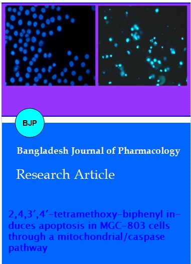
Morphological changes were observed by fluorescence microscopy. Nuclear morphological changes were observed by Hoechst 33258 staining. In control group, cells were round in shape and stained homogeneously. After treatment with 150 µM TMBP for 36 hours, blabbing nuclei and granular apoptotic bodies appeared (Figure 2A).
To further verify apoptosis induced by TMBP, the Cell Death Detection ELISA kit was employed to quantify cytoplasmic histone-associated-DNA-fragments by measuring optical density. The results showed that TMBP induced apoptotic cell death at various concentrations, which the effect of 100 uM is stronger than others (Figure 2B). In addition, DNA fragmentation became obvious after 50, 100, 200, 400 µM TMBP treatment for 36 hours and 150 µM for 24, 36 hours on agarose gel electrophoresis (Figure 2C), which further confirmed TMBP may induce cell apoptosis.
Figure 2A: TMBP-induced apoptotic morphological changes. MGC-803 cells were incubated in the medium alone for 36 hours (A) or in the medium containing 150 mM TMBP for 36 hours (B)
Figure 2B: Pro-apoptotic effect of TMBP determined by ELISA quantification of DNA fragmentation expressed as optical density (OD) values. MGC-803 cells were incubated in the medium alone or in the medium containing 50, 100, 200 and 400 mM TMBP for 36 hours and determined as instruction of manufacturer (astands for p<0.01 compared with 0 µM)
Figure 2C: DNA fragmentation induced by TMBP in MGC-803 cells
A: MGC-803 cells were treated with TMBP 0, 50, 100, 200, 400 µM for 36 hours, respectively. B: MGC-803 cells were treated with TMBP 150 µM for 0, 12, 24 and 36 hours, respectively. DNA was isolated by agarose gel electrophoresis and analyzed by ethidium bromide staining
It is generally assumed that after the loss of the outer mitochondrial membrane integrity and the release of cytochrome C from the mitochondria to the cytosol, the cells are committed to apoptosis (Martindale and Holbrook, 2002). To address the possibility that TMBP-induced apoptosis could be related to contributions from the mitochondrial pathway, MGC-803 cells were treated with various concentrations of TMBP for 36 hours. The ratio of cells with lower fluorescent intensities increases with increasing concentration of TMBP in a dose-dependent manner (Figure 3A).
Next, the effect of TMBP on the cytochrome C release from mitochondria into the cytosol was examined by western blot analysis. After incubation with TMBP, the release of cytochrome C from mitochondria was increased (Figure 3B).
Figure 3A: The TMBP induced concentration-dependent loss of mitochondrial membrane potential (ΔΨm) in MGC-803 cells.
Cells were stained with Rhodamine-123 dye and flow cytometric analyses of ΔΨm in MGC-803 cells treated with TMBP at indicated concentrations (A: 0 uM, B: 50 uM, C: 100 uM, D: 200 uM, D: 400 uM) for 36 hours was done using flow cytometry. Data are representative one of three similar experiments
Figure 3B: Increase of cytoplasmic cytochrome C in TMBP-treated MGC-803 cells. The cells were treated with 0, 50, 100, 200, 400 uM TMBP for 36 hours. Cytochrome C (15 KDa; Upper) was analyzed by western blot. GAPDH (36 KDa; Lower) was used as an equal loading control
Mitochondrial Bcl-2 family is a series of proteins that regulate apoptosis and the Bcl-2 protein is an important regulator of the apoptotic cascade and promotes cell survival. We measured the expressions of Bcl-2 by western blot analysis. After incubation with TMBP, expression of Bcl-2 protein was decreased (Figure 4).
Figure 4: Decrease in the level of anti-apoptotic protein Bcl-2. The cells were treated with 0, 50, 100, 200, 400 uM TMBP for 36 hours. Bcl-2 (21 KDa; Upper) was analyzed by western blot. GAPDH (36 KDa; Lower) was used as an equal loading control
Caspases are a family of cysteine proteases that play a central role during the executional phase of apoptosis (Vaux and Korsmeyer, 1999). To explore whether TMBP induces apoptosis by activation of caspase-3 and caspase-9, fluorogenic peptide substrates were used to detect specific caspase activity. As shown in Figure 5A, the marked activation of caspase -3 and 9 was observed after treated with 50 or 100 μM TMBP for 36 hours.
To further confirm the activation of caspase-3 and caspase-9, specific antibodies were used to detect the expressions of procaspse-3 and 9 by Western blot analysis. After incubation with indicated concentrations of TMBP, expression of procaspse-3 and 9 protein was decreased, indicating caspase-3 and 9 activation (Figure 5B). Additionally, Western blot analysis was performed to examine the expressions of caspase-3 substrate, PARP. Figure 5C showed that the expression of minor PARP (85 kDa) protein after cleavage was up-regulated by TMBP, indicating the activation of caspase-3.
To further explore whether the activation of casapse-3, 9 was required for induction of apoptosis by TMBP. MGC-803 cells were co-treated with caspase inhibitors and TMBP. To accomplish this, MGC-803 cells were pretreated for 1 hour with 100 µM caspase-3 inhibitor (Z-DEVD-FMK) and caspase-9 inhibitor (Z-LEHD-FMK). The cells were then treated with 150 uM TBMP for 36 hours. As depicted in Figure 5D, incubation with the inhibitor of caspase-3 and 9 significantly blocked TMBP-triggered apoptosis in MGC-803 cells. There observations indicated that activation of caspase-3, 9 might play a crucial role in TMBP-induced apoptotic death in MGC-803 cells.
Figure 5A: Increase of caspase-3 (Upper) and -9 (Lower) activities of TMBP-treated MGC-803 cells. The cells were treated with 0, 50, 100, 200, 400 uM TMBP for 36 hours (astands for p<0.05 compared with 0 M,bstands for p<0.01 compared with 0 μM, cstands for p<0.001 compared with 0 uM)
Figure 5B: Decrease in expression of procaspase-3 and procaspase 9 in TMBP-treated MGC-803 cells. The cells were treated with 0, 50, 100, 200, 400 uM TMBP for 36 hours. Procaspase 3 (35 KDa; Upper) and procaspase-9 (47 KDa; Middle) were analyzed by western blot. GAPDH (36 KDa; Lower) was used as an equal loading control
Figure 5C: The expression of PARP in TMBP-treated MGC-803 cells. The cells were treated with 0, 50, 100, 200, 400 μM TMBP for 36 hours. PARP (116 and 85 KDa; Upper) were analyzed by western blot. GAPDH (36 KDa; Lower) was used as an equal loading control
Figure 5D: Caspase-3, 9 inhibitors inhibited TMBP-induced apoptosis. MGC-803 cells were treated with 150 uM TMBP in the presence or absence of caspase-3, 9 inhibitors (100 uM). After 36 hours incubation, the inhibitory ratio was measured by MTT assay. Data represent the means ± SD of three independent experiments
Discussion
Protosappanin A was reported to possess antioxidant activity and inhibitory activity (Hu et al., 2008; Reddy et al., 2003). Twelve intermediates biphenyl compounds were produced in the course of the synthesis of protosappanin A. However, little has been reported about the biological activity of these compounds. In the present study we have investigated the inhibitory effects of biphenyl compounds on MGC-803 cells and it was clearly shown that some biphenyl compounds possess a cytotoxic activity against MGC-803 cells. Among twelve biphenyl compounds examined TMBP exhibited significantly stronger growth inhibitory effect in MGC-803 cells and inhibited the growth of MGC-803 cells in a concentration- and time-dependent manner. In addition, we did observe TMBP-induced typical chromatin condensation and DNA fragmentation shown by Hoechst 33258 staining and ethidium bromide staining, respectively.
Programmed cell death (PCD) is essential for normal development and maintenance of tissue homeostasis in multicellular organisms. While it is now evident that PCD can take many different forms, apoptosis is probably the most well-defined cell death program (Shemarova, 2010). The family of cysteine proteases known as caspases plays a central role in apoptosis, contributing to a signaling cascade between death promoting stimuli and the cleavage of protein substrates which contribute to the characteristic apoptotic morphology (Cohen, 1997).
Elevation in the activities of caspases observed in the present study suggests that apoptosis of cancer cells induced by TMBP is mediated through activation of these proteases. These proteases act on specific substrates leading to the degradation of PARP (Lazebnik et al., 1994). PARP (116 kDa), a DNA repair enzyme, is probably best characterized caspase substrate, which is cleaved during apoptosis to a 24 kDa and a 85 kDa fragment representing the N-terminal DNA-binding domain and the C-terminal catalytic subunit, respectively. During apoptosis, PARP is selectively cleaved by several caspases, especially by caspase-3 (Lazebnik et al., 1994; Kaufmann et al., 1993). Detection of a 85 kDa or 24 kDa caspase cleavage fragment of PARP was shown to be a hallmark of apoptosis. In this paper, PARP minor 85 kDa fragment expression was increased in a dose dependent manner, indicating the activation of caspase-3. In addition, procaspase-3 protein level was decreased, verifying the activation of caspase-3.
The activated caspases might target the permeabilized mitochondria. In recent years, it has become clear that mitochondria have significant roles to play not only in routine energy metabolism, but also in cell death. The roles in apoptosis involve a phenomenon called the mitochondrial permeability, an increase in permeability that seems to be linked to the opening of a channel termed MPT pore which is thought to underlie the loss of mitochondrial membrane potential (ΔΨm). Activation of these MPT pores permits the redistribution of molecules across the inner mitochondrial membrane, thus disrupting the ΔΨm of this organelle (Ly et al., 2003). Consequently, cytochrome C expression was more in cytosol which might be due to its release from mitochondria as an apoptotic event. Moreover, the loss of mitochondrial membrane potential ΔΨm) is facilitated through opening of mitochondrial permeability transition pores (PTP) largely due to Bax translocation, which in turn, conduct the leakage of cytochrome C and pro-apoptotic proteins from mitochondria to the cytosol (Bhushan et al., 2007). Translocation of these proteins impairs mitochondrial functions and brings the cells to a "point of no return" to enter apoptosis via activation of caspases. Furthermore, cytochrome C, which interacts with Apaf-1 and procaspase-9 to form the apoptosome, which then leads to activation of other down-stream caspases like caspase-6, particularly caspase-3. In this study, an obvious loss of ΔΨm and the release of cytochrome C were found after treatment with various concentrations of TMBP for 36 hours, suggesting the opening of MPT. Moreover, the activation of caspase-9 indicated by its activity increasing and procaspase-9 protein decreasing strongly suggests an involvement of a mitochondrial pathway.
The Bcl-2 protein family members are potent regulators of the mitochondrial changes in apoptosis. Their importance is often illustrated by eminent descriptors such as"the lords of death", "proteins in control", "gatekeepers and gatecrashers" or "bodyguards and assassins" (Fleischer et al., 2003; Gelinas and White, 2005). The main protagonists are suggested to be anti-apoptotic Bcl-2 and pro-apototic Bax. If the concentration of Bcl-2 is enough to complex with at least half of Bax, then apoptosis is prevented (Burlacu, 2003). In this paper, the Bcl-2 protein in TMBP-treated MGC-803 cell was down-regulated, which further verified that the mitochondrial pathway of cell death might be involved in TMBP-induced MGC-803 cells death.
Conclusion
TMBP possesses in vitro cytotoxicity against human gastric cancer MGC-803 cell line. TMBP induces apoptosis through a mitochondrial/caspase pathway, as evidenced down-regulation of Bcl-2, the loss of mitochondrial membrane potential, the release of mitochondrial cytochrome C and activation of caspase-3 and 9.
References
Ameisen JC. On the origin, and nature of programmed cell death: A timeline of four billion years. Cell Death Differ. 2002; 9: 367-93.
Bhushan S, Kumar A, Malik F, Andotra SS, Sethi VK, Kaur IP, Taneja SC, Qazi GN, Singh J. A triterpenediol from Boswellia serrata induces apoptosis through both the intrinsic and extrinsic apoptotic pathways in human leukemia HL-60 cells. Apoptosis 2007; 12: 1911-26.
Burlacu A. Regulation of apoptosis by Bcl-2 family proteins. J Cell Mol Med. 2003; 7: 249-57.
Cohen GM. Caspases: The executioners of apoptosis. Biochem J. 1997; 326: 1-16.
Fleischer A, Rebollo A, Ayllon V. BH3-only proteins: The lords of death. Arch Immunol Ther Exp. 2003; 51: 9-17.
Fulda S, Debatin KM. Extrinsic versus intrinsic apoptosis pathways in anti-cancer chemotherapy. Oncogene. 2006; 25: 4798-811.
Gelinas C, White E. BH3-only proteins in control: Specificity regulates MCL-1 and BAK-mediated apoptosis. Genes Dev. 2005; 19: 1263-68.
Ghobrial IM, Witzig TE, Adjei AA. CA. Targeting apoptosis pathways in cancer therapy. Cancer J Clin. 2005; 55: 178-94.
Han YH, Kim SZ, Kim SH, Park WH. Pyrogallol inhibits the growth of lung cancer Calu-6 cells via caspase-dependent apoptosis. Chem-Biol Interact. 2009; 177: 107-14.
Harada H, Grant S. Apoptosis regulators. Rev Clin Exp Hematol. 2003; 7: 117-38.
Hu J, Yan XL, Wang W, Wu H, Hua L, Du LJ. Antioxidant activity in vitro of three constituents from Caesalpinia sappan L. Tsinghua Sci Tech. 2008; 13: 474-79.
Ji YB, Qu ZY, Zou X. Juglone-induced apoptosis in human gastric cancer SGC-7901 cells via the mitochondrial pathway. Exp Toxicol Pathol. 2011; 63: 69-78.
Kaufmann SH, Desnoyers S, Ottaviano Y, Davidson NE, Poirier GG. Specific proteolytic cleavage of poly (ADP-ribose) polymerase: An early marker of chemotherapy-induced apoptosis. Cancer Res. 1993; 53: 3976-85.
Lazebnik YA, Kaufmann SH, Desnoyers S, Poirier GG, Earnshaw WC. Cleavage of poly (ADP-ribose) polymerase by a proteinase with properties like ICE. Nature 1994; 371: 346-47.
Lou C, Yang G, Cai H, Zou M, Xu Z, Li Y, Zhao F, Li W, Tong L, Wang M, Cai B. 2′, 4′-Dihydroxychalcone-induced apoptosis of human gastric cancer MGC-803 cells via down-regulation of survivin mRNA. Toxicol in vitro. 2010; 24: 1333-37.
Ly JD, Grubb DR, Lawen A. The mitochondrial membrane potential (deltapsi(m)) in apiotosis; An update. Apoptosis 2003; 8: 115-28.
Martindale JL, Holbrook NJ. Cellular response to oxidative stress: Signaling for suicide and survival. J Cell Physiol. 2002; 192: 1-15.
Niranjan Reddy VL, Ravikanth V, Jansi Lakshmi VV, Suryanarayan Murty U, Venkateswarlu Y. Inhibitory activity of homoisoflavonoids from Caesalpinia sappan against Beauveria bassiana. Fitoterapia 2003; 74: 600-02.
Pilatova M, Varinska L, Perjes P, Sarissk M, Mirossa L, Solar P, Ostro A, Mojzis J. In vitro antiproliferative and antiangiogenic effects of synthetic chalcone analogues. Toxicol in Vitro. 2010; 24: 1347-55.
Shen Y, Du X. The preparation and application of biphenyl compound, China Patent 200810070623.0. 2008.
Shemarova IV. Signaling mechanisms of apoptosis-like programmed cell death in unicellular eukaryotes. Comp Biochem Phys B. 2010; 155: 341-53.
Vaux DL, Korsmeyer SJ. Cell death in development. Cell 1999; 96: 245-54.
Wu J, Hou JB, Zhang MM, Zou YP, Yu B. Protosappanin A, an immunosuppressive constituent from a Chinese herb, prolongs graft survival and attenuates acute rejection in rat heart allografts. Transplant Proc. 2008; 40: 3719-22.