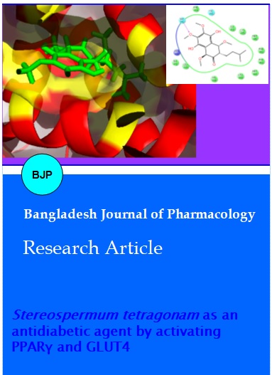
Stereospermum tetragonam as an antidiabetic agent by activating PPARγ and GLUT4
Abstract
Present study evaluates the antidiabetic activity of S. tetragonam LC-MSMS experiments showed the presence of two novel molecules C1 and C2, which were further taken for in silico study against PPARγ. Cell culture studies with A431 cells in the presence of crude aqueous extract showed the elevated level of PPARγ and GLUT4 and also confirmed using in silico studies. Thus, the present study proves the mecode of action of S. tetragonam as an antidiabetic drug.
Introduction
Diabetes mellitus (DM) is a major health problem and one of the common endocrine metabolic disorders throughout the world which has caused morbidity and mortality due to microvascular and macrovascular complications. Metabolic disorder of DM arises due to relative or absolute deficiency of insulin secretion (Ozkol et al., 2013). Glucose transporters (GLUT) are the transmembrane proteins responsible for the transport of glucose and other substrates into the cells on the basis of specific inducers (Thorens, 2001). GLUT4 is one of the most important isoforms of GLUT in skeletal muscles and adipocytes, which facilitates the entry of glucose to these hexose utilizing cells (Wood and Trayhurn, 2003). Peroxisome proliferator-activated receptor (PPAR) is a member of nuclear receptor superfamily present in adipocytes, muscle and liver. PPARγ triggers cellular assimilation of lipids through anabolic pathways (Semple et al., 2006). The agents like glitazone-class antidiabetic agents act on PPARγ. PPARγ is highly expressed in adipocytes to facilitate glucose and lipid uptake and in turn stimulates glucose oxidation, decreases free fatty acid level and ameliorates insulin resistance in type 2 DM.
Recent studies on Stereospermum tetragonum showed promising antidiabetic activity in alloxan- and streptozotocin-induced diabetic model. Two active principles were isolated and identified as an iridoid type glycoside and the other one was a lapachol like compound (Bino Kingsley et al., 2013). The aim of the present study is to determine the active principles and evaluate their efficiency to activate of PPARγ and GLUT4 against diabetes mellitus.
Materials and Methods
Aqueous extraction of dry powder
To prepare water extract, the powder S. tetragonam was extracted with distilled water (5 g/100 mL) by stirring for 4 hours and then filtering through filter paper (Whatman No. 1). This process was repeated thrice with the residue. The combined filtrate was freeze-dried in a lyophilizer.
Isolation of an active fraction (AF)
The water extract of S. tetragonum root powder was precipitated with absolute ethanol (1:1 v/v) and separated in to precipitate and soluble fractions. Then the soluble fraction of the root extract further purified for the isolation of active principles using thin layer chromatography and characterized using FT-IR and NMR which was already published from our group.
Qualitative analysis of components using electro spray ionization mass spectrometry (ESI-MS/MS)
Qualitative identification of compound C1 and C2 present in the active fraction of S. tetragonam, crude aqueous extract of S. tetragonam was analyzed using UHPLC/ESI attached to a Q-TOF mass detector (micrOTOF-Q II, Bruker, Germany). First phytomolecules were separated using RP C18 column (150 x 2.1 mm with internal diameter of 3.0 µm 120Angstrom Acclaim®, USA). For better elution gradient solvent system was performed using acetonitrile (A) and milliQ water (1% acetic acid) starting at 0.2 min at 1% ACN and 99% water (1% acetic acid) to 75% ACN at 16th min, this was brought to 100% ACN at 19th min to 5% ACN at 21st min and was maintained at same condition till run ends at 23rd min, with flow rate of 200 µL/min. UV absorbance was detected at arbitrary at 325 nm. Negative mode of ionization was selected at following parameters: Nebulizer 30.5 psi with 6.0 l/min N2 flow, m/z range: 50-1000 m/z, Capillary voltage 4500 V, dry heater temperature at 280°C. and analyzed using Data analysis software provided by the manufacturer. Based on the MS and its corresponding MSMS fragmentation pattern, C1 and C2 were identified.
Molecular docking
The structures considered for the study were obtained from the isolation of active fraction and from spectral studies. 1,4a,5,7a-tetrahydro-5-hydroxy-7-(hydroxymethyl)-1-(tetrahydro-6-(hydroxy-methyl)-3,4,5-trimethoxy-2H-pyran-2-yloxy)cyclopenta [c]pyran-4-carboxylic acid (C1) and 5,8-dihydro-7-isopentyl-2,3,5,8-tetramethoxynaphthalene-1,4,6-triol (C2) were used as ligands and draw the structures using CHEMDRAW (Version 11). The structure of PPARγ was obtained from the protein databank. Hydrogen atoms were added to the protein consistent with pH 7.0 using the protein preparation wizard in the Schrodinger suite (Madhavi Sastry et al., 2013). Further, the protein's hydrogen bond network was also optimized using the wizard. The so-prepared structure was then subject to energy minimization and the termination condition for minimization was fixed as the step when the root mean square deviation of the heavy atoms in the structure relative to the starting structure exceeded 0.3 Angstrom. This process also ensures that the hydrogen atoms are placed in optimized geometries. The protein thus prepared was used for docking of the ligands as described below. Potential binding sites in PPARγ were predicted using the SiteMap tool in the Schrodinger suite (Halgren, 2007; Halgren, 2009). Five different binding sites were identified in PPAR. Of these predicted sites, site 3 had the highest score. Hence binding of a ligand to site 3 is more likely to result in the activation of this receptor. Receptor grid was then generated for site 3 using Glide module (v 5.8) of the Schrodinger suite. The grid box and center were set to default. LigPrep module (version 2.5) of the Schrodinger suite was used to generate conformers of the ligands. The ligands were then docked using the extra precision mode in the Glide module (Friesner et al. 2004; Friesner et al., 2006; Halgren et al., 2004) of the Schrodinger suite.
Cell culture assay
A431 human skin carcinoma cells (NCCS, INDIA) were maintained in Dulbecco's Modified Eagle Medium (DMEM) containing high glucose level of 4.5 g/L, L-glutamine, sodium pyruvate and sodium bicarbonate. 20% Fetal calf serum (FCS) and 1% penstrep antibiotics were added in the working media solution. A431 cells line are well known for their expression study on GLUT4 and PPAR gamma.
Determination of cell GLUT4 and PPAR gamma
Cell lysate were made and the resultant supernatant was used for the estimation of GLUT4 and PPAR gamma expression levels using ELISA kit (Qiagen QY-03235). Briefly, at 70% confluence, cells were trypsinized and 0.2 x 10⁶ cells were seeded and grown to 70% confluence in a 24-well tissue culture plate conditioned with 5% CO2 atmosphere at 37°C. Cells were then treated with 20, 30 and 40 µg/mL of active fraction of S. tetragonam for 48 hours followed by cell lysis using tween 80, resulted mixture was then mixed and centrifuged for 10 min at 3,000 rpm. Supernatant was collected and used as test sample for ELISA. Assay was performed by the addition of test samples at different concentration into respective wells which are already coated with anti-GLUT4 and anti-PPAR gamma antibodies, followed by the addition of secondary antibody conjugated to horseradish peroxidase except for the blank wells; this was then incubated for 60 min at 37°C. Each wells were then washed thrice with 1x PBS (washing buffer), followed by the addition of chromogen solution (o-phenylenediamine dihydrochloride) with gentle shake and incubated at dark condition for 10 min at room temperature. This reaction was terminated with the addition of 3 N HCl (stop solution). Absorbance were read at 450 nm using Microplate Reader (Epoch, BioTek, USA) within 15 min after reaction termination. Background absorbance obtained with blank well was subtracted from all values of other wells. Estimation of GLUT4 and PPAR gamma in the test samples were determined according to standard graph generated using various standard concentrations and their corresponding OD values.
Result and Discussion
C1 and C2 were identified using LC-MSMS analysis on the active fraction of S. tetragonam. C1 has shown a m/z value of 431 [M-H]- with major MS2 peaks as 283, 187, 255, 298, 255, 160, 227, 269. Similarly C2 showed m/z value of 366 [M-2H]- with major MS2 peaks were 338, 237, 160, 215, 310, 265, 293, 187. MSMS fragmentation pattern of C1 and C2 proves the proposed structure of these molecules (Figure 1).
Figure 1: LC/MS/MS pattern for novel two active principles present in the aqueous extract of S. tetragonam
Active fraction has shown its antidiabetic activity by elevating the expression level of PPAR gamma and GLUT4 as seen through ELISA detection of these proteins in the A431 treated cell lysate (Figure 2). PPAR gamma activation is essential for the gene level activation of glucose uptake related proteins specifically GLUT4 transporter. Increase level of PPAR gamma leads to the increase expression level of GLUT4 in the cell membranes which in turn increase the uptake of blood glucose and utilizes it within the cell as energy metabolism or for storing it as glycogen for further usage. Many synthetic drugs have shown binding affinity followed by activation of peroxisome proliferator-activated receptor (PPAR-gamma) (Marx and Hombach, 2001; McCarthy and Elmendorf, 2007). PPAR related proteins are well known for its role in lipid homeostasis through fatty acid catabolism and are present at high levels in the adipose tissues, liver and in skin epithelial cells. Specifically PPAR-gamma is highly expressed in adipose tissues during adipogenesis. Although binding of ligands to PPAR-gamma is required for its transcriptional activation, natural occurring ligands against PPAR proteins are not yet scientifically explored which can be used as an eco-friendly antidiabetic drug. Present study has clearly demonstrated that C1 and C2 require minimum binding energy to bind with PPAR-gamma. Present studies provide scientific evidence that C1 and C2 are involved in the activation of PPAR-gamma which in turn provides antidiabetic activity through activation of GLUT4 (Semple et al., 2006; Thorens, 2001).
Figure 2: Influence of different concentrations aqueous extract of S. tetragonam on the activity of PPARγ (A) and GLUT4 (B). Values are expressed as mean ± SD; n = 4; ap<0.05
The activation of PPARγ is further confirmed using in silico studies. It indicates that both C1 and C2 strongly interact with PPARγ through different residues (Figure 3 and 4). Among the two C1 exhibited maximum number of hydrophobic interactions compared to C2. The XP Glide score for both the compounds includes -4.7 Kcal/mol for C1 and -4.0 Kcal/mol for C2 clearly suggested that both the compounds show better interaction with PPARγ which may probably activate PPARγ signaling cascades thereby increasing the levels of GLUT4. This is already confirmed through ELISA experiments.
Figure 3: Probable interaction of C1 with PPARγ predicted using molecular docking studies. A: surface view; B: ligand interaction diagram
Figure 4: Interaction of C2 compound with PPARγ predicted using in silico studies. A: Surface view; B: Ligand interaction diagram
Conclusion
The present study evaluated the molecular mechanism for the antidiabetic potential of Indian herb, S. tetragonam using ELISA technique and molecular docking. LC/MS/MS technique results indicated that the aqueous extract of S. tetragonam contains two new compounds (C1 and C2). The cell culture studies indicated that the extract dose-dependently increase the level of PPARγ and GLUT4 compared to control which in turn express the levels of thereby increasing glucose metabolism.
Contribution
First two authors contributed equally in this study.
Acknowledgement
The authors wish to acknowledge SASTRA University for financial and infrastructural support.
References
Friesner RA, Murphy RB, Repasky MP, Frye LL, Greenwood JR, Halgren TA, Sanschagrin PC, Mainz DT. Extra precision Glide: Docking and scoring incorporating a model of hydrophobic enclosure for protein-ligand complexes. J Med Chem. 2006; 49: 6177-96.
Halgren T. New method for fast and accurate binding-site identification and analysis. Chem Biol Drug Design. 2007; 69: 146-48.
Halgren TA. Identifying and characterizing binding sites and assessing druggability. J Chem Inf Model. 2009; 49: 377-89.
Halgren TA, Murphy RB, Friesner RA, Beard HS, Frye LL, Pollard WT, Banks JL. Glide: A new approach for rapid, accurate docking and scoring. 2. Enrichment factors in database screening. J Med Chem. 2004; 47: 1750-59.
Madhavi Sastry G, Adzhigirey M, Day T, Annabhimoju R, Sherman W. Protein and ligand preparation: Parameters, protocols, and influence on virtual screening enrichments. J Comput Aided Mol Des. 2013; 27: 221-34.
Ozkol H, Tuluce Y, Dilsiz N, Koyuncu Ä°. Therapeutic potential of some plant extracts used in Turkish traditional medicine on streptozocin-induced type 1 diabetes mellitus in rats. J Membrane Biol. 2013; 246: 47-55.
Postic C, Shiota M, Magnuson MA. Cell-specific roles of glucokinase in glucose homeostasis. Recent Prog Horm Res. 2001; 56: 195-217.
Semple RK, Chatterjee VK, O'Rahilly S. PPAR gamma and human metabolic disease. J Clin Invest. 2006; 116: 581-89.
Thorens H-GJB. The extended GLUT-family of sugar/polyol transport facilitators: Nomenclature, sequence characteristics, and potential function of its novel members. Molec Membrane Biol. 2001; 18: 247-56.
Wood IS, Trayhurn P. Glucose transporters (GLUT and SGLT): expanded families of sugar transport proteins. Bri J Nutri. 2003; 89: 3-9.