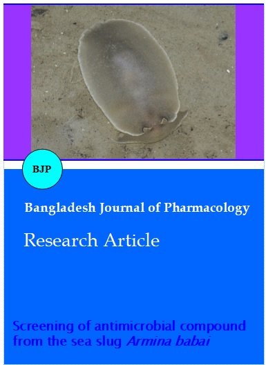
Screening of antimicrobial compound from the sea slug Armina babai
Abstract
Different solvents such as acetone, butanol, ethanol, hexane and methanol of sea slug Armina babai was evaluated for its biomedical properties. Most potent extracts were purified using silica gel column and the active fractions were characterized by TLC, SDS-PAGE and FTIR. Maximum zone of inhibition was recorded against Pseudomonas sp., and minimum zone of inhibition was recorded against Proteus mirabilis by butanol extracts. TLC profiling with Rf value 0.82 indicating the presence of amino acids and peptides. Total protein was estimated as 21.36% with molecular weight range between 13 and 72 kDa in SDS-PAGE. The FT-IR spectrum of fractions, obtained from sea slug, reveals characteristic functional groups range between 465.8 and 3423.8 cm-1. Thus the obtained results indicate the presence of potent antimicrobial compounds in sea slug A. babai may pave the way to explore the potential development of new compounds to be launched in the pharmaceutical filling.
Introduction
Marine invertebrates are a rich source of structurally novel and biologically active metabolites. Among them, Sea slugs are the most primitive invertebrates considering as the major rich phyla with novel bioactive compounds. They are soft bodied and slow moving and thus would be highly vulnerable to predators if not for the possession of a variety of defenses. Sea slugs are constantly exposed to relatively high concentrations of bacteria, viruses and fungi of which many of them are harmful. The survival of these organisms depends on efficient antimicrobial mechanisms to protect themselves against microbial infections and fouling. These include cryptic coloration behavior, large size, ability to produce copious mucus and most notably chemical metabolites (Johnson and Willows, 1999; Wagele and Klussmann-Kolb, 2005). Screening of organic extracts from marine organisms is a common approach to identify compounds of biomedical importance. Sea slug with both rich variety of secondary metabolites and great biomedical potential represent the most intensively studied group of mollusks in natural product chemistry. In this report, we describe the screening of organic solvent extracts of sea slug Armina babai, collected from the coastline of south India for antibacterial and antifungal activities and partially purify and characterize the antibacterial compound with the aim potentially useful therapeutic activities.
Materials and Methods
Collection and extraction
Live specimens of A. babai were collected from the Parangipettai, Southeast coast of India. Specimens collected were transported to the laboratory then homogenized in a blender and extracted using different solvents viz., acetone, butanol, ethanol, hexane and methanol at room temperature. The homogenate of different extracts were centrifuged at 5,000 rpm for 15 min; then the supernatants were collected and then filtered through Whatman 1 filter paper. The solvent from combined filtrates was evaporated at reduced pressure and temperatures and the residue obtained i.e. the crude extract of different solvents was used for primary screening. Crude extracts showing significant activity were further partially purified using Silica gel column. For the purpose, butanol extract was dissolved in 5% aqueous buatnol and then partitioned successively with n-butanol. Organic layer was separated and concentrated under vacuum to obtain respective fractions.
Microbial pathogens
List of pathogens includes 10 bacterial strains viz., Escherichia coli (-ve), Klebsiella oxytoca (-ve), K. pneumoniae (-ve), Proteus mirabilis (-ve), Pseudomonas sp (-ve), P. aeruginosa (-ve), Salmonella paratyphi (-ve), S. typhi (-ve), Staphylococcus aureus (+ve) and Vibrio cholerae (-ve) as well as 10 fungal strains viz., Alternaria alternate, Aspergillus flavus, A. niger, Candida albicans, C. tropicalis, Epidermophyton floccosum, Mucor sp., Penicillium sp., Rhizopus and Trichophyton rubrum. These clinical bacterial and fungal strains were obtained from the Department of Medical Microbiology, Raja Muthiah Medical College and hospital, Annamalai University, Annamalai nagar.
Antimicrobial assay
Antibacterial activity of different extracts was assessed using standard disc-diffusion assay on nutrient agar as described earlier (Rodrigues et al., 2004). Briefly crude extracts were dissolved in the respective solvents and loaded onto sterile paper discs, Each test organism was inoculated on respective agar plates (HiMedia) and spread to form a lawn of individual pathogens. Crude extracts loaded discs a well as positive and negative control discs for comparison were also placed on the plates. Positive control discs comprised of streptomycin (10 µg/disc) while negative control discs were discs with the respective solvents. Plates were incubated at 37°C for 24 hours until culture developed a confluent film. Similar procedure was followed for antifungal activity and Ketoconazole was used as positive control (100 µg/disc). Antimicrobial activity was expressed in terms of diameter zone of inhibition and it was measured in millimeter using scale recorded. Both the antibacterial and antifungal assays were repeated twice and each assay was performed in duplicate.
Minimum inhibitory concentration (MIC)
The MIC of A. babai extracts were determined against the test pathogens by a standard agar dilution method. Briefly, to the tubes containing 9 mL melted media, serial dilutions of the extract were added to 10 mL and poured into a sterile plate. Sample concentrations in the agar plates were in the range of 20-200 µg/mL. Equivalent amounts of sterile distilled water and sterile solvents were used as controls. Ten μL of each overnight grown pathogen was inoculated into each agar dilution plate. The inoculated plates were incubated overnight at 37°C. The MIC was defined as the lowest concentration of the samples which prevented this change and exhibited complete inhibition of bacterial growth (Ngameni et al., 2009).
Thin layer chromatography (TLC)
5 uL of fractionated extracts were applied on 5 x 10 precoated silica gel 60 TLC plate (Merck) of 254 uniform thickness of 0.2 mm by using Hamilton Syringe. Plates were developed in n-butanol: acetic acid: water (BAW) (5:1:4) as solvent system to resolve different components of these extracts. The plates were sprayed with ninhydrin (1.5 gm ninhydrin was dissolved in 100 mL of ethanol) and the plate was air dried and kept in an oven at 45°C for 5-10 min to develop purple to pink colored spots. The retention factor (Rf) was calculated by using the formula the distance traveled by the compound divided by the distance traveled by the solvent.
Molecular mass determination
SDS PAGE): The protein concentration of the A. babai was determined based on the method of Bradford, (1976) with bovine serum albumin (BSA) as standard. The protein profile of fractionated extract was analyzed using sodium dodecylsulphate-polyacrylamide gel electrophoresis (SDS-PAGE) as described by Laemmli, (1970). SDS-PAGE standard markers (High range, Genei, Bangalore) were included to estimate the molecular mass of proteins. The separated peptides were stained in 0.1% (v/v) Coomassie blue R-250 (Coomassie brilliant blue G 0.025% and acetic acid 10%) and destained in 40% methanol and 10% acetic acid for 30 min.
Fourier transforms infrared spectroscopy (FTIR) spectral analysis
Approximately 5 mg of sample was mixed with 100 mg of dried potassium bromide (kbr) was subjected to a pressure of 5 x 10⁶ pa and made into clear pellet of 3 mm diameter and 1 mm thickness. Absorbance spectra were recorded using Nicolet Avatar-360 FTIR Spectrometer equipped with a KBr beam splitter and an air-cooled DTGS detector, Department of Chemistry, Annamalai University. The absorption of light intensity of the peak was calculated using the base line method. The frequencies for all sharp bands were accurate to 0.01 cm-1 (Abu et al., 1991).
Results
Different solvent extracts of A. babai were active against the tested pathogens in primary screening as well as comparison with the respective standards (streptomycin/ketoconazole) but to varying degrees (Table I). Maximum zone of inhibition (16 ± 0.2 mm) was recorded against Pseudomonas sp., and minimum zone of inhibition (4 ± 0.9 mm) was recorded against Proteus mirabilis by butanol extracts. Acetone extracts showed a moderate activity on the bacterial pathogenic strains with the maximum zone of inhibition (9 ± 1.4 mm) against V. cholerae and the minimum against E. coli (3 ± 1.6 mm).
Table I: Antimicrobial activity of various solvent extracts from A. babai against the various pathogens
| Tested organism | Different extracts of A. babai | Std | ||||
|---|---|---|---|---|---|---|
| Acetone | Butanol | Ethanol | Hexane | Methanol | ||
| E. coli | + | ++ | + | - | - | +++ |
| K. oxytoca | + | + | - | + | - | ++ |
| K. pneumoniae | - | + | - | - | - | ++ |
| P.mirabilis | - | + | - | - | - | + |
| Pseudomonas sp | - | ++++ | - | + | - | +++ |
| P. aeruginosa | ++ | +++ | + | - | + | ++ |
| S. paratyphi | + | + | + | ++ | ++ | - |
| S. typhi | + | ++ | - | + | + | + |
| S. aureus | + | + | - | - | ++ | - |
| V. cholerae | ++ | + | - | - | - | + |
| A. alternate | - | - | - | - | - | - |
| A. flavus | - | - | - | - | - | ++ |
| A. niger | - | - | - | - | - | ++ |
| C. albicans | - | - | - | - | - | - |
| C. tropicalis | - | - | - | - | - | - |
| E. floccosum | - | - | - | - | - | + |
| Mucor sp | - | - | - | - | - | + |
| Pencillium sp | - | - | - | - | - | + |
| Rhizopus sp | - | - | - | - | - | + |
| T. rubrum | - | - | - | - | - | - |
| - = No activity, + = weak activity (inhibition zone 1-5 mm), ++ = moderate activity (inhibition zone 5-10 mm), +++ = good activity (inhibition zone 10-15 mm), ++++ = highly active (inhibition zone above 15 mm). Std = Streptomycin for bacteria/ Ketoconazole for fungi | ||||||
The crude ethanol extracts exhibited poor activity and only three strains (E. coli, P. aeruginosa, S. paratyphi) inhibited at the minimum zone. The hexane extracts showed a good activity with the maximum zone of inhibition (9 ± 0.5 mm) against S. paratyphi and minimum level (2 ± 1.5 mm) against S. typhi. Similarly the crude methanol extracts exhibited good activity on the selected pathogens. All the solvent extracts of A. babai was insensitive against all the fungal strains used. As the evident from the results of primary screening, crude butanol extracts appeared to be quite promising for their ability to inhibit the selected pathogenic bacteria. In secondary screening all the fractions of A. babai were more active against all the bacterial strains and also on few fungal strains but to varying degrees (Figure 1).
Figure 1: Antimicrobial spectrum of fractions of A.babai against pathogens
MIC of the A. babai extracts was further examined against pathogens. The results indicated that the extract with butanol extract possessed the strongest antibacterial activity followed by the methanol, acetone, hexane and ethanol extracts (Table II). No extracts showed the activity against the fungal strains even greater concentrations (500 µg/mL). Hence the results were not provided.
Table II: The agar dilution method was used to determine MIC of the extract with different organic solvents from A. babai against different bacterial strains*
| Microorganisms | MIC for A. babai extracts (µg/mL) | ||||
|---|---|---|---|---|---|
| Acetone | Butanol | Ethanol | Hexane | Methanol | |
| E. coli | 200 | 50 | 200 | >200 | 25 |
| K. oxytoca | 200 | 100 | >200 | 200 | >200 |
| K. pneumoniae | >200 | 100 | >200 | >200 | 100 |
| P. mirabilis | >200 | 100 | >200 | >200 | >200 |
| Pseudomonas sp | >200 | 25 | >200 | 200 | 200 |
| P. aeruginosa | 150 | 50 | 200 | >200 | 100 |
| S. paratyphi | >200 | 200 | 200 | >200 | >200 |
| S. typhi | >200 | 150 | >200 | >200 | 200 |
| S. aureus | 150 | 200 | >200 | 100 | 50 |
| Vibrio cholerae | >200 | 200 | >200 | 200 | 100 |
| *No bacterial growth in the negative control plate | |||||
TLC profiling was done for both crude and fractions of A. babai in BAW solvent system. The development of purple to pinkish colored spots on the TLC plate was observed when sprayed with location reagent (nin-hydrin reagent). The plate showed pink spots indicating the presence of amino acids and peptides (Figure 2). The Rf value was calculated to the fractions showing broad band as 0.8. Total protein concentration of A.
Figure 2: TLC profile of A. babai extracts
babai was estimated as 21.4% of dry weight .The electrophoretic profiles obtained from the extracts displayed the largest protein concentration. SDS-PAGE analysis revealed that the extracts of A. babai showed the molecular weight of 13, 18, 20, 46 and 72 kDa (Figure 3). The FT-IR spectrum of fractions, obtained from sea slug, reveals characteristic functional groups range between 465.8 and 3423.8 cm-1. Sample of the 6 major peaks were at 2860.3, 1544.8, 1446.6, 1404.2, 1117.8 and 465.8 cm-1, whereas the spectra of the sample of A. babai showed all peaks with very close values at 3423.8, 3074.0, 2964.4, 2925.4, 1638.5, 1237.7, 1047.5, 674.0 and 615.0 cm-1. The OH stretching frequency appeared at 3423.8 cm-1 and CH stretching frequency in O-CH3 group appeared at 2964.4 cm-1.
Figure 3: Molecular determination of A. babai extracts (L1- Protein marker; L2- Extracts)
The OH stretch group antisymmetry frequency appeared at 3074.0 cm-1 and CH3 symmetry deformation at 2860.3 cm-1.Substituted Benzene ring stretches appears at 2057 cm-1with the NH stretch in secondary amide (polypeptides) appeared at 1638.5 cm-1 in C-H stretch-ing C-CH3 group appeared at 1544.8 cm-1 and the C-O stretch on tertiary amide (Amide-I) appeared at 1446.6 and 1404.2 cm-1. The C-O-C stretching frequency appeared at 1237.7 cm-1 and the alcohol C-H bend frequency at 1117.8 cm-1. The cyclic compound carbon ring frequency appeared at 1047.5 cm-1. The N H deformation frequency of secondary amide (II) appeared 674.0 cm-1 and the amide (III) group C-N stretch at 465.8 cm-1 (Figure 4). The FT-IR spectrum of A. babai confirms the presence of primary amine group, secondary amide, aromatic-compounds and aliphatic–alkyl group.
Figure 4: FTIR spectra of the A. babai fractionated sample
Discussion
In recent years, great attention has been paid to study the bioactivity of natural products for their potential pharmacological utilization. Secondary metabolites clearly seem to play a role in the biotic interactions with potential predators throughout all systematic groups of Opisthobranchia (Cimino et al., 2004) and the effect of their secondary metabolites on pathogens is hardly investigated. In this study the secondary metabolites were partially purified from slug A. babai collected from the southern coast of India and evaluated for its biological activity. The results of primary screening revealed that the solvent extracts showed moderate activity against the tested pathogens except the fungal strains. Generally, extraction with a predominantly solvent, improved activity because of biologically active components that were extracted by solvents (Voultsiadou, 2010). Thus, the bacterial strains were more sensitive to extracts than the fungal pathogens.
Wagele et al. (2006) noted the presence of homogenously staining violet glands as a characteristic of Dendronotoidea. In their survey on glandular structures in Opisthobranchia, they observed these glands for nine (out of 17 investigated) members of the Dendronotoidea. These glands are absent in any members of the Aeolidoidea and "Arminoidea". Defensive mechanism is one such adaptation which is well studied in most of invertebrates. The antibacterial assay have been reported long back in several molluscan species such as Crassostrea virginica (oyster), Mytilus edulis and Geukensia demissa (mussel) (Anderson and Beaven, 2001), Dicathais orbita (muricid mollusks) (Benkendorff et al., 2001), Dolabella auricularia (sea hare) (Vennila et al., 2011). Significant antimicrobial activity was observed from the polysaccharide extracted from cuttle bone and methanolic extract of body tissue of Sepia parshadi, against ten human pathogenic bacteria and five fungi (Ramasamy et al., 2011b).
In our present investigation the butanol extracts showed a wide range of activity against the pathogens in crude as well as fractions thus the results can be attributed to solvent-extract component affinity, i.e. the solubility of slug active compounds in solvents will determine the composition of the extract. Similar results were observed in polar solvent Octopus methanol extracts with 100% concentration, the highest inhibition zone was observed against E. coli in Octopus dolfusii extract and lowest inhibition zone against S. pneumonia in O. dolfusii extract respectively (Ramasamy et al., 2011a). The crude and purified sample of glycosaminoglycans (GAGs) from Euprymna berryi showed activity against five pathogenic bacteria and four fungal strains (Shanmugam et al., 2008). The activity was higher in fractions and lower in crude extracts; but activity was absent in negative control. Moderate antibacterial and antifungal activity were also reported from the extracts of various bivalve mollusks (Prem Anand and Patterson Edward, 2002) and also from the aqueous ink extract of the cephalopods L.duvaceli and S. pharaonis against nine human pathogens (Patterson Edward and Murugan, 2000). The butanol extracts of A. babai were analyzed by TLC chromatography displaying different secondary metabolite patterns. In particular, the extract was characterized by the presence of broad positive spot at Rf 0.8 along with usual lipids and sterols and a minor polar compound at Rf 0.7. The protein concentration was carried out in the current study resulting about 21.4% of the total dry weight of the animal and the molecular weight existed from 13, 18, 20, 46 and 72 kDa were relatively less distinct to the protein concentration profiling of the ink sample of O. vulgairs (Vennila et al., 2011).
The FT-IR spectroscopy is complementary to the CD-spectroscopy method according to the conformational studies of peptides and proteins (Baginska et al., 2008). Therefore, sometimes they may be used simultaneously. However, in the FT-IR spectroscopy in water solution to have a good signal-to-noise ratio, the concentrations of the peptide is usually from 4 to 30 mg/mL and it should be marked, in current studies for CD-spectroscopy the concentration of peptide is relatively low (0.2 mM). The second derivative spectrum gives a negative peak for every band maximum or shoulder in the absorption spectrum whereas the fourth derivative spectrum gives a positive peak for every band maximum. Second derivative spectra allow the identification of various secondary structures present in the protein.
Most of the peak positions are easily found in the second derivative spectra. An improved method for carrying out second derivative analysis has established the utility of the method for obtaining quantitative as well as qualitative determination of alpha-helix, beta-sheet, random and turn structures. A curve fitting procedure can be applied to calculate quantitatively the area of each component representing a type of secondary structure. The results showed the second derivative spectrum of peptides as well as those of amyloid-β-peptide and amide I and II. In the present investigation, solvent extract of A. babai muscles samples evaluated against 10 different pathogenic bacteria and multi drug resistant bacterial strains and the positive active muscle extract was further subjected to TLC studies. It is evident from the active fractions of positive that antimicrobial activity observed in the extracts presence of peptides and molecular weight analysis showed different molecular weight protein of proteins present after electrophoresis band were detected in the gel which represented proteins and Fourier transforms infrared spectroscopy analysis reveals the presence of bioactive compounds signals at different ranges.
Conclusion
This is the first report of a screening of bioactive compound in A. babai. The preliminary results obtained in these experiments pave the road to explore the potential development of new compounds to be launched in the pharmaceutical market filling a tremendous gap, as day by day new multi resistant microorganisms emerges.
Acknowledgement
Authors are very thankful Dean and Director of Centre of Advanced Study in Marine Biology, Annamalai University for providing facilities and encouragement.
References
Abu GO, Weiner RM, Rice J, Colwell RR. Properties of an extracellular adhesive polymer from the marine bacterium Shewanella colwelliana. Bio foul. 1991; 3: 69-84.
Anderson RS, Beaven AE. Antibacterial activities of oyster (Crassostrea virginica) mussel (Mytilus edulis and Geukensia demissa) plasma. Aquat Living Resour. 2001; 14: 343-49.
Bagińska K, Makowska J, Wiczk W, Kasprzykowski F, Chmur-zyński L. Conformational studies of alanine-rich peptide using CD and FTIR spectroscopy. J Peptide Sci. 2008; 14: 283–89.
Benkendorff K, Bremner JB, Davis AR. Indole derivatives from the egg masses of Muricid molluscs. Molecules 2001; 6: 70-78.
Bradford MM. A rapid and sensitive method for the quantitation of microgram quantities of protein utilizing the principle of protein-dyebinding. Anal Bio chem. 1976; 72: 248-54.
Cimino G, Fontana A, Cutignano A, Gavagnin M. Biosynthesis in opisthobranch molluscs: General outline in the light of recent use of stable isotopes. Phytochem Rev. 2004; 3: 285-307.
Johnson PM, Willows AOD. Defense in sea hares (Gastropoda, Opisthobranchia, Anaspidea): multiple layers of protection from egg to adult. Mar Fresh Behav Physiol. 1999; 32: 147–80.
Laemmli UK. Cleavage of structural proteins during the assembly of the head of bacteriophage T4. Nature 1970; 227: 680-85.
Ngameni B, Kuete V, Simo IK, Mbaveng AT, Awoussong PK, Patnam R, et al. Antibacterial and antifungal activities of the crude extract and compounds from Dorstenia turbinata (Moraceae). S Afr J Bot. 2009; 75: 256-61.
Patterson Edward JK, Murugan A. Screening of cephalopods for bioactivity. Phuket. Marine Biological Center Space Weather Prediction Center Publications, 2000; 21: 253-56.
Prem Anand T, Patterson Edward JK. Antimicrobial activity in the tissue extracts of five species of cowries Cyprea sp. (Mollusca: Gastropoda) and an ascidian Didemnum psammathodes (Tunicata: Didemnidae). Indian J Mar Sci. 2002; 25: 239-08.
Ramasamy P, Subhapradha N, Srinivasan A, Shanmugam V, Jayalakshmi K, Shanmugam A. In vitro evaluation of antimicrobial activity of methanolic extract from selected species of Cephalopods on clinical isolates. Afr J Microbiol Res. 2011a; 5: 3884-89.
Ramasamy P, Vino AB, Saravanan R, Subhapradha N, Shanmugam V, Shanmugam A. Screening of antimicrobial potential of polysaccharide from cuttlebone and methanolic extract from body tissue of Sepia prashadi Winkworth, 1936. Asian Pac J Trop Biomed. 2011b; 1: S244-48.
Rodrigues E, Tilvi S, Naik CG. Antimicrobial activity of marine organisms collected off the coast of South East India. J Exp Mar Biol Ecol. 2004; 309: 121-27.
Shanmugam A, Amalraj T, Palpandi C. Antimicrobial activity of sulfated mucopolysaccharides [heparin and heparin–like glycosaminoglycans (GAGs)] from cuttlefish Euprymna berryi, Sasaki, 1929. Trends Applied Sci Res. 2008; 3: 97-102.
Vennila R, Kumar RK, Kanchana S, Arumugam M, Balasubramanian T. Investigation of antimicrobial and plasma coagulation property of some molluscan ink extracts: Gastropods and cephalopods. Afr J Biochem Res. 2011; 5: 14-21.
Voultsiadou E. Therapeutic properties and uses of marine invertebrates in the ancient Greek world and early Byzantium. J Ethnopharmacol. 2010; 130: 237-47.
Wägele H, Ballesteros M, Avila C. Defensive glandular structures in opisthobranch mollusks-from histology to ecology: In Gibson RN, Atkinson RJA, Gordon JDM, editors. Oceanography and Marine Biology: An Annual Review CRC Press; 2006; 44: 197-276.
Wägele H, Klussmann-Kolb A. Opisthobranchia (Mollusca, Gastropoda): More than just slimy Slug, shell reduction and it implications on defence and foraging. Front Zool. 2005; 2: 1-18.