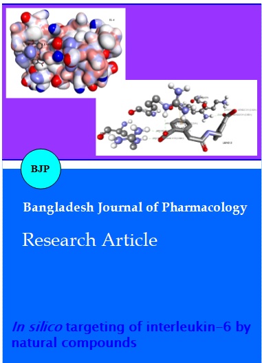
In silico targeting of interleukin-6 by natural compounds
Abstract
Rheumatoid arthritis an autoimmune disorder affecting many tissues and organs, is a challenging prospect in terms of treatment. Our work is looking into finding lead compound of natural origin against IL-6, an important cytokine having a profound role in pathogenesis of rheumatoid arthritis. For this purpose we have targeted the active site (ARG 179) of IL-6 using IBS database having 48531 natural compounds. Computer aided drug designing methods of virtual screening and in silico ADME/Tox helped us to limit our study of molecular docking to selected few for an atomic insight into their binding modes. The molecular docking analysis helped us to propose five possible drugs, out of which one compound (Chem. ID 10465) margaric acid is an edible fatty acid and exhibited good binding affinity with the active site of IL-6, thus making it a good starting point for developing drug for treatment of arthritis.
Introduction
Early mortality, severe disability and structural damages are the outcome of a chronic inflammatory autoimmune disorder, rheumatoid arthritis (Klareskog et al., 2009; Scott et al., 2010). Although a lot of progress has been achieved in controlling the disease activity by using drugs known as disease modifying antirheumatic drugs (DMARDs), but still challenges are there in early diagnosis and treatment (McInnes at al., 2011). The etiology of rheumatoid arthritis is a mystery till date and it is the pathogenesis that has been deciphered to a lot of extent (Misato et al., 2011).
IL-6 a highly up-regulated (Houssiau et al., 1988; Madhok et al., 1993) cytokine is a key molecule with a pivotal role in the pathogenesis of rheumatoid arthritis (Misato et al., 2011). Its role in synovitis (Maruotti et al., 2006) Joint damage (Gravallese et al., 2000), autoimmunity (Zhou et al., 2007), anemia (Hochberg et al., 1988) and hypolepidemia (Hashizume et al., 2010) makes it a potential target in treatment of rheumatoid arthritis. The argentine (ARG) at the 179th position of IL-6 plays an important role (Fontaine et al. 1992), targeting this particular site in IL-6 will impair its normal functioning. To achieve this we resorted to computer aided drug discovery, which has become an important tool in drug discovery (Alvarez, 2004; Chikan et al., 2013).
The present study targeted crystal structure of IL-6 bearing accession code of 1ALU (Somers et al., 1997), to obtain the selected few lead compounds of natural origin. To achieve our goal we used multistep structure-based virtual screening of around fifty thousand natural compounds obtained from Inter Bio Screen (IBS) database. Virtual screening which is incorporated in the major pipeline of drug discovery in most pharmaceutical companies has become an indispensible tool for identifying active lead compounds. The Absorption, distribution, metabolism, elimination (ADME) and Toxicology (Tox) are also the properties that have been identified as the major cause of failure of major drug screening projects (Davis et al., 2004) therefore ADME/Tox data validation is an important aspect (Van et al., 2003).
Materials and Methods
The crystal structure of human IL-6 protein (PDB ID: 1ALU) was obtained from protein databank (PDB). SPDB viewer at the default cut off RMSD (Root Mean Square Deviation) value of 0.5 using OPLS 2001 force field was used for energy minimization of the PDB structure. The modus operandi that followed this step is illustrated in Figure 1.
Figure 1: Step wise illustration of the Modus operandi in use
Virtual screening (VS)
Virtual screening has become a necessary tool in cutting short lead compounds and has fitted itself in the pipeline of drug discovery in most pharmaceutical companies. We used ArgusLab (Thompson et al., 2004) to perform the VS of the ligand dataset. The software was set to high precision screening using ArgusDock docking engine. The ligands were set to flexible mode and were targeted against the amino acid ARG at 179 position with a grid box of diameter 15 X 15 X 15. Natural compounds from IBS Database, having 48531 natural compounds were used for the VS of IL-6 inhibitors. From the initial dataset, a total of 500 compounds were selected based on their calculated binding affinity with the protein IL-6 for further in silico ADME/Tox analysis.
Rule of Five and ADME/T screening
The subset of 500 compounds was screened for Lipinski Rule of five violations (Lipinski, 2004) with the objective of increasing the success rate of compounds reaching further stages of the development. The subset of compounds was also subjected to in silico prediction (Muanz et al., 2013). A total of fifty top compounds which satisfied the criteria were chosen for further analysis.
Molecular docking
AutoDock 4.2 is used for molecular docking (Morris et al., 2009). The software was used to find the best binding mode using binding free energy evaluations and number of physical interactions. AutoDock calculates the energy values by the characterization of internal energy of ligand, torsional free energy and intermolecular energy consisting of van der Walls energy, hydrogen bonding energy, desolvation energy, and electrostatic energy.
Result and Discussion
Virtual screening followed by in silico ADME/Tox analysis of IL-6 and IBS database was successful in limiting our search to top 50 compounds. From the initial data set of around fifty thousand natural products selected for the study, subsets of five hundred lead-like natural products were selected using ArgusLab based on the feature of binding energy. The VS was followed by ADME/Tox analysis using Lazar Toxicity Predictions online server to further limit the size of initial dataset. The tool was pivotal in checking their mutagenic and carcinogenic property, the shortlisted fifty compound were selected on their drug likeness (Lipinski et al., 2012) and their observance of ADME and related properties of typical drugs (Zhao et al., 2001, van de Waterbeemd et al., 1998). For drug likeness molecular weight (MW), number of hydrogen bond donor (HBD), number of hydrogen bond acceptor (HBA) and total polar surface area (tpsa) ,which all fall under Lipinski’s rule of five (RO5) were calculated. Table I shows the RO5 data of top five compounds. These properties have an important role in the final outcome and analyzing them using by in silico means not only saves time but also is economically more viable. The permissible range of RO5 are MW<500 Dalton, clog p 4.5, HBD<5, HBA <10, which is associated with 90% of orally active drugs that have achieved phase II clinical status (Lipinski, 2004). Our lead compounds are in compliance with the RO5 properties se by Lipinski.
Table I: Lipinski Rule of five and toxicolgy report
| Name | ChemId | MW (Dalton) | clogp | HBD | HBA | Mutagenicity | Carcinogenecity |
|---|---|---|---|---|---|---|---|
| Lead 1 | 10465 | 270.5 | 4.7 | 1 | 2 | No | No |
| Lead 2 | Not found | 313.3 | 1.9 | 3 | 6 | No | No |
| Lead 3 | 10518237 | 299.3 | 2.8 | 5 | 6 | No | No |
| Lead 4 | Not found | 423.5 | 3.1 | 1 | 7 | No | No |
| Lead 5 | Not found | 340.4 | 2.0 | 3 | 6 | No | No |
clinical status (Lipinski, 2004). Our lead compounds are in compliance with the RO5 properties se by Lipinski.
The initial database contained 48,531 natural products selected for the virtual screening was condensed based on binding free energy obtained by ArgusLab, the initial dataset was shortened almost 99% of the initial size to mere five hundred compounds. Further screening ensured that only 0.1% of total compounds i.e. fifty compounds of natural origin were available for further in silico studies using AutoDock 4.2 tool. The proposed target site was optimized for performing the docking studies. Among the top 50 compounds only 5 compounds were found to physically interact with Arg179.
The final shortlisted natural compounds obtained by using AutoDock 4.2 tool into the optimized target site (Figure 2) of the protein were analyzed for interaction using Discovery Studio 3.5 client software, the interactions are described in Table II. All five lead compounds were found to form hydrogen bond with ARG179. The interactions (covalent or non covalent), the number of hydrogen bonds and number of Pi–-Pi interactions has been displayed in Figure 3 for all the selected lead compounds.
Figure 2: The solid surface structure of Il-6 with lead 5 binding pocket
Figure 3: 2D docked conformation of five lead compounds with IL-6
Table II: AutoDock analysis of five lead natural products
| Name | IUPAC Name | ΔG Kcal/mol |
Ligand binding pocket | Interactions |
|---|---|---|---|---|
| Lead 1 | Heptadecanoic acid | -1.5 | GLN175,LEU178,ARG179, ARG182 | 1 Hydrogen bond |
| Lead 2 | 2-(2-((3-carboxybenzyl)amino)-2-oxoethyl)benzoic acid | -5.4 | LYS171, GLN175,ARG179, ARG182 | 5 Hydrogen bond 2 Pi bonds |
| Lead 3 | 2-(2-(2-carboxyphenyl)acetamido)benzoic acid | -5.4 | GLN175,LEU178,ARG179, ARG182 | 4 Hydrogen bond |
| Lead 4 | (S)-methyl 2-(3-(7-methoxy-4-methyl-2-oxo-2H-chromen-6-yl)propanamido)-3-phenylpropanoate | -3.9 | ASP26, ARG30, LYS171, GLN175, SER176, LEU178, ARG179, ARG182 | 2 Hydrogen bond 5 Pi bonds |
| Lead 5 | 2-(2-((3-(isopropylcarbamoyl)phenyl)amino)-2-oxoethyl)benzoic acid | -5.2 | LEU33, CYS73, PHE74, GLN75, LEU174, GLN175, SER176, LEU178, ARG 179, ARG 182. | 2 Hydrogen bond 1 Pi bonds |
| The ligand binding pocket and the hydrogen bond formation was calculated using Discovery Studio 3.5 software.The bold amino acids represent the one which are involved in forming interaction with the ligand | ||||
The first lead compound is famous as Margaric acid having PubChem ID 10465 showed lowest binding energy of -1.5 kcal/mol. The complex exhibited a lone hydrogen bond (Figure 4A) with the key residue ARG179 of IL-6.The hydrogen bond interaction of 2.72 Ǻ is between the O2 of the Lead 1 and N1 atom of ARG179. The binding pocket of this complex constitutes of GLN175, LEU178, ARG179 and ARG182.
Figure 4: 3D mapping of lead compounds with their respective hits where (A, B, C and E) represent the lead compounds in order
The complex between lead 4 and IL-6 is rich in Pi–-Pi interactions (Figure 4D), four of the six Pi–-Pi interactions is between the lead 4 and ARG179 with a distance ranging from 3 to 5 . The ΔG of -3.9 Kcal/mol makes it the forth least free energy complex forming two hydrogen bond interaction , the interaction with ARG179 is of 3 Ǻ between O18 of lead and NE of ARG179. The binding groove consists of ASP26, ARG30, LYS171, GLN175, SER176, LEU178, ARG179 and ARG182.
The final complex to form an interaction with ARG179 is Lead 5, with ΔG of -5.2 Kcal/mol the complex is third in line in terms of free energy. The complex is forming both hydrogen bond and Pi–-Pi interactions. The NH2 of ARG179 is forming a hydrogen bond with O14 of lead compound with a distance of 2.55 Ǻ. The lone Pi–-Pi interaction is between PHE74 of IL-6 and lead 5. The binding pocked of the lead comprises of LEU33, CYS73, PHE74, GLN75, LEU174, GLN175, SER176, LEU178, ARG 179 and ARG 182.
All the five compounds are of natural origin having tremendous potential in blocking IL-6 activity and are providing an opportunity for further in vitro and in vivo analysis. Our approach of computer aided drug designing has helped us in limiting our focus to mere 0.01% of the total volume of Inter Bio Screen database of around fifty thousand compounds (Figure 5).
Figure 5: 3D point plot of four properties (HBA, HBD, clogP, and tpsa) of the compounds at different stages of drug discovery, showing the journey from ~ 50000 compounds to top five in accordance with Lipinski's parameters
Out of the five compounds only lead 1 source could be confirmed, this particular compound is known as margaric acid a fatty acid found in fat and milk fat of ruminants (Cooke et al., 1957). Margeric acid is also found in the contents of imitation butter famously known as margarine. As fatty acids are already reported to have anti inflammatory effects (Khalil et al., 2000) makes lead 1 the most significant of the five shortlisted compounds. This particular selected natural compound we feel holds an edge over the selected four other compounds and can be crucial in blocking considerably elevated Il-6 in the serum of rheumatoid arthritis patients.
Contribution
First two authors contributed equally in this study.
References
Alvarez JC. High-throughput docking as a source of novel drug leads. Curr Opin Chem Biol. 2004; 8: 365-70.
Chikan NA, Bhavaniprasad V, Anbarasu K, Shabir N, Patel TN. From natural products to drugs for epimutation computer-aided drug design. Appl Biochem Biotechnol. 2013; 170: 164-75.
Cooke NJ, Hansen RP, Shorland FB. Occurrence in butterfat of n-heptadecanoic acid (margaric acid). Nature 1957; 179: 98.
Davis AM, Riley RJ. Predictive ADMET studies, the challenges and the opportunities. Curr Opin Chem Biol. 2004; 8: 378-86.
Fontaine V, Savino R, Arcone R, de Wit L, Brakenhoff JP, Content J, Ciliberto G. Involvement of the Arg179 in the active site of human IL-6. Eur J Biochem. 1993; 211: 749-55.
Gaffen SL. The role of interleukin-17 in the pathogenesis of rheumatoid arthritis. Curr Rheumatol Rep. 2011; 11: 365-70.
Gravallese EM, Manning C, Tsay A, Naito A, Pan C, Amento E, Goldring SR. Synovial tissue in rheumatoid arthritis is a source of osteoclast differentiation factor. Arthritis Rheum. 2000; 43: 250-58.
Hashizume M, Yoshida H, Koike N, Suzuki M, Mihara M. Overproduced interleukin 6 decreases blood lipid levels via up-regulation of very-low-density lipoprotein receptor. Ann Rheum Dis. 2010; 69: 741-46.
Hochberg MC, Arnold CM, Hogans BB, Spivak JL. Serum immunoreactive erythropoietin in rheumatoid arthritis: Impaired response to anemia. Arthritis Rheum. 1988; 31: 1318-21.
Houssiau FA, Devogelaer JP, Van Damme J, de Deuxchaisnes CN, Van Snick J. Interleukin-6 in synovial fluid and serum of patients with rheumatoidarthritis and other inflammatory arthritides. Arthritis Rheum. 1988; 31: 784-88.
Khalil MH, Marcelletti JF, Katz LR, Katz DH, Pope LE. Topical application of docosanol- or stearic acid-containing creams reduces severity of phenol burn wounds in mice. Contact Dermatitis 2000; 43: 79-81.
Klareskog L, Catrina AI, Paget S. Rheumatoid arthritis. Lancet 2009; 373: 659-72.
Lipinski CA, Lombardo F, Dominy BW, Feeney PJ. Experimental and computational approaches to estimate solubility and permeability in drug discovery and development settings. Adv Drug Deliv Rev. 2001; 46: 3-26.
Lipinski CA. Lead-and drug-like compounds: The rule-of-five revolution. Drug Discov Today Technol. 2004; 1: 337-41.
Madhok R, Crilly A, Watson J, Capell HA. Serum interleukin6 levels in rheumatoid arthritis: Correlation with clinical and laboratory indices of disease activity. Ann Rheum Dis. 1993; 52: 232-34.
Maunz A, Gütlein M, Rautenberg M, Vorgrimmler D, Gebele D, Helma C. lazar: A modular predictive toxicology frame-work. Front Pharmacol. 2013; 4: 38.
McInnes IB, Schett G. The pathogenesis of rheumatoid arthritis. N Engl J Med. 2011; 365: 2205-19.
Morris GM1, Huey R, Lindstrom W, Sanner MF, Belew RK, Goodsell DS, Olson AJ. AutoDock4 and AutoDock Tools 4: Automated docking with selective receptor flexibility. J Comput Chem. 2009; 30: 2785-91.
Scott DL, Wolfe F, Huizinga TW. Rheumatoid arthritis. Lancet 2010; 376: 1094-108.
Somers W, Stahl M, Seehra JS. 1.9 A crystal structure of interleukin 6: Implications for a novel mode of receptor dimerization and signaling. EMBO J. 1997; 16: 989-97.
Szekanecz Z, Besenyei T, Paragh G, Koch AE. Angiogenesis in rheumatoid arthritis. Autoimmunity 2009; 42: 563-73.
Thompson MA. ArgusLab 4.0. 1. Planaria software LLC, Seattle, WA, 2004.
Van de Waterbeemd H, Camenisch G, Folkers G, Chretien JR, Raevsky OA. Estimation of blood-brain barrier crossing of drugs using molecular size and shape, and H-bonding descriptors. J Drug Target. 1998; 6: 151-65.
Van de Waterbeemd H, Gifford E. ADMET in silico modelling: Towards prediction paradise? Nat Rev Drug Discov. 2003; 2: 192-204.
Zhao YH, Le J, Abraham MH, Hersey A, Eddershaw PJ, Luscombe CN, Butina D, Beck G, Sherborne B, Cooper I, Platts JA. Evaluation of human intestinal absorption data and subsequent derivation of a quantitative structure-activity relationship (QSAR) with the Abraham descriptors. J Pharm Sci. 2001; 90: 749-84.
Zhou L, Ivanov II, Spolski R, Min R, Shenderov K, Egawa T, Levy DE, Leonard WJ, Littman DR. IL-6 programs T(H)-17 cell differentiation by promoting sequential engagement of the IL-21 and IL-23 pathways. Nat Immunol. 2007; 8: 967-74.