Phenotypic characterization of drug resistance and tumor initiating cancer stem cells from human bone tumor osteosarcoma cell line OS-77
Abstract
The cancer stem cell theory suggest that presence of small subpopulation of cancer stem cells are the major implication in the cancer treatment and also responsible for tumor recurrence. Based on Hoechst 33342 dye exclusion technique, we have identified about 3.3% of cancer stem like side population (SP) cells from human osteosarcoma OS-77 cell line whose prevalence is significantly reduced to 0.3% after treatment with verapamil. The sphere formation assay revealed that osteosarcoma SP cells are highly capable to form tumor spheres (sarcospheres). Further by immunocytochemistry and RT-PCR, we show that OS-77 SP cells have enhanced expression of stem cell surface markers such as CD44, Nanog and ATP-binding cassette (ABC) transporter gene (ABCG2) which contributes to self-renewal and drug resistance, respectively. Our findings help to designing a novel therapeutic drug which could effectively target the cancer stem cells and prevent the tumor relapse.
Introduction
An aggressive human bone tumor in children and young adolescents is "Osteosarcoma". It is an eighth most common malignancy which contributed to 2.4% of all pediatric malignancies. After diagnosis the mean survival rate was estimated as less than 5 years (Marina et al., 2004, Damron et al., 2007) and almost 20% of the patients are at the metastatic stage while diagnosis. According to the recently proposed CSC (cancer stem cell) theory, the presence of small population of tumor initiating cancer stem cells are involved in multidrug resistance, treatment failure and tumor recurrence. These cancer stem cells are highly drug and apoptosis resistance and express stem cell surface genes such as Oct3/4A and Nanog (Wu et al., 2007; Gibbs et al., 2005). Therefore, elucidating the molecular mechanism and signaling pathways involved in cancer stem cells mediated tumorgenesis is essential to target the cancer stem cells inorder to eliminate the tumor relapse (Jordan et al., 2006; Hemmati et al., 2003; Li et al., 2007).
One of the valuable method suggested to isolate cancer stem cells is the FACS based Hoechst 33342 dye exclusion (Goodell et al., 1996) and the cancer cells which efflux Hoechst 33342 dyes are named as side population (SP) cells. These SP cells are considered as enriched cancer stem cells since they shares all the characteristic features of cancer stem cells such as highly tumorigenic, express stem like genes (CD133, CD44, CD34, CD29, CD24) and resistant to chemotherapeutic drugs. These SP cells also over express the ATPase binding cassette (ABC) transporters such as ABCB1 (MDR1), ABCC1, ABCG2 (BCRP1), etc which contribute to multidrug resistance etc. Hence, the characterization of SP cells helps to identify the cancerous stem cells and substantiates the mechanism of oncogenesis and drug resistance (Kondo et al., 2004). Consequently, in the present study, we made an attempt to isolate and characterize the cancer stem like SP cells from human osteosarcoma cell line OS-77. The isolated cancer stem like SP cells are further analyzed for the expression of stem cell surface markers such as Oct-3/4A, CD44, and Nanog.
Materials and Methods
Cell lines and culture conditions
Human Osteosarcoma (OS) samples (aggressive primary human osteosarcoma; stage-III; with lung metastasis) were collected at the time of surgery. After isolation, the samples were washed extensively in PBS solution containing antibiotics and incubated overnight in DMEM/F12 (GIBCO) containing penicillin (500 U/mL), streptomycin (500 ug/mL), and amphotericin B (1.25 ug/mL) (GIBCO). Enzymatic digestion was performed using collagenase (1.5 mg/mL) (GIBCO) and hyaluronidase (20 ug/mL) in PBS for 1 hour. Cells were cultured in DMEM with 10% FBS, supplemented with antibiotics and maintain in T-75 flasks at 37°C in a humidified 5%CO2 and 95%air atmosphere. Cells on becoming 90% confluent are removed from culture flask using trypsin-EDTA (0.25% - 53 mM EDTA) washed, cells suspended in 10% DMEM. Cell count was taken using hemocytometer.
FACS analysis
Cells were cultured in DMEM with 10% FBS, supplemented with antibiotics and maintain in T-75 flasks at 37°C in a humidified 5%CO2 and 95%air atmosphere. Cells on becoming 90%confluent are removed from culture flask using trypsin-EDTA (0.25% - 53 mM EDTA) washed, cells suspended in 10% DMEM. Cell count was taken using hemocytometer.
Study groups
Control - cells labeled with Hoechst 33342 dye alone (n=5); Drug treated - cells treated with verapamil drug and Hoechst 33342 dye (n=5). Cells were counted by hemocytometer ad approximately 106 cells/mL of 10% DMEM were labeled with Hoechst 33342 stock (sigma)-bis-benzimide (5 uL/mL) either with dye alone or in combination with drug (ABC transporter inhibitor verapamil - 0.8 uL/mL). Further, the cells were counter stained with PI (propidium iodide) 2 ug/mL. The cells were sorted using a flow cytometer and the sorted cells were cultured and maintained in DMEM/F-12 supplemented with 10% FBS. The Hoechst 33342 emission was first split by using a 610 nm dichroic short-pass filter, and the red and the blue emissions were collected through 670/30 and 450/65 nm band-pass filters, respectively).
Sarcosphere formation assay
We performed sphere formation assay according to the protocol described by (Gibbs et al., 2005). Cells were plated at a density of 60,000 cells/well in ultra-low attachment six-well plates containing serum-free DMEM/F12 medium supplemented with N2, epidermal growth factor (10 ng/mL) and human basic fibroblast growth factor (10 ng/mL). The culture was analyzed for sphere formation everyday till day 7 and pictures were taken by inverted phase contrast microscopy. After 7 days of culturing, the total number of spheres (sarcospheres) were generated by both FACS sorted SP and non-SP cells were quantified.
Immunofluorescent staining
The sorted SP and non-SP cells of OS-55 were fixed in BD Cytofix solution and incubated for 20 min at 4°C. After 20 min blocking in donkey serum, cells were incubated with goat anti-Oct3/4A antibody (sc-8628; Santa Cruz Biotechnology, Inc., USA) for overnight at 4°C, and subsequently incubated with Rhodamine red conjugated donkey anti-goat antibody (1:200; Jackson Immunoresearch Laboratories). For CD44 and Nanog immunofluorescence analysis, cells were stained with fluorescein isothiocyanate (FITC) conjugated anti-human Nanog or CD44 antibody (1:5; eBioscience, CA). The cells were observed under confocal microscope and all the pictures were analyzed and processed by adobe photoshop CS4.
Real-time reverse transcription (RT)-polymerase chain reaction (PCR)
Total RNA was extracted and Complementary DNA was prepared using Reverse Transcriptase kit (Fermentas). Real-time RT-PCR analysis was subsequently performed by using IQ Supermix with SYBR-Green (Bio-Rad). The sequences of human specific primers were used as follows: ABCG2: (forward - TCA ATC AAA GTG CTT CTT TTT TAT G and reverse TTG TGG AAG AAT CAC GTG GC); Oct3/4A: (forward - TGG AGA AGG AGA AGC TGG AGC AAA A and reverse - GGC AGA TGG TCG TTT GGC TGA ATA GAC C); Nanog (forward - TCC TCC TCT TCC TCT ATA CTA AC; reverse - CCC ACA AAT CAC AGG CAT AG); actin: (forward - GCG GGA AAT CGT GCG TGA CAT T; reverse - GGC AGA TGG TCG TTT GGC TGA ATA). PCR products were electrophoresed on a 1.2% agarose gel and stained with ethidium bromide.
Statistical analysis
One-way analysis of variance (ANOVA) and student t-test was performed to determine the significant difference between the treatment and control groups. A probability level of p<0.01 was considered as statistically significance.
Results
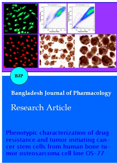
We investigated the human osteosarcoma cell line OS-77 for the presence of cancer stem like SP cells by FACS based Hoechst 33342 dye exclusion assay. We have identified about 3.3% of side population cells from OS-77 which exclude the Hoechst 33342 dye (Figure 1, left panel). However, after treatment with verapamil, an inhibitor of ABC transporter protein, the presence of SP cells was significantly reduced to 0.3% (Figure 1, right panel). This is a confirmatory test for the presence of ABC transporter protein, which has shown to be involved in multi-drug resistance properties of SP cells in several solid tumors. After carefully sorted SP and non-SP cells, we have performed sphere formation assay in order to determine the capacity of self-renewal. The OS-77 SP cells are able to form tumor spheres (sarcospheres) on the day 3 (D3) itself and they attain maximum size on day 6 (D6) (Figure 2A). Interestingly, the total number of tumor spheres formed by SP cells is significantly higher than non-SP cells (Figure 2B). Further, our immunofluorescence assay revealed that enhanced signal for the presence of stem cell surface markers such as CD44, Nanog and Oct-4 (Figure 3), whereas the signal level is very less or null in non-SP cells. This enhanced expression is further confirmed by RT-PCR analysis, which showed that mRNA level of Nanog and Oct-3/4A, are significantly elevated in SP cells (Figure 4). In addition, the mRNA expression of ABC transporter protein ABCG2 are significantly elevated in SP cells than non-SP cells (Figure 4) which clearly suggest that over expression of ABCG2 contributed to chemotherapetutic drug resistance in SP cells. Taken together, these data suggest that existence of cancer stem like SP cells in human osteosarcoma cell line OS-77 might be a major implication in eradication of tumor refractory and thus involved in formation of bone tumor recurrence, invasion and metastasis.
Figure 1: A representative image for dot plots analysis by FACS. Live cells were selected against propidium iodide staining. Graph (left panel) showing 3.3% of SP cells from human osteosarcoma cell line OS-77. Graph on right panel showing reduced population of SP cells (0.3%) after treatment with verapamil (ABC transporter inhibitor)
Figure 2: A) Representative phase contrast images of monoclonal spheres formed from OS-77 osteosarcoma SP cells. The sarcospheres were generated rapidly on day 3 (D3) itself and the size of the spheres were increased on day 6 (D6). The scale bar represents 25 um. B) Quantification graph for clonogenic formation efficiency showing that sorted SP cells of OS-77 generated significantly higher number of sarcospheres than non-SP cells. The bar represents the standard deviation and ap<0.01
Figure 3: Representative immunofluorescent staining images of sorted OS-77 SP cells showing immunofluorescence positive to CD44 (A), Nanog (B) and Oct3/4A (C). D. Representative image for immunofluorescence negative of non-SP cells for the above mentioned proteins. Very week fluoresence intensity or null expression was observed in non-SP cells. The scale bar represents 50 um.
Figure 4: RT-PCR analysis for expression of stem cell genes in SP and non-SP cells. The elevated mRNA expression of ABC transporter gene ABCG2 and stem cell surface genes such as Oct-3/4A and Nanog was detected in SP cells when compared to non-SP cells
Discussion
Cancer stem cells are highly self-renewal, possess differentiaiton potential and express stem cell surface genes (Jordan et al., 2006; Pardal et al., 2003; Chang 2006). In specific, the ABC transporter protein like ABCG2 is highly expressed in cancer stem cells are mainly involved in chemotherapy resistance. Consequently, the current conventional treatment strategies are able to kill most of the neoplastic cells within a tumor by leaving the cancer stem cells unaffected. Emerging data in different types of cancers supports that cancer stem cells survive conventional anticancer therapies that target only the rapidly dividing cells (Kondo et al., 2004). Hence the elimination of cancer stem cells is an essential goal for eradicating tumor cell of origin and for providing long term disease free survival. In such case, understanding the molecular mechanism of tumorigenesis of cancer stem cells would be more valuable to design a novel anticancer therapy.
The cancer cells which exclude Hoechst 33342 dye are called as side population (SP) cells which shares all the remarkable features of cancer stem cells which includes chemotherapy and apoptosis resistance, expressing stem cell genes and high potent of self-renewal (Cho and Clarke, 2008). In the current study, we showed that human osteosarcoma OS-77 cells contain 3.3% of cancer stem like SP cells which was reduced to 0.3% upon treatment with verapamil. These data together with the elevated ABCG2 mRNA level from RT-PCR analysis confirms the contribution of over expression of ABC transporters in multidrug resistance in bone tumor treatment. The sorted SP cells can able to generate sarcospheres frequently and therefore they are highly self-renewal when compared to non-SP cells. Similarly, it has been previously reported that established human osteosarcoma cell lines OS99-1 and MG63 cells are able to form more sarcospheres frequently (Wang et al., 2009).
Oct3/4A, CD44 and Nanong are the major factors shown to be involved in maintenance of self-renewal, tumorigenesis and tumor invasion (Chang et al., 2006; Lee et al., 2006; Okita et al., 2007). The significant role of these proteins in self-renewal and tumor invasion are well demonstrated that in several human cancers (Ezeh et al., 2005; Looijenga et al., 2003). In addition, the expression of these proteins can be used as a marker for the diagnosis of cancer metastasis (Cheng, 2004). Increased expression of Oct3/4A, Oct3/4B and Nanog genes was already reported in human osteosarcoma OS99-1 (Wang et al., 2009) and they are involved in metastasis of tumor. Similarly, our data also showed that human osteosarcoma OS-77 cancer stem like cells have enhanced expression of Oct3/4A and Nanog by immunofluorescence and RT-PCR analysis. The stem cell surface protein CD44 was characterized as a family of cell surface proteoglycans and glycoproteins involved in tumor invasion, metastasis, chemo- and radiotherapy resistance (Wu et al., 2007). Recent studies in nasopharyngeal carcinoma also showed that increased expression of CD44 is responsible for tumorigenesis and metastasis (Su et al., 2011). In line with these findings, we also observed SP cells are highly immuno positive to CD44 when compared to non-SP cells.
Conclusion
In summary, our data clearly illustrated that existence of cancer stem like cells in human osteosarcoma cell line OS-77 which might be involved in tumor recurrence and metastasis. Further, the differences in molecular mechanism and signaling pathways between normal and bone tumor stem cells need to be addressed clearly to provide new therapeutic targets with the eventual goal of eliminating residual disease and recurrence.
References
Chang CC. Recent translational research: Stem cells as the roots of breast cancer. Breast Cancer Res. 2006; 8: 103-05.
Cheng L. Establish a germ cell origin for metastatic tumors using OCT4 immunohistochemistry. Cancer 2004; 101: 2006-10.
Cho RW, Clarke FM: Recent advances in cancer stem cells. Curr Opin Genet Dev. 2008: 18: 48-53.
Damron TA, Ward WG, Stewart A. Osteosarcoma, chondrosarcoma and Ewing’s sarcoma: National Cancer Data Base Report. Clin Orthop Relat Res. 2007; 459: 40-47.
Ezeh UI, Turek PJ, Reijo RA, Clark AT. Human embryonic stem cell genes OCT4, NANOG, STELLAR and GDF3 are expressed in both seminoma and breast carcinoma. Cancer 2005; 104: 2255-65.
Gibbs CP, Kukekov VG, Reith JD, Tchigrinova O, Suslov ON, Scott EW. Stem-like cells in bone sarcomas: Implications for tumorigenesis. Neoplasia 2005; 7: 967-76.
Goodell MA, Brose K, Paradis G, Conner AS, Mulligan RC. Isolation and functional properfies of murine hematopoietic stem cells that are replicating in vivo. J Exp Med. 1996; 183: 1797-806.
Hemmati HD, Nakano I, Lazareff JA, Masterman-Smith M, Geschwind DH, Bronner-Fraser M. Cancerous stem cells can arise from pediatric brain tumors. Proc Natl Acad Sci USA. 2003; 100: 15178-83.
Jordan CT, Guzman ML, Noble M. Cancer stem cells. N Engl J Med. 2006; 355: 1253-61.
Kondo T, Setoguchi T, Taga T: Persistence of a small subpopulation of cancer stem-like cells in the C6 glioma cell line. Proc Natl Acad Sci USA. 2004; 101: 781-86.
Lee J, Kim HK, Rho JY, Han YM, Kim J. The human OCT-4 isoforms differ in their ability to confer self-renewal. J Biol Chem. 2006; 281: 3554-65.
Li C, Heidt DG, Dalerba P, Burant CF, Zhang L, Adsay V. Identification of pancreatic cancer stem cells. Cancer Res. 2007; 67: 1030-37.
Looijenga LH, Stoop H, deLeeuw HP, De Gouveia Brazao CA, Gillis AJ, vanRoozendaal KE, van Zoelen EJ, Weber RF, Wolffenbuttel KP, van Dekken H, Honecker F, Bokemeyer C, Perlman EJ, Schneider DT, Konenen J, Sauter G, Oosterhuis JW. POU5F1 (OCT3/4) identifies cells with pluripotent potential in human germ cell tumors. Cancer Res. 2003; 63: 2244-50.
Marina N, Gebhardt M, Teot L, Gorlick R. Biology and therapeutic advances for pediatric osteosarcoma. Oncologist 2004; 9: 21-22.
Okita K, Ichisaka T, Yamanaka S. Generation of germline-competent induced pluripotent stem cells. Nature 2007; 448: 313-17.
Pardal R, Clarke MF, Morrison SJ. Applying the principles of stem-cell biology to cancer. Nat Rev Cancer. 2003; 3: 895-902.
Su J, Xu XH, Huang Q, Lu MQ, Li DJ, Xue F, Yi F, Ren JH, Wu YP. Identification of cancer stem-like cd44 cells in human nasopharyngeal carcinoma cell line. Arch Med Res. 2011; 42: 15-21.
Wang L, Park P, Lin YC. Characterization of stem cell attributes in human osteosarcoma cell lines. Cancer Biol Ther. 2009; 8: 543-52.
Wu C, Wei Q, Utomo V, Nadesan P, Whetstone H, Kandel R. Side population cells isolated from mesenchymal neoplasms have tumor initiating potential. Cancer Res. 2007; 67: 8216-22.