Wnt5a silencing inhibits the osteosarcoma migration by suppressing PI3K/AKT signaling pathway
Abstract
Osteosarcoma is one of the most common malignant bone tumors in children and adults. Till this date, molecular mechanism behind the growth and invasiveness of osteosarcoma is poorly understood. Wnt5a plays an important role in the oncogenesis and cancer metastasis. Results show that Wnt5a has a remarkable influence on the U2OS and SaOS-2 cancer cell migration. The cell migration was dependent on the concentration of Wnt5a with low concentration (25 ng/mL) has almost no effect on the cell migration while it has remarkable influence at 100 ng/mL. The results revealed that Wnt5a can control the osteosarcoma cell migration via PI3K/Akt signalling pathways. We showed that Wnt5a activated the PI3K (p-Tyr458) and Akt phosphorylation (p-Ser473) immediately after the ligand incubation. More importantly, we demonstrated that chemotherapeutic drug like doxorubicin can effectively suppress the activity of Wnt5a ligand and thereby can inhibit the osteosarcoma cell growth and tumorigenesis. This lay the foundation for therapeutic application of Wnt5a towards the treatment of osteosarcoma.
Introduction
Osteosarcoma is the most prevalent type of malignant bone cancer, occurs mainly in children and young adults (Picci et al., 2010). It is the second most cause of cancer related death in adult's age group (Yang et al., 2011). Osteosarcoma metastasizes in lungs after moving from distal femur and proximal tibia part of bones. When limb amputation was the only treatment option, less than 10% of people survived from osteosarcoma during the year 1970 (Yang et al., 2010). However, introduction of multidrug chemotherapy increased the survival rate to 60-70%. Generally, multidrug chemotherapy was started after the surgical intervention in patients. Therefore, surgery and conventional chemotherapy became standard treatment protocol in OS (Geller et al., 2010; Ferguson et al., 2001). No substantial improvement in osteosarcoma therapy has been observed despite great strides in osteosarcoma treatment. Most of the chemotherapeutic drug administered via oral and intravenous route are carries high risk of short term and long term adverse effects (Patel et al., 2002; Maran et al., 2013). Moreover, high systemic concentration of drug will result in high toxicity and narrow therapeutic index of the respective anticancer drugs. This could be due to the lack of potential biomarker and non-specific targeting of anticancer drugs towards the normal and cancer cells. In this perspective, an alternative therapeutic approach is desirable (Thomas, 2010). Therefore, molecular signaling pathways or biomarkers could be considered as a key in the treatment of osteosarcoma.
In the aforementioned perspective, Wnt signalling plays an important role in the development of osteoblastogenesis and tumorigenesis (Nusse, 2005). Osteoprogenitor cell proliferation/progression has been reported to increase with the Wnt signalling along with the prevention of apoptosis in osteoblasts. It has been reported that Wnt ligands and Wnt receptors are overexpressed in osteosarcoma while at the same time Wnt inhibitor was found in lowest level in bone malignancies (Bodine and Komm, 2006). In Wnt/β-catenin signaling pathways, Wnt ligands binds with the Frizzled receptors and low-density lipoprotein receptor-related protein 5/6 (LRP5/6) coreceptors leading to the activation of disheveled (Dsh) that releases β-catenin from the axin-adenomatous polyposis coli (APC)-glycogen synthase kinase-3β(GSK-3β) complex (Bodine, 2008; Gaur et al., 2005). Wnt5a has been classified in a non-canonical Wnt group. Wnt signalling activates Ca2+ pathways leading to the ultimate activation of activate protein kinase C (PKC) and Ca2+/calmodulin kinase II. Besides, it can activate various intracellular proteins including c-Jun N-terminal kinase (JNK), extracellular signal-regulated kinase (ERK), and Akt (McQueen et al., 2011).
Materials and Methods
Cell culture: U2OS and SaOS-2 human osteosarcoma cell lines were procured from American Tissue Culture Collection (ATCC), Rockville, MD. U2OS and SaOS-2 cells were cultured in McCoy's 5A medium. Prior to that, growth medium was prepared by adding 10%fetal bovine serum (FBS) and 1%penicillin-streptomycin in the rest of the media. The cells were maintained in a sophisticated incubator at ambient conditions (37°C and 5%CO2). The mouse anti-beta-actin antibody was purchased from KangChen Biotech, Shanghai, China. The rabbit anti-Akt antibody, rabbit anti-phospho-Akt (p-Ser473) antibody, rabbit anti-PI3K p85 antibody, rabbit anti-phospho-PI3K p85 (Tyr458) (Cell Signaling Technology, Danvers, MA), mouse anti-beta-catenin antibody, rabbit anti-phospho-β-catenin (p-Ser33) antibody were purchased from Santa Cruz Biotechnology, Santa Cruz, CA.
Cell migration analysis: Boyden chamber was used to determine the cell migration. The Boyden chamber is consists of two chambers which are separated by a polycarbonate membrane. U2OS and SaOS-2 cells were seeded in tissue culture plates and allowed to reach 90%confleuncy. The cells were detached by means of trypsinization. The cells were then centrifuged and made into a cell suspension. The serum free media was used to make the cell suspension which contains 5 µg/mL of BSA. Approximately, 6 x 10⁴ cells were placed on to the wells containing the membrane at the bottom. Now, Wnt5a is added to the compartments located up and downside of chamber. The cells were allowed to migrate for 24 hours at 37°C in the Boyden chamber. Followed by this, culture medium was removed along with the floating cells and membranes were detached from the chamber. The membranes were stained with 0.5% crystal violet. The extent of cell migration in the membrane was visualized using a light microscope. Number of cells migrated on to the membrane can also be counted.
Immunoblot analysis: The cells were seeded on to well plates and allowed to incubate for 24 hours. siRNA duplexes which is particular for Akt were incubated with U2OS and SaOS-2 cancer cells in order to start transfection. Lipofectamine 2000 reagent was used in serum free media as per the manufacturer's protocol. It was allowed to transfect for 48 hours, following which protein levels in each cell was evaluated using Western blotting.
The transfected cells were washed two times with PBS and lysed using lysis buffer. The lysis buffer was maintained at pH 7.4 and consists of 50 mmol/L Tris, 150 mmol/L NaCl, 1%Triton X-100, 1%sodium deoxycholate, 0.1%SDS, 1 mmol/L sodium orthovanadate, 1 mmol/L sodium fluoride, 1 mmol/L EDTA, 1 mmol/L PMSF, and 1% cocktail of protease inhibitors. The cell lysates were collected and centrifuged at 10,000 rpm for 20 min at 4°C. The proteins were separated using electrophoresis technique in a 10% SDS-acrylamide gel phase. This was blotted on a nitrocellulose membrane. The membranes were then blocked overnight in Tris buffer containing 0.1% (v/v) Tween 20 and 5% (w/v) fat-free dry milk. This was incubated with primary anti body which is followed by a secondary antibody. The antibodies used includes mouse anti-beta-actin antibody, rabbit anti-Akt antibody, rabbit anti-phospho-Akt (p-Ser473) antibody, rabbit anti-PI3K p85 antibody, rabbit anti-phospho-PI3K p85 (Tyr458), mouse anti-beta-catenin antibody, rabbit anti-phospho-beta-catenin (p-Ser33) antibody. The protein bands were determined by incubition with horseradish peroxidase-conjugated antibodies and images were captured using an X-ray film detector. The bands were analyzed using the software provided by the manufacturer.
BrdU incorporation assay: The cells were seeded in a well plate and allowed to attach and grow overnight. Cells were incubated with Wnt3a conditioned medium and followed by doxorubicin for a specified time. Cells were trypsinized and harvested and incubated with 10 µM of 5'-bromo-2-deoxyuridine (BrdU) for 3 hours. The cells were fixed with cold ethanol/HCL and BrdU labelling/detection kit (Roche Diagnostics GmbH, Mannheim, Germany) was employed to determine BrdU as per the instruction of manufacturer's.
Statistical analysis: For all analyses, p<0.05 was considered statistically significant. Two-tailed, unpaired Student's t-test was used to calculate statistical difference/analysis using Microsoft excel or graph pad prism. The data are expressed as mean ± standard deviation (SD) and performed in triplicate.
Result and Discussion
Wnt5a signalling pathway activates a range of physiological and pharmacological activities including the cellular proliferation, migration, invasion, cell cycle disruption, and suppresses the cell apoptosis in multiple cancers which includes osteosarcoma (Logan and Nusse, 2004). Wnt5a is the main ligand that activates the Wnt signalling pathways. Specifically, animal with abnormal Wnt signalling pathways frequently develops facial abnormalities, dwarfism, dysmorphic ribs and shortened limbs/tails (Klaus and Birchmeier, 2008). It has also been reported that lack of Wnt pathways would also result in abnormal distal lung morphogenesis, dysmorphic vertebrae, and complete absence of genital tubercle. This indicates how important Wnt signalling in the development and growth of critical organs (Weeraratna et al., 2002). In addition, Wnt-mediated cell signalling plays a unique role in the cancer proliferation and cancer differentiation. It has been reported that that Wnt5a promotes the cancer metastasis and proliferation in various malignant tissues including breast, melanoma, gastric and liver cancer (Zhu et al., 2012). Keeping all this facts in mind, present study was designed to investigate the effect of Wnt on osteosarcoma cancer cells including U2OS and SaOS-2 cancer cells. In addition, role of PI3K/Akt signaling pathway in Wnt pathway has been investigated.
Cell migration is a clear indication of the ligands influence on the cell proliferation and differentiation. The effect of Wnt5a on U2OS and SaOS-2 osteosarcoma cancer cells has been carried out after treating the individual cells with recombinant Wnt5a. The cell migration was evaluated using Boyden chamber assay. It can be clearly seen that Wnt5a has a remarkable influence on the U2OS cell migration. The concentration of Wnt was varied from 25 to 100 ng/mL to observe the concentration dependent effect on the cell migration capacity. As can be seen (Figure 1), low concentration (25 ng/mL) has almost no effect on the cell migration capacity while when the concentration of Wnt increased up to 50 ng/mL, the cell migration significantly inhibited. Specifically, appreciable cell migration inhibition was observed at around 75-100 ng/mL. Similar trend was observed in SaOS-2 cancer cells although the level of migration was different between both the cell lines. Followed by, number of cells per field was calculated and as expected the trend was similar to that of migration rate (Figure 1). The number of cells was almost equal to control at lowest concentration however it increased for the increase in the concentration of the Wnt. This result clearly indicates that Wnt5a is an effective stimulator of osteosarcoma cells.
Figure 1: Effect of Wnt5a on osteosarcoma cell migration. Relative cell migration rate of U2OS (A) and SaOS-2 (B) osteosarcoma cells. Number of cells per field (C) U2OS (D) SaOS-2 cells. The cells were incubated with increasing concentration of Wnt5a ligand from 25 ng/mL to 100 ng/mL
Phosphatidylinositol-3 kinases (PI3Ks) enzyme family is involved in the vital cellular functions of the body including cell growth proliferation, differentiation, cell survival and intracellular trafficking. These pharmacological activities have been important for the development of cancers. Therefore in the present study, molecular mechanisms behind the role of Wnt on cancer growth have been investigated. The downstream signalling pathway in U2OS and SaOS-2 cells has been studied in detail (Figure 2). Firstly, PI3K activation state has been studied. Phosphorylated-PI3K p85 is considered as the important biomarker of PI3K activation state. For this, cells were starved from serum for 24 hours then treated with highest concentration of Wnt5a at which it showed the maximum cell migratory ability. The cells were collected at a specified time interval of 15, 30, and 60 min and SDS-PAGE and western blot experiments were conducted. The results clearly showed that basal phosphorylation has been increased at increasing the incubation time. Specifically, basal phosphorylation was elevated after 15 min of Wnt5a stimulation, while it increased for 30 min incubation and decreased thereafter in both the cancer cell lines (Almeida et al., 2005).
Figure 2: Effect of Wnt5a on PI3K activation of osteosarcoma cells. U2OS (A) and SaOS-2 (B) cells were treated with 100 ng/mL of Wnt5a ligand and collected at specific time intervals. SDS-PAGE and western blot analysis were performed to estimate p-PI3K p85 (p-Tyr458) and total PI3K p85
Moreover, it is well known that PI3K is the important activator of Akt in the physiological systems (Kawasaki et al., 2007). Therefore, we have next investigated the response of Akt for the Wnt incubation (Figure 3). For this, cells were starved from serum for 24 hours then treated with highest concentration of Wnt5a at which it showed the maximum cell migratory ability. phosphorylated-Akt (p-Ser473) is considered as the important biomarker of Akt activation state. Consistent with the PI3K results, basal phosphorylation has been increased at increasing the incubation time. Specifically, basal phosphorylation was elevated after 15 min of Wnt5a stimulation, while it increased for 30 min incubation and decreased thereafter in both the cancer cell lines.
It is well-known that Wnt5a activates protein kinase C (PKC) and affect the tumor progression process in the cancer cells. It has been reported that Wnt5a inhibits the cell apoptosis by activating Akt and ERK pathways (Ko et al., 2014). We showed that Wnt5a activated the PI3K (p-Tyr458) and Akt phosphorylation (p-Ser473) immediately after the ligand incubation. Therefore it can be expected that by blocking Akt or PI3K will actively control the osteosarcoma cell proliferation.
Figure 3: Effect of Wnt5a on Akt activation of osteosarcoma cells. U2OS (A) and SaOS-2 (B) cells were treated with 100 ng/mL of Wnt5a ligand and collected at specific time intervals. SDS-PAGE and western blot analysis were performed to estimate p-Akt (p-Ser473) and total Akt
We next investigated the effect of DOX on the proliferation of osteosarcoma cells. For this, we have treated the cells with both Wnt5a ligand as well as chemotherapeutic drug. As can be seen (Figure 4A, B), Wnt5a has a remarkable influence on the U2OS and SaOS-2 cell proliferation. Wnt5a remarkably increased the cell proliferation comparing to that control or untreated cells. Importantly, when DOX was exposed to the same cells, it resulted in decrease in the basal conditions. The results clearly indicate that DOX abolished the stimulatory effect of Wnt5a ligand in the cells.
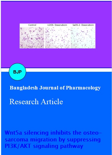
The cell migration is an important factor in the development of osteosarcoma tumorigenesis and metastasis. Therefore, we have investigated the effect of DOX on the cell migration in an identical condition. As seen clearly (Figure 4C), comparing to control cells (untreated), DOX markedly inhibited the cell migration potential of cells.
Figure 4: Effect of doxorubicin on cell apoptosis and cell migration. BrdU assay was performed in U2OS (A) and SaOS-2 (B). Cells were treated with Wnt5a and doxorubicin and DNA replication was evaluated by BrdU incorporation (C) effect of DOX on the U2OS cell migration. Boyden's chamber was used to evaluate the cell migration level
Overall, our results show that PI3K/Akt signaling plays a central role in cell viability-promoting phenotypes such as inhibition of cell death and cell cycle progression. We showed that Wnt5a could influence U2OS and SaOS-2 cell progression and proliferation by activating PI3K/Akt pathways. This PI3K/Akt-dependent activation of cell cycle progression plays an important role in the osteosarcoma proliferation in the body (Kawasaki et al., 2007; Ko et al., 2014). We have further showed the influence of chemotherapeutic agent on the cell proliferation and Wnt5a therapeutic action.
Conclusion
Results revealed that DOX remarkably halted the stimulatory effects of Wnt ligands. Consistently, it remarkably suppressed the migration of both the osteosarcoma cell lines.
Acknowledgement
One of the authors would like to thank Dr. Bao for his immense help.
References
Almeida M, Han L, Bellido T, Manolagas SC, Kousteni S. Wnt proteins prevent apoptosis of both uncommitted osteoblast progenitors and differentiated osteoblasts by beta-catenin-dependent and –independent signaling cascades involving Src/ERK and phosphatidylinositol 3-kinase/AKT.J Biol Chem. 2005; 280: 41342-51.
Bodine PV, Komm BS. Wnt signaling and osteoblastogenesis. Rev Endocr Metab Disord. 2006; 7: 33-39.
Bodine PV. Wnt signaling control of bone cell apoptosis. Cell Res. 2008; 18: 248-53.
Chamberlain GR, Tulumello DV, Kelley SO. Targeted delivery of doxorubicin to mitochondria. ACS Chem Biol. 2013; 8: 1389-95
Gabizon A, Shmeeda H, Barenholz Y. Pharmacokinetics of pegylated liposomal doxorubicin: Review of animal and human studies. Clin Pharmacokinet. 2003; 42: 419-36.
Gaur T, Lengner CJ, Hovhannisyan H, Bhat RA, Bodine PV, Komm BS, Javed A, van Wijnen AJ, Stein JL, Stein GS, Lian JB. Canonical WNT signaling promotes osteogenesis by directly stimulating Runx2 gene expression. J Biol Chem. 2005; 280: 33132-40.
Geller DS, Gorlick R. Osteosarcoma: A review of diagnosis, management, and treatment strategies. Clin Adv Hematol Oncol. 2010; 8: 705-18.
Ferguson WS, Goorin AM. Current treatment of osteosarcoma. Canc Invest. 2001; 19: 292-315.
Kawasaki A, Torii K, Yamashita Y, Nishizawa K, Kanekura K, Katada M, Ito M, Nishimoto I, Terashita K, Aiso S, Matsuoka M. Wnt5a promotes adhesion of human dermal fibroblasts by triggering a phosphatidylinositol-3 kinase/Akt signal. Cell Signal. 2007; 19: 2498-506.
Klaus A, Birchmeier W. Wnt signaling and its impact on development and cancer. Nat Rev Cancer. 2008; 8: 387-98.
Ko YB, Kim BR, Nam SL, Yang JB, Park SY, Rho SB. High-mobility group box 1 (HMGB1) protein regulates tumor-associated cell migration through the interaction with BTB domain. Cell Signal. 2014; 26: 777-83.
Kuhl M, Sheldahl LC, Park M, Miller JR, Moon RT. The Wnt/Ca2+ pathway: A new vertebrate Wnt signaling pathway takes shape. Trends Genet. 2000; 16: 279-83.
Logan CY, Nusse R. The Wnt signaling pathway in development and disease. Annu Rev Cell Dev Biol. 2004; 20: 781-810.
Maran A, Dadsetan M, Buenz CM, Shogren KL, Lu L, Yaszemski MJ. Hydrogel-PLGA delivery system prolongs 2-methoxyestradiol-mediated anti-tumor effects in osteosarcoma cells. J Biomed Mat Res. 2013; 9: 101A.
McQueen P, Ghaffar S, Guo Y, Rubin EM, Zi X, Hoang BG. The Wnt signaling pathway: Implications for therapy in osteosarcoma. Expert Rev Anticancer Ther. 2011; 11: 1223-32
Nusse R. Wnt signaling in disease and in development. Cell Res. 2005; 15: 28-32.
Patel SJ, Lynch Jr JW, Johnson T, Carroll RR, Schumacher C, Spanier S, Scarborough M. Dose-intense ifosfamide/doxorubicin/cisplatin based chemotherapy for osteosarcoma in adults. Am J Clin Oncol. 2002; 25: 489-95
Picci P, Mercuri M, Ferrari S, Alberghini M, Briccoli A, Ferrari C, Pignotti E, Bacci G. Survival in high-grade osteosarcoma: Improvement over 21 years at a single institution. Ann Oncol. 2010; 21: 1366-73.
Thomas DM. Wnts, bone and cancer. J Pathol. 2010; 220: 1-4.
Veeman MT, Axelrod JD, Moon RT. A second canon: Functions and mechanisms of beta-catenin-independent Wnt signaling. Dev Cell. 2003; 5: 367–77.
Yamanaka H, Moriguchi T, Masuyama N, Kusakabe M, Hanafusa H, Takada R, Takada S, Nishida E. JNK functions in the non-canonical Wnt pathway to regulate convergent extension movements in vertebrates. EMBO Rep. 2002; 3: 69-75.
Yang J, Yang D, Sun Y, Sun B, Wang G, Trent JC, Araujo DM, Chen K, Zhang W. Genetic amplification of the vascular endothelial growth factor (VEGF) pathway genes, including VEGFA, in human osteosarcoma. Cancer 2011; 117: 4925-38.
Yang J, Cogdell D, Yang D, Hu L, Li H, Zheng H, Du X, Pang Y, Trent J, Chen K, Zhang W. Deletion of the WWOX gene and frequent loss of its protein expression in human osteosarcoma. Cancer Lett. 2010; 291: 31-38.
Weeraratna AT, Jiang Y, Hostetter G, Rosenblatt K, Duray P, Bittner M, Trent JM. Wnt5a signaling directly affects cell motility and invasion of metastatic melanoma. Cancer Cell. 2002; 1: 279-88.
Zhu Y, Tian Y, Du J, Hu Z, Yang L, Liu J, Gu L. Dvl2-dependent activation of Daam1 and RhoA regulates Wnt5a-induced breast cancer cell migration. PloS One. 2012; 7: e37823.