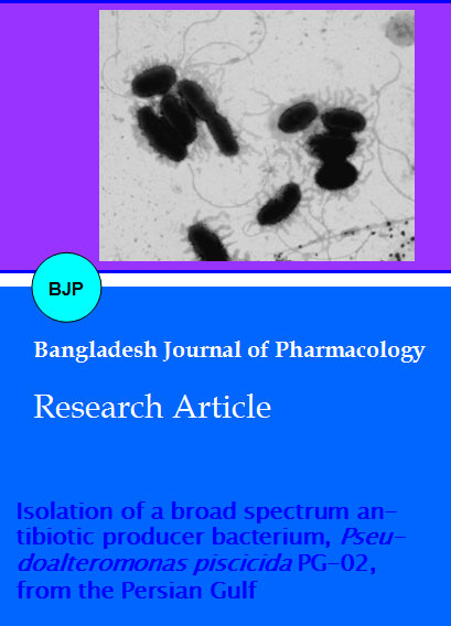
Isolation of a broad spectrum antibiotic producer bacterium, Pseudoalteromonas piscicida PG-02, from the Persian Gulf
Abstract
The aim of this study was to isolation of marine antibiotic-producing bacteria from the Persian Gulf, as an untapped source for searching new natural drugs. Bioactivity of ethyl acetate extract prepared from marine bacterial isolates was evaluated against 21 human pathogenic bacteria. GCI (Growth Curve Interference) parameter was determined for the obtained marine antibiotic compound against MRSA (Methicillin resistant Staphylococcus aureus). Finally, identification of intended isolate was done based on phenotipically and phylogenetically characters. Overall, 49 bacterial colonies were isolated but only one isolate from coastal sediment identified as Pseudoalteromonas piscicida PG-02 showed antibacterial activity. It was effective against all of tested bacteria except Listeria monocytogenes (clinical), L. monocytogenes (ATCC 19112), Proteus mirabilis (clinical) and Klebsiella pneumoniae (clinical). GCI parameter was obtained with concentration just near to MIC (GCI: 20 mg/mL). In conclusion, this marine bacterium can give hopes for treatment of diseases caused by multidrug resistant (MDR) bacteria.
Introduction
Antibiotic usually assumed as secondary metabolite with special chemical structure that produced during the idiophase of microbial growth (Das et al., 2006). Now-a-days, the rapid spread of multidrug-resistant (MDR) bacteria, reemergent infections, and newly emerging infectious diseases have become a serious global public health threat which especially affects economic development. Furthermore, with respect to the spread of methicillin-resistant Staphylococcus aureus (MRSA), the most common causative agent of infections among hospitalized patients, development of new classes of antibiotic is urgently needed. So, in addition to engineered drugs using combinatorial chemistry strategies, search for new natural sources for drugs such as microbes from extreme ecological niches can be a good choice for pharmaceutical companies for antibiotic drug discovery.
Oceans as the largest ecosystem on earth with an enormous diversity of different life forms which cover 70% of the earth's surfaces represent a potential source for finding new bioactive compounds (Das et al., 2006). The sea is an immense and practically unexploited source of potentially useful biologically active substances. Marine bacteria are often under extreme conditions such as temperature, pressure, salinity and depletion of micronutrients, so, in order to survive and maintain their biological activities, bacteria must prompt some special metabolic pathways to produce biologically active compounds that in some cases these secondary metabolites might be structurally unique substances and serve as new pharmaceutical agents that not found in terrestrial sources (Carvalho and Fernandes, 2010; Oku et al., 2003). Today, several different antibiotics have been reported from marine bacteria which among them Streptomyces, Pseudoalteromoas, Alteromonas, Bacillus and Pseudomonas are most famous (Darabpour et al., 2010). The Persian Gulf is a relatively shallow and semi-enclosed body of water that has some special ecological conditions such as having high degree of temperature so that its average sea water temperature may rise to 35.8°C and due to high evaporation, its salinity can increased as high as 4% and so on (Agah et al., 2009). The objective of present work was to isolation of potent antibiotic producing bacteria from water and sediment samples collected from different stations of Persian Gulf, as an unexplored source for searching new natural drugs.
Materials and Methods
Sample collection
Different samples including coastal water, surface water, deep water, coastal sediment, bed sediment and mangrove forest sediment were collected from 21 study sites in some northern area of Persian Gulf, during February 2008 to November 2009 (Figure 1 and Table I). Water samples were harvested by strelized-niskin bottle (using 70% ethanol prior to sampling) and subsequently samples were collected in sterilized glass bottles. Sediments were collected by sterilized van veen grab into sterilized plastic bags. All of the samples were kept at 4°C till to delivering to the laboratory.
Figure 1: Map of sampling sites (1: Bahrakan port, 2: Mahshahr port, 3: Bushehr port, 4: Qeshm island) in the Persian Gulf
Table I: The geographical characteristics and physicochemical properties of the major location of sampling sites in the Persian Gulf
| Temperature (°C ) | pH | Salinity (g/L) | Longitude | Latitude | Location Name | Station No. |
|---|---|---|---|---|---|---|
| 16.9 | 7.2 | 40 | 49°34' | 30°15' | Bahrakan port | 1 |
| 24 | 8 | 50 | 49°13' | 30°33' | Mahshahr port | 2 |
| 30.7 | 8.1 | 43.5 | 50°51' | 28°59' | Bushehr port | 3 |
| 30 | 8.4 | 39.5 | 55°23' | 26°40' | Qeshm Island | 4 |
Isolation of bacteria from marine samples
For isolation of marine bacteria from water samples, 8 uL from each sample was inoculated into Marine Agar 2216 (MA, Highmedia). Sediment samples were serially diluted using sterilized sea water (1:10 v/v ratio) and spread on the entire surface of marine agar 2216. Also, isolation of marine bacteria from both water and sediment samples was performed using pour plate technique. After incubation at 30°C for 3 days, developed colonies were purified by repeatedly subculturing on marine agar and finally, bacterial colonies with distinct characteristics such as pigmentation, size, opacity, elevation, margin and surface appearance (yeon et al., 2005) were chosen for further process.
Production medium and preparation of extract
For screening of the antibiotic producing bacteria, pure colonies were transferred to Erlenmeyer flasks containing Marine Broth (MB, Laboratorios CONDA) medium and were incubated at 30°C on a rotatory shaker (140 rpm) to produce antibiotic compounds. After 2-5 days, the broth culture was centrifuged at 10,000 rpm for 15 min at 4°C, and then supernatant was extracted using same volume of ethyl acetate. Solvent was removed at 37°C.
Selected human pathogens
A broad spectrum of human pathogenic bacteria including Standard (reference) and clinical isolates was selected for this study. Gram-positive bacterial species were Staphylococcus aureus (ATCC 6538), Staphylococcus epidermidis (ATCC 12228), Bacillus subtilis (ATCC 12711), Listeria monocytogenes (ATCC 19112), MRSA (Methicillin Resistant Staphylococcus aureus) (clinical), Staphylococcus epidermidis (clinical), Bacillus anthracis (clinical), Bacillus cereus (clinical), Bacillus pumilus (clinical), L. Monocytogenes (clinical), and Streptococcus pyogenes (clinical). Gram negative bacterial species were Escherichia coli (ATCC 11303), Pseudomonas aeruginosa (ATCC 27853), Salmonella typhi (ATCC 19430), P. aeruginosa (clinical), E. coli (clinical), Proteus mirabilis (clinical), Brucella melitensis (clinical), S. typhi (clinical), Bordetella bronchiseptica (clinical) and Klebsiella pneumoniae (clinical). Standard strains were prepared from Iranian Research Organization for Science and Technology (IROST); and clinical species were originally isolated from clinical specimens and identified by standard biochemical reactions.
Screening for antibacterial activity
Freshly grown colonies of tested bacterial pathogens were used to inoculate Muller Hinton Broth (MHB, Merck) medium at 37°C for 5-8 hours until turbidity of the suspension was adjusted to the Mc Farland 0.5 turbidity standard (108 cfu/mL) (Mulks et al., 1990; Asthana et al., 2009). Antibacterial activity was assessed two times by disc diffusion method. A total of 0.1 mL of bacterial suspension was poured on each plate containing Muller Hinton Agar (MHA, Merck). The lawn culture was prepared by sterile cotton swab and allowed to remain in contact for 1 min. The dried crude extract was dissolved in ethyl acetate to a concentration of 50 mg/mL. The sterile filter paper discs (6 mm diameter) (Kanoh et al., 2008) were saturated by 35 uL of this concentration of each extract and then were placed on lawn cultures. The Petri dishes were subsequently incubated at 37°C for 24 hours and the inhibition zone around each disc was measured in mm. Nine different antibiotics including vancomycin 30 ug, erythromycin 15 μg, methicillin 5 ug, carbenicillin 100 μg, nitrofurantoin 300 ug, penicillin 10 ug, oxacillin 1 ug, nafcillin 1 ug and streptomycin 10 ug were used as control. All these synthetic antibiotic discs were produced by Difco.
MIC and MBC determination
The MIC (Minimal Inhibitory Concentration) and MBC (Minimal Bactericidal Concentration) of ethyl acetate extract from the most effective bacterial strain were determined against two somewhat important and more sensitive bacteria. MIC was determined by the macro broth dilution assay method (Motamedi et al., 2010). In the tube dilution assay, standard bacterial suspension and different concentrations of extract (5, 10, 20, 40, 80, 160, and 200 mg/mL) were added to tubes containing 1 mL MHB medium. These tubes were incubated at 37°C for 24 hours. The first tube in the above series without sign of visible growth was considered as the MIC. MBC was determined by culturing one standard loop from tubes with no apparent growth on MHA and subsequent incubation at 37°C for 24 hours. The least concentration that inhibited colony formation on agar was considered as MBC for the extract.
Kinetics of growth and antibiotic production
Kinetic of growth and antibiotic production was studied in a batch culture system in a 250 mL Erlenmeyer containing 100 mL marine broth and at 30°C on a rotatory shaker (140 rpm). Kinetics of growth or biomass accumulation measured according to measuring of optical density (OD) at 600 nm. At first, the best antibiotic producing bacterium was grown in marine broth. Serial samples were harvested from production medium at 2 hours intervals from 0 till 6 hours and 4 hours intervals from 6 hours till entering stationary and death phases of growth. Supernatants were harvested from these samples by centrifugation at 8,000 rpm at 4°C for 10 min and subsequently were sterilized by filtration 0.45 µm and then used for antibacterial assay (Shibl and Sowaygh, 1980). Antibiotic activity of samples was assessed against MRSA using disc diffusion method.
Determination of growth curve interference (GCI)
GCI is a parameter which defined as the lowest concentration of an antibiotic compound that modifies the growth curve of tested pathogen compared with a control without antibiotic (Castillo et al., 2008). Tested pathogen in this study was MRSA. At first, four 250 mL flasks containing 15 mL MHB medium was prepared, and then freshly colonies of MRSA from a 24 hours culture on Nutrient Agar (NA, Merck) was inoculated in medium until turbidity of the suspension adjusted to the Mc Farland 0.5 turbidity standard. Also, 15 mL of crude extract from marine isolate at four concentrations equal to and below the MIC was prepared. A flask containing 30 mL MHB with the Mc Farland 0.5 turbidity of MRSA but without extract was used as control. Finally, each of the prepared extracts were added to one of the flasks and then incubated at 37°C on a rotatory shaker (110 rpm); biomass accumulation measured (every 1 hour) according to measuring optical density (OD) at 600 nm until entering stationary and death phases of growth. Related curves were drawn and GCI was determined.
Morphological and biochemical characterization of antibiotic producer bacterium
The isolated marine bacterial strain with antibacterial activity was identified based on morphological and biochemical characters according to the methods described in Bergey's manual for systematic of bacteriology (Garrity et al., 2006). Definite identification was done based on 16S rRNA gene sequencing.
Identification based on 16 S rRNA gene sequencing
In order to genomic DNA extraction, a single colony of intended bacterium was sub-cultured in MB and incubated at 30°C for 48 hours. The genomic DNA was extracted by a commercial kit (Genfanavaran, Iran), according to the manufacture instruments. Full length sequence of 16S rRNA genes was amplified from the isolated genomic DNA using the following universal bacterial 16S rRNA primers: Forward strand: 5´-CCGAATTCGTCGACAACAGAGTTTGATCCTGGCTCAG and reverse strand: CCCGGGATCCAAGCTTA-CGGTTACCTTGTTACGACTT-5 (Papizadeh et al., 2010). Polymerase chain reaction (PCR) was performed in a 50 uL reaction mix (mixture) containing 50 mM MgCl2, 10 mM each deoxynucleoside triphosphate (dATP, dTTP, dGTP and dCTP) dNTP, 10x PCR buffer, 10 umol/μL of each primer, and 5 U of Taq polymerase. The PCR thermal profile was consisted of denaturation at 94°C for 5 min followed by 30 cycles of amplification, each cycle consisting of a denaturation at 94°C for 1 min, annealing at 62°C for 40 sec, and chain elongation at 72°C for 2.5 min. A final elongation step at 72°C for 20 min was included. The Electrophoresis of PCR product was done on 1% agarose gel for 55 min at 95 v/cm in order to confirmation of the 16S rRNA amplification (Figure 2). PCR product was sequenced by Genfanavaran biotech corp. Alignment of sequences was done by CLC Main Workbench 6.0 software program. The obtained sequences were compared to those available in the Genbank by BLAST searches (http://www.ncbi.gov/blast). Finally, phylogenetic tree was constructed by CLC Main Workbench 6.0 software program using the neighbor-joining algorithm (Saitou and Nei, 1987). Also, the obtained sequence from intended antibiotic producer bacterium was compared to the sequence of its closest phylogenetic neighbor strain by chimera check/pintail program (http://www.bioinformatics-toolkit.org/Web-Pintail/).
Figure 2: Agarose gel electrophoresis of amplified 16 S rRNA fragment (1500 bp) of PG-02 isolate
Morphological observation of antibiotic producer isolate
Surface morphology study of intended bacterium was done using scanning electron microscopy (SEM). A single colony of antibiotic producer bacterium from 24 hours culture on marine agar was dissolved in 5 mL sterile distilled-water and subsequently 2 uL from this suspension was harvested and dried at room temperature. The prepared sample was coated by silver for 7 min and surface morphology was studied by Leo1455 Vp scanning electron microscopy.
Results
In the present research we isolated a total of 49 bacterial strains from marine samples collected from Persian Gulf (Table II). The majority of isolates were obtained from sediment samples. Among all of isolates, only one bacterium from the coastal sediment collected from seashore of Bushehr port showed antagonistic activity against pathogenic bacteria and designated as PG-02. The prepared raw extract from broth culture of this bacterium was effective against most of the tested pathogens.
Table II: Number of bacterial strains isolated from different samples collected from the northern regions of Persian Gulf
| Coastal water | Surface water | Deep water | Coastal sediment | Bed sediment | Mangrove forest sediment | Total | |
|---|---|---|---|---|---|---|---|
| Bahrakan port | - | 1 | 2 | 2 | 4 | - | 9 |
| Mahshahr port | - | 2 | 3 | - | 3 | - | 8 |
| Qeshm island | 2 | 1 | 3 | 4 | 1 | 2 | 13 |
| Bushehr port | 2 | 2 | 4 | 4 | 7 | - | 19 |
On the basis of obtained results from paper disc diffusion test (Table III), the antibiotic substance produced by indented marine bacterium has a broad-spectrum antibacterial activity as that among 21 tested bacterial pathogenic strains only L. monocytogenes (clinical), L. monocytogenes (ATCC 19112), P. mirabilis (clinical) and K. pneumoniae (clinical) showed reistance to the raw extract of PG-02 strain. Among Gram positive bacteria, S. aureus (ATCC 6538), S. epidermidis (ATCC 12228) and Str. pyogenes presented the most susceptibility to this antibiotic compound and about of Gram-negative bacteria, E. coli (ATCC 11303), S. typhi (ATCC 19430) and B. bronchiseptica (clinical) were the most susceptible strains. Also, tested Bacillus species exhibited a good level of sensitivity to antibiotic produced by PG-02. All of the examined clinical bacteria were multidrug resistant (MDR). Clinical S.aureus strain was resistant to methicillin so named MRSA. Antibacterial activity of the raw extract of PG-02 against MRSA in comparsion to synthetic antibiotics was remarkble. P. aeruginosa (clinical) and E. coli (clinical) were the most resistant strains to synthetic antibiotics while the growth inhibition effect of intented natural antibiotic was considerable. Among tested bacteria, only S. aureus (ATCC 6538) and B. subtilis (ATCC 12711) were sensitive to penicllin. On the other hand, all of tested pathogens were resistant to oxacillin.
Table III: Antibacterial activity of ethyl acetate extract of Pseudoalteromonas piscicida PG-02 against some clinical and standard pathogens compared with commercial antibiotics
| Concentration of extract (mg/mL) |
Antibiotic disc | |||||||||
|---|---|---|---|---|---|---|---|---|---|---|
| Bacterial species | 50 | VA | CB | P | FM | MT | S | NF | E | OX |
| Gram-positive bacteria | ||||||||||
| B. pumilus | 16 | 15 | 13 | R | 17 | 22 | 16 | R | 15 | R |
| B. anthracis | 15 | 20 | 16 | R | 21 | 23 | 14 | R | 21 | R |
| B. cereus | 14 | 19 | 15 | R | 18 | 23 | 15 | R | 15 | R |
| S. aureus | 16 | 22 | 31 | R | 23 | R | 12 | R | 16 | R |
| S. epidermidis | 15 | 25 | 18 | R | 22 | 13 | 13 | R | 14 | R |
| Str. pyogenes | 20 | 30 | 18 | R | 26 | 23 | 18 | R | 25 | R |
| L. monocytogenes | R | 17 | 12 | R | 16 | R | 15 | R | 16 | R |
| S. aureus ATCC | 19 | 18 | 26 | 12 | 23 | 13 | 16 | 10 | 28 | R |
| S. epidermidis ATCC | 18 | 15 | 31 | R | 28 | 15 | 18 | R | 30 | R |
| B. subtilis ATCC | 16 | 24 | 19 | 13 | 23 | 18 | 21 | 13 | 23 | R |
| L. monocytogenes ATCC | R | R | 16 | R | 14 | 11 | 18 | 12 | 19 | R |
| Gram-negative bacteria | ||||||||||
| E. coli | 15 | R | 22 | R | 20 | R | R | R | R | R |
| S. typhi | 13 | R | 28 | R | 21 | R | R | R | R | R |
| P. mirabilis | R | 24 | 28 | R | 10 | 14 | R | R | R | R |
| B. bronchiseptica | 21 | R | 13 | R | 17 | 30 | 18 | R | 17 | R |
| K. pneumoiae | R | 13 | 26 | R | 18 | R | R | R | R | R |
| P. aeruginosa | 13 | R | 19 | R | R | R | R | R | R | R |
| B. melitensis | 15 | R | 27 | R | 28 | R | 11 | R | R | R |
| E. coli ATCC | 22 | 18 | 31 | R | R | R | 18 | R | 20 | R |
| P. aeruginosa ATCC | 15 | R | 25 | R | R | R | R | R | 14 | R |
| S. typhi ATCC | 16 | 10 | 32 | R | 20 | R | 10 | R | R | R |
| *(6 mm) diameter disc; R: Resistant | ||||||||||
MIC and MBC values for ethyl acetate extract of PG-02 broth culture against MRSA were 40 and 70 mg/mL, respectively. Meanwhile, MIC and MBC values against B. bronchiseptica were the same (15 mg/mL).
On the basis of obtained results from growth curve of PG-02 and disc diffusion test (Figure 3), production of antibiotic by PG-02 was occurred after about 64 hours of growth when the culture entered in the late stationary phase and extended to death phase and remained stable with minor fluctuations in the antibacterial activity. This means that intended antibiotic is produced mainly during the period of growth cessation. The maximum antibiotic effect against MRSA was observed from the sample harvested at 76 hours age culture.
Figure 3: Results of Co-variation between cell growth of Pseudoalteromonas piscicida PG-02 and its antibiotic production
Figure 4 revealed that GCI parameter for antibiotic component produced by PG-02 against MRSA was obtained with concentration immediately blow MIC (70 mg/mL) so that at this concentration, growth curve of MRSA after 10 hours was not reached to stationary phase compared with control. Nonetheless, also growth curve of MRSA was affected partially at 60 and 50 mg/mL compared to control.
Figure 4: Determination of GCI parameter for Pseudoalteromonas piscicida PG-02 broth culture extract against MRSA
The obtained results from biochemical diagnostic tests (Table IV) presented that antibiotic producer isolate is a Gram negative rod (Figure 5), motile, aerobe and with oxidative metabolism that utilizes D-glucose and maltose as carbon source. PG-02 isolate was capable to growth at NaCl 8%. Therefore, it was assumed as a moderately-halophilic bacterium. On the basis of BLASTn search of Genbank, the isolate PG-02 showed the most similarity to Pseudoalteromonas piscicida strain 1314 (nucleotide identity: 98%) so it was finally identified as Pseudoalteromonas piscicida PG-02. The phylogenetic tree shows a relatively wide phylogenetic heterogeneity of Pseudoalteromonas piscicida PG-02 with other Pseudoalteromonas species (Figure 6). Also, the mean of the observed %differences between Pseudoalteromonas piscicida PG-02 and 1314 (Figure 7) was 0.20, and is roughly equivalent to evolutionary distance. The Genbank accession number for the 16S rRNA gene sequence of strain PG-02 is JF509137.
Figure 5: Scanning Electronic Microscope micrographs of PG-02 isolate in order to study of its morphology
Table IV: Morphological and biochemical characteristics of Pseudoalteromonas piscicida PG-02
| Parameter | Character | Parameter | Character |
|---|---|---|---|
| Cell shape | Rod | Growth at 4°C | - |
| Pigmentation | Orange | Growth at 42°C | - |
| Gram reaction | Negative | Growth at 8% NaCl | + |
| Catalase | + | O-F test | + |
| Oxidase | + | Acid from: | |
| Motility | + | D-glucose | + |
| Indol production | - | L-arabinose | - |
| Citrate utilization | + | D-mannitol | - |
| Urease | - | Maltose | + |
| Nitrate reduction | + | Xylose | - |
| Gelatin hydrolysis | - | Sensitivity to : | |
| TSI | Alk/Alk | Penicillin | - |
Figure 6: Phylogenetic relationships of Strain PG-02 and some related Pseudoalteromonas species on the basis of 16S rRNA gene sequence analysis. Numbers at branch nodes are bootstrap values (expressed as a percentage of 1000 replicates) based on the neighbour-joining algorithm. Bar represents approximately 0.7% nucleotide sequence difference
Figure 7: Variation in % difference between P. piscicida 1314 (1372 nt) and P. piscicida PG-02 (1248 nt) determined with a 300 base sliding window, moving 25 bases at a time along the sequences' length
Discussion
Antibiotic resistance among pathogenic bacteria is usually of greater magnitude in developing countries that is very important in national and international economical, political and scientific aspects. For instance, MRSA particularly is responsible for the largest outbreak of hospital-acquired infection (HAI) that the world has ever seen, so there is a continued need to develop new antibiotics. Unfortunately, because of the ability of bacteria in exchanging of genetic information and new mutations this problem has been more complicated (Tenover, 2006). With respect to enormous biodiversity in the marine environment so that in the case of microorganisms, sea water is composed of 78 million microscopic cell per ounce or the bottom, which mimic the soil, contain more than 1 billion cells in the volume of an ordinary cube of sugar this environment as a rich resource can be exploited for developing new natural pharmaceutical products especially antibiotics (Darabpour et al., 2010). Furthermore, existence of a given the intense microbial competition for resources such as space and nutrients between marine bacteria especially those that inhabited in sediment introduce a powerful selection pressure that can cause excretion of metabolites by these microorganisms for help mediate microbe-microbe interaction that occur on the order of micrometers, it is likely that many of these metabolites were structurally unique and bioactive secondary metabolites (Teasdale et al., 2009; Balcazar et al., 2010). Rosnfeld and Zobell, in 1947, were reported the first document of antibiotic producing marine bacterium (McCarthy et al., 1994). In this study, of 49 marine isolates only one bacterium that was isolated from sediment showed antibacterial activity; so, this result can confirm the hypothesis mentioned above about antibiotic producer marine bacteria that imply on particle-inhabited bacteria had broader-spectrum ability in antibiotic production than free-live bacteria (Long et al., 2001). This isolate is a Gram negative that belongs to γ-proteobacteria phylum, various studies have been shown that Gram negative bacteria (most abundant bacteria in the marine environment) especially those that belong to γ-proteobacteria have a particular potential for antibiotic production (Long et al., 2003; Shankar et al., 2010). The antibiotic compound produced by PG-02 presented a very good level of antibacterial activity against tested human pathogenic bacteria including both Gram positive and negative bacteria so that it was effective against 81% of tested bacteria (Table III). Three of the most important tested clinical pathogens including S. aureus (clinical), E. coli (clinical) and P. Aeruginosa (clinical) showed a high level of resistance to synthetic antibiotics while antibacterial effect of intended antibiotic compound was considerable compared to these antimicrobials.
MRSA as major nosocomial pathogen exhibited resistance to 50% of tested synthetic antibiotics. Also, L. monocytogenes (ATCC and clinical strains) which is responsible for Listeriosis (sever foodborne illness) was resistant to intended natural antibiotic and more of the synthetic antibiotics, so this strain must be focused with respect to its high level of resistance. Also, antistaphylococcal and anti-bacillus activity of the antibiotic produced by Pseudoalteromonas piscicida PG-02 was valuable. All of tested bacteria were resistant to oxacillin, so it seems that gradually we should exit this antibiotic from the usual antibiotic list. MIC and MBC values for ethyl acetate extract of P. piscicida PG-02 broth culture against B. bronchiseptica were same, so antibiotic compound existed in extract can act as bactericidal agent against this pathogenic bacterium (Motamedi et al., 2010). Because of urgently need for developing new antibacterial agents against MRSA; we have focused on antibacterial activity of the intended antibiotic against this rebellious pathogenic bacterium. Results of kinetic of growth and antibiotic production for P. piscicida PG-02 (Figure 3) revealed that production of antibiotic against MRSA was begun in the middle stages of stationary phase and extended in death phase; so it can be concluded that this antibiotic as a secondary metabolite will be produced during secondary metabolism and also production of antibiotic will be occurred parallel to the biomass accumulation; that is to say, kinetic of antibiotic production by P. piscicida PG-02 is growth-associated (Isnansetyo et al., 2009).
Based on the obtained results from GCI parameter (Figure 4), antibacterial activity of indented antibiotic compound against MRSA was maintained with sub-MIC concentration, this outcome has a pharmaceutical importance especially for high-risk patients. At first, the morphological characters and biochemical tests of PG-02 indicate that this strain might be one member of Pseudomonas genus but based on homology obtained from 16S rRNA, this strain belongs to Pseudoalteromonas genus and piscicida species; however, because 98%sequence similarity (is not 100%) and some particular biochemical and morphological characters (compared to those that mentioned in Bergey's manual of systematic bacteriology; Garrity et al., 2006) such as inability to use of ammonia but ability to nitrate reduction, this strain can be considered as a novel species in the genus Pseudoalteromonas and this hypothesis must be confirmed by further phenotypic characterization and DNA-DNA hybridization.
Conclusion
The antibacterial compound produced by Pseudoalteromonas piscicida PG-02 is a broad spectrum antibiotic that has a good level of inhibitory effect against most of the human pathogenic bacteria especially MDR bacteria such as MRSA, E. coli and P. aeruginosa. Considering MIC, MBC and GCI parameters, this marine antibiotic can be used as an anti-MRSA compound with high efficiency.
Acknowledgments
The authors wish to thank the vice chancellor for research of Shahid Chamran University, Ahvaz, Iran, for the research grant and financial support.