Overexpression of phosphorylated MARCKS in the nicotine-derived nitrosamine ketone-induced lung cancer mice
Abstract
Lung cancer is the most frequently occurring lethal cancers in men and women population. The aim of the present study is to observe the overexpression pattern of phosphorylated MARCKS in the nicotine-derived nitrosamine ketone (NNK)-induced lung cancer mice. Pathogen-free female A/J mice were used for the present experiment to induce lung cancer by the carcinogen namely, NNK. At different time intervals namely, 5th, 6th and 7th month after NNK injection, lung tissue samples were collected. Immunohistochemistry in accordance with the immunoblotting techniques were used to confirm the overexpression of phosphorylated MARCKS in the NNK induced lung cancer mice model. The present study concludes that the phosphorylated MARCKS was overexpressed in the NNK induced lung cancer mice during the early stages of lung cancer and it may be used as a tool to detect the lung cancer in the initial stages.
Introduction
Lung cancer is the most frequently occurring cancer other than breast and colorectal cancers (Dedhia and Parker, 2011). Worldwide, lung cancer is the most common and lethal cancers of men and women in North America, Europe, and East Asia. Over one million people were reported each year for lung cancer, which illustrates the severity of the disease. Right now, only 15% of lung cancers are detected in the early stages and localized, at the same time, most of them are well advanced during initial diagnosis itself (Dedhia and Parker, 2011).
MARCKS is a PIP2-associated acidic protein that is unusually rich in alanine, glycine, proline and glutamic acid. It is a rod-shaped protein containing three distinct domains namely; a) An N-terminal myristoylated domain that mediates binding to membranes; b) A highly conserved MH2 domain of unknown function, and; c) A basic effector domain containing the PKC phosphorylation sites, which in terms has the calmodulin and actin-binding sites (Erusalimsky et al., 1991; Harlan et al., 1991; Seykora et al., 1991; Stumpo et al., 1989). It was reported that the phosphorylation of MARCKS by protein kinase C results in the detachment of MARCKS from the membrane and suppress PIP2 by sequester manner (Gambhir et al., 2004; McLaughlin and Murray, 2005). At the same time it was also identified that in lungs, MARCKS especially phosphorylated MARCKS, plays a crucial role in controlling mucin secretion and inflammation (Eckert et al., 2010; Green et al., 2011; Li et al., 2001; Park et al., 2007; Singer et al., 2004). In addition, it was reported that phosphorylated MARCKS specifically regulate the cancer migration and metastasis (Chen et al., 2014; Chen and Rotenberg, 2010; Ghoul et al., 2006; Reddy et al., 2010; Techasen et al., 2010). In contrast, very little is known about the importance of phospho-MARCKS and its relevance in lung cancer. Based upon the importance of phosphorylated MARCKS in lung cancer and the literature survey a hypothesis was raised, that the phosphorylated MARCKS was overexpressed in the lung cancer tissues. In order to answer the hypothesis, following experiments were designed. In the current experiment, 4-(methylnitrosamino)-1-(3-pyridyl)-1-butanone (NNK) was used to induce lung cancer in A/J mice. NNK is a class 1 IARC carcinogen (Cogliano et al., 2004), originated from the nicotine during curing of tobacco. NNK is activated in the lung via alpha-carbon hydroxylation by cytochrome P450 (CYP), hemoglobin, and lipoxygenases (LOX) (Bedard et al., 2002).
Materials and Methods
Animals
Pathogen-free female A/J mice were chosen and purchased for the current project. Animals were housed in filter-top metal cages and maintained in a 12/12 hour light/dark cycle. Water and food were made available for the mice 24 hours and they are handled according to the international ethical regulation. The animals were observed twice a day and subjected to weighing once a week.
NNK Treatment
NNK (4-(methylnitrosamino)-1-(3-pyridyl)-1-butanone) were purchased from Sigma Alrich. Pathogen-free A/J mice of same age group were injected intraperitoneally with NNK at the concentration of 80 mg/kg/day in 0.1 mL PBS for three alternate days. Control animals received an equal volume of PBS. The NNK treatments produced multiple lung tumors in all the NNK-treated A/J mice, but none in the control group. The lung tumors were observed, which is visible with the naked eye. During 5th, 6th and 7th month after NNK injection the lung tissue samples were collected for the experiments.
Immunohistochemistry
Lung tissue samples were collected from the control and NNK induced lung cancer mice at different time intervals namely, 5th, 6th and 7th month after NNK injection. Lung tissues were formalin-fixed and paraffin-embedded using the standard protocol. The tissue sections (7 µm) were deparaffinized and hydrated. Antigens were retrieved by tri-sodium citrate treatment (pH 6.0). Endogenous peroxides and non-specific immune staining were blocked by hydrogen peroxide and normal serum, respectively. The sections were then incubated overnight at 4ºC with the monoclonal anti-phosphorylated-MARCKS antibody. After incubation with primary antibody, tissue sections were washed and incubated with secondary antibodies conjugated with HRP. The washed slides were developed with DAB substrate. The prepared slides were counterstained, mounted with DPX and observed under a Nikon Ti-S fluorescent microscope.
Immunoblotting
Protein samples were prepared from the NNK treated and control mice lung tissues (at different time intervals namely, 5th, 6th and 7th month after NNK injection) and resolved in the 8% SDS-PAGE. The resolved protein samples from the SDS-PAGE gel was transferred to the PVDF membrane, blocked with 4% BSA for 1 hour and incubated with primary antibody (anti-phosphorylated MARCKS and anti-Akt antibodies (from Sigma) for overnight at 4ºC. For loading controls, anti-beta-actin (from Abcam) was used. The nonspecific binding of the primary antibody was washed out with a 1X TBST buffer. The secondary antibody conjugated with HRP was used in the dilution concentration of 1:10000. The washed membrane was developed (as per manufacturer's instruction) with the substrate DAB/H2O2 (from Amresco) to get the brown colored product, which appeared on the membranes.
Results
Pathogen-free female A/J mice were chosen for the experiments. Animals were handled as per the regulations mentioned in the materials and methods. In order to induce the lung tumor, pathogen free female A/J mice of same age group were injected intraperitoneal with NNK at the concentration of 80 mg/kg/day in 0.1 mL PBS for three alternate days. Control animals were received an equal volume of PBS. The animals were observed twice a day and subjected to weighing once a week. After 5th, 6th and 7th month of interval, the lung tissue samples were collected for the experiments. Lung tumors were observed in all the NNK-treated A/J mice, but none in the control group. The presence of lung cancer in all the NNK-induced pathogen-free A/J mice, were confirmed by the pathologist based on the number of adenomas.
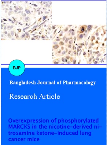
Animals were killed based on the standard protocol and the organs were dissected. Lung tissue samples were collected from the control and NNK induced lung cancer mice at different time intervals namely, 5th, 6th and 7th month after NNK injection. The lung tissue samples were collected and fixed in 10% neutral buffered formalin and processed for immunohistochemistry. In order to identify the overexpression of phosphorylated MARCKS in the NNK induced lung cancer mice, immunohistochemistry was performed as per the protocol mentioned in the materials and methods, along with control animal. The data Figure 1A shows the expression of phosphorylated MARCKS in the control A/J mice at 5th month, whereas Figure 1B shows the overexpression pattern of Phosphorylated MARCKS in the NNK induced lung cancer A/J mice at 5th month after NNK injection. Similarly, the control lung tissue sample along with the NNK induced A/J mice at 6th month were shown in Figure 2A and 2B, respectively. In addition, the control lung tissue sample along with the NNK induced A/J mice at 7th month were shown in the Figure 3A and 3B, respectively.
Figure 1: Immunohistochemistry of control and NNK induced lung cancer tissues at 5th month. A. Immunohistochemistry of control lung tissues (A/J mice) stained with anti-phosphorylated MARCKS antibody. B. Immunohistochemistry of NNK induced lung cancer tissues (A/J mice) stained with anti-phosphorylated MARCKS antibody. The positive signals were identified by brown color and the sections were counterstained with heamotoxylin at 60X. The percentages of phosphorylated MARCKS positive cells were shown in the figures
Figure 2: Immunohistochemistry of control and NNK induced lung cancer tissues at 6th month. A. Immunohistochemistry of control lung tissues (A/J mice) stained with anti-phosphorylated MARCKS antibody. B. Immunohistochemistry of NNK induced lung cancer tissues (A/J mice) stained with anti-phosphorylated MARCKS antibody. The positive signals were identified by brown color and the sections were counterstained with heamotoxylin. The percentages of phosphorylated MARCKS positive cells were shown in the figures
Figure 3: Immunohistochemistry of control and NNK induced lung cancer tissues at 7th month. A. Immunohistochemistry of control lung tissues (A/J mice) stained with anti-phosphorylated MARCKS antibody. B. Immunohistochemistry of NNK induced lung cancer tissues (A/J mice) stained with anti-phosphorylated MARCKS antibody. The positive signals were identified by brown color and the sections were counterstained with heamotoxylin. The percentages of phosphorylated MARCKS positive cells were shown in the figures
To confirm and validate the immunohistochemistry data, immunoblotting was performed. Protein samples were prepared from the lung tissues of NNK induced lung cancer mice along with its control (at different time intervals namely, 5th, 6th and 7th month after NNK injection) and resolved in the 8% SDS-PAGE. The results of immunoblot were shown in the Figure 4. The data (Lane - 1, 3 and 5) shows the expression of phosphorylated MARCKS in the control A/J mice at 5th, 6th and 7th month after PBS injection. In contrast, Lane - 2, 4 and 6 show the overexpression pattern of phosphorylated MARCKS in the NNK induced lung cancer A/J mice at 5th, 6th and 7th month after NNK injection. The data confirms the overexpression of phosphorylated MARCKS in the NNK induced lung cancer A/J mice. Immunohistochemistry data was validated with the immunoblot analysis.
Figure 4: Immunoblotting of control and NNK induced lung cancer tissues samples with anti-phosphorylated MARCKS, anti-Akt and anti-beta-actin antibodies at different time intervals. Lane 1, 3 and 5 represents control lung tissue samples at 5th, 6th and 7th months, respectively; Lane 2, 4 and 6 represents NNK induced lung cancer tissue samples at 5th, 6th and 7th months, respectively
Discussion
Pathogen-free female A/J mice were chosen for the experiments and to induce lung cancer by the carcinogen namely, NNK. It is a potent tobacco-specific carcinogen, which requires the activation of the cytochrome P450 enzyme system for its tumorigenic activity. But it was reported that the inhibitors of the enzyme system reduce NNK towards lung tumorigenesis (Wang et al., 2003). In addition, it was reported that the NNK stimulates the release of growth factors (acetylcholine and serotonin) from the normal lung as well as from the lung cancer cells (Song et al., 2003). Thus, NNK has the capacity to induce the normal lung tissue into the cancerous one. In the present experiment, all the NNK injected animals shows the properties of lung cancer without any variation in the weight when compared to the control. In addition, pathologist confirmed that the NNK injected mice has lung cancer, based upon the presence of adenomas.
To identify the expression pattern of phosphorylated MARCKS during the NNK-induced lung cancer and control A/J mice, lung tissue samples were collected from the animals at different time intervals namely, 5th, 6th and 7th month after NNK injection, and allowed for immunohistochemistry analysis. The control and the NNK-induced lung cancer tissue at 5th, 6th and 7th month were shown in the Figure 1-3, respectively. The data illustrates the expression of phosphorylated MARCKS, which was observed in control as well as NNK induced lung cancer tissues. By careful observation on the control and NNK induced lung cancer tissues, it was identified that the expression of phosphorylated MARCKS was higher in later when compared to control. The data suggested that the phosphorylated MARCKS was overexpressed in the NNK induced lung cancer mice. In addition, the data suggest that during the initial stages of NNK induced lung cancer, phosphorylated MARCKS expressed highly. The data confirms the hypothesis that phosphorylated MARCKS was overexpressed during lung cancer. To support the data, it was reported that the phosphorylated MARCKS specifically regulate the cancer migration and metastasis (Chen et al., 2014; Chen and Rotenberg, 2010; Ghoul et al., 2006; Reddy et al., 2010; Techasen et al., 2010).
Further to validate the data, the immunoblot was performed. The data illustrates that both control and the NNK induced lung cancer tissues at 5th, 6th and 7th month shows positive for phosphorylated MARCKS. Rather, careful observation on the intensity of the data confirms that the expression of phosphorylated MARCKS was higher in NNK-induced lung cancer tissues when compared to control. Hence, it was confirmed that the phosphorylated MARCKS was overexpressed in the NNK-induced lung cancer mice. In addition, the immunohistochemistry data was cross-checked and validated by the immunoblot data.
Early detection of lung cancer is the key and it may also provide better treatment options, for potential better survival rates against the disease. Moreover, various basic researches turn on the search of new efficient markers to detect the lung cancer in the initial stages.
Conclusion
The current experiment shows that the overexpression of phosphorylated MARCKS was observed during the early stage of lung cancer and it may be useful to detect the lung cancer in the initial stages.
Acknowledgements
The authors were thankful for the gift of A/J mice, which is granted from the leading research group and the support is the backbone of the project. Also, authors are privileged to thank institutional review board approval committee and ethical committee for the successful completion of this project.
Ethical Committee
The study was approved by the institutional review board approval committee and ethical committee.
References
Bedard LL, Smith GB, Reid KR, Petsikas D, Massey TE. Investigation of the role of lipoxygenase in bioactivation of 4-(methylnitrosamino)-1-(3-pyridyl)-1-butanone (NNK) in human lung. Chem Res Toxicol. 2002; 15: 1267-73.
Chen C-H, Statt S, Chiu C-L, Thai P, Arif M, Adler KB, Wu R. Targeting myristoylated alanine-rich C Kinase substrate phosphorylation site domain in lung cancer: Mechanisms and therapeutic implications. Am J Respir Crit Care Med. 2014; 190: 1127-38.
Chen X, Rotenberg SA. PhosphoMARCKS drives motility of mouse melanoma cells. Cell Signal. 2010; 22: 1097-103.
Cogliano V, Straif K, Baan R, Grosse Y, Secretan B, Ghissassi FE. Smokeless tobacco and tobacco-related nitrosamines. Lancet Oncol. 2004; 5: 708.
Dedhia HV, Parker JE. A new era for lung cancer detection. Comm Oncol. 2011; 8: 442-44.
Eckert RE, Neuder LE, Park J, Adler KB, Jones SL. Myristoylated alanine-rich C-kinase substrate (MARCKS) protein regulation of human neutrophil migration. Am J Respir Cell Mol Biol. 2010; 42: 586-94.
Erusalimsky J, Brooks S, Herget T, Morris C, Rozengurt E. Molecular cloning and characterization of the acidic 80-kDa protein kinase C substrate from rat brain: Identification as a glycoprotein. J Biol Chem. 1991; 266: 7073-80.
Gambhir A, Hangyás-Mihályné G, Zaitseva I, Cafiso DS, Wang J, Murray D, Pentyala SN, Smith SO, McLaughlin S. Electrostatic sequestration of PIP2 on phospholipid membranes by basic/aromatic regions of proteins. Biophy J. 2004; 86: 2188-207.
Ghoul A, Serova M, Benhadji K, Cvitkovic E, Faivre S, Philips E, Calvo F, Lokiec F, Raymond E. Protein kinase C α and δ are members of a large kinase family of high potential for novel anticancer targeted therapy. Targeted Oncol. 2006; 1: 42-53.
Green TD, Crews AL, Park J, Fang S, Adler KB. Regulation of mucin secretion and inflammation in asthma: A role for MARCKS protein? Biochimica et Biophysica Acta (BBA)-General Subjects. 2011; 1810: 1110-13.
Harlan DM, Graff J, Stumpo D, Eddy R, Shows T, Boyle JM, Blackshear P. The human myristoylated alanine-rich C kinase substrate (MARCKS) gene (MACS). Analysis of its gene product, promoter, and chromosomal localization. J Biol Chem. 1991; 266: 14399-405.
Li Y, Martin LD, Spizz G, Adler KB. MARCKS protein is a key molecule regulating mucin secretion by human airway epithelial cells in vitro. J Biol Chem. 2001; 276: 40982-90.
McLaughlin S, Murray D. Plasma membrane phosphoinositide organization by protein electrostatics. Nature 2005; 438: 605-11.
Park JA, Crews AL, Lampe WR, Fang S, Park J, Adler KB. Protein kinase Cδ regulates airway mucin secretion via phosphorylation of MARCKS protein. Am J Pathol. 2007; 171: 1822-30.
Reddy M, Fernandes M, Salgia R, Levine R, Griffin J, Sattler M. NADPH oxidases regulate cell growth and migration in myeloid cells transformed by oncogenic tyrosine kinases. Leukemia 2010; 25: 281-89.
Seykora JT, Ravetch JV, Aderem A. Cloning and molecular characterization of the murine macrophage" 68-kDa" protein kinase C substrate and its regulation by bacterial lipopolysaccharide. Proc Natl Acad Sci USA. 1991; 88: 2505-09.
Singer M, Martin LD, Vargaftig BB, Park J, Gruber AD, Li Y, Adler KB. A MARCKS-related peptide blocks mucus hypersecretion in a mouse model of asthma. Nature Med. 2004; 10: 193-96.
Song P, Sekhon H, Proskocil B, Blusztajn J, Mark G, Spindel E. Synthesis of acetylcholine by lung cancer. Life Sci. 2003; 72: 2159-68.
Stumpo DJ, Graff JM, Albert KA, Greengard P, Blackshear PJ. Molecular cloning, characterization, and expression of a cDNA encoding the" 80-to 87-kDa" myristoylated alanine-rich C kinase substrate: A major cellular substrate for protein kinase C. Proc Natl Acad Sci USA. 1989; 86: 4012-16.
Techasen A, Loilome W, Namwat N, Takahashi E, Sugihara E, Puapairoj A, Miwa M, Saya H, Yongvanit P. Myristoylated alanineâ€rich C kinase substrate phosphorylation promotes cholangiocarcinoma cell migration and metastasis via the protein kinase Câ€dependent pathway. Cancer Sci. 2010; 101: 658-65.
Wang H, Tan W, Hao B, Miao X, Zhou G, He F, Lin D. Substantial reduction in risk of lung adenocarcinoma associated with genetic polymorphism in CYP2A13, the most active cytochrome P450 for the metabolic activation of tobacco-specific carcinogen NNK. Cancer Res. 2003; 63: 8057-61.