Neuroprotective effects of pterostilbene against isoflurane-induced apoptosis through regulating the JNK and PI3K/Akt pathway in neonatal rats
Abstract
Increasing numbers of children undergo surgery and are exposed to anesthesia that raises concerns regarding the safety of frequently employed anesthetics. Isoflurane, often used in pediatric anesthesia, has been reported to cause neurodegeneration in animal models. The study investigates the effectiveness of pterostilbene on neurodegeneration caused by isoflurane. Separate groups of neonatal mice were administered with pterostilbene at 10, 20 or 40 mg/kg from post natal day 1 (P1) to P15. On P7, rats received isoflurane at 0.75% for 6 hours. Control rats received no anesthesia or pterostilbene. Neuroapoptosis following isoflurane exposure were markedly reduced by pterostilbene , further pterostilbene down-regulated the expressions of caspase-3, Bad, phospho-JNK and phospho-c-Jun and as well improved the expressions of Bcl-xL, JNK, phospho-Bad and phospho-Akt. Pterostilbene enhanced the performance of rats in Morris water maze tests. The observations suggest that pterostilbene was able to effectively reduce isoflurane-induced neurodegeneration.
Introduction
Isoflurane is a commonly used volatile anesthetic in pediatric surgeries (Istaphanous and Loepke, 2009). However prolonged exposure to volatile anesthetics causes neuronal apoptosis and degeneration in developing brains leading to learning and memory deficits (Satomoto et al., 2009; Brambrink et al., 2010; Kong et al., 2011; Li et al., 2013a,b). Recent investigations in children less than 4 years of age, exposed to anaesthesia more than once, present to have greater risks of developing cognitive disabilities (DiMaggio et al., 2011; Ing et al., 2012) thus raising serious concerns on possible detrimental effects of anesthetics. This has forced researchers to exploit possible protective strategies.
Jevtovic-Todorovic et al. (2003) demonstrated that during synaptogenesis the developing brains is more vulnerable to neurotoxic insults. Studies have proposed activation of gamma-aminobutyric acid and inhibition of N-methyl-D-aspartate receptors in neurodegeneration (Jevtovic-Todorovic et al., 2003; Olney et al., 2004). Isoflurane-induced neuronal apoptosis and degeneration has been observed to occur via JNK pathway and also through disruption of intracellular calcium homeostasis and hyperactivation of inositol 1,4,5-trisphosphate (IP3) receptors (Li et al.,2013b; Wei et al., 2008; Yang et al., 2008; Zhao et al., 2011). Isoflurane-induced Ca2+ overload activates mitochondrial apoptosis pathway (Wei et al., 2005; Yon et al., 2005) and also causes activation of c-Jun N-terminal kinase (Brambrink et al., 2010).
Protein kinase B (Akt) that plays a major role in regulating neuronal survival, on activation, inhibits apoptosis through inactivating Bad (Luo et al., 2003; Song et al., 2005). Studies have showed a potential link between JNK and Akt signalling (Fornoni et al., 2008; Yeste-Velasco et al., 2009). Thus compounds that can regulate JNK/Akt signalling pathway could offer neuroprotection.
Bai et al. (2013) reported that reseveratrol, a phytoalexin offers protection against isoflurane-induced neurotoxicity via regulation of Akt pathway. Pterostilbene, an analog of resveratrol, found in blueberries and in several types of grapes (Roupe et al., 2006; Lin et al., 2009; McCormack and McFadden, 2012) possess various pharmacological activities including anti-inflammatory, antioxidant and anticancer activities (Remsberg et al., 2008). Chang et al. (2012) reported that pterostilbene supplementation improved cognition in a mouse model of Alzheimer's disorder. Considering the protective effects of pterostilbene, we investigated its effects on isoflurane-induced neonatal rats.
Materials and Methods
Animals
This study was approved by the animal care committee and was performed in accordance with the National Institutes of Health Guide for the Use of Laboratory Animals. Pregnant female Sprague-Dawley rats (Guangdong Medical Laboratory Animal Co., China, permission number: SCXK2011-0029) were closely monitored for the day of delivery that was considered as postnatal day (P0) and the pups were carefully observed.
Separate groups of pups were administered with pterostilbene at 10, 20 or 40 mg/kg b.wt, orally from day P1 and continued till P15. On P7, rat pups (15-18 g) were exposed to 0.75% isoflurane for 6 hours [approximately 0.3 MAC in P7 rats as determined by Orliaguet et al. (2001)] in 30% oxygen or air in a temperature-controlled chamber as described by Li et al. (2013b). The pups were exposed to isoflurane after 1 hour of pterostilbene administration on P7. At the end of the exposure, some rats pups (n=6) were sacrificed immediately and their hippocampi were used for western blot studies and TUNEL assay. The rest of the rats (n=6) were maintained under standard experimental conditions till P30 with oral supplementation of pterostilbene, till P15.
Reagents and chemicals
Isoflurane (0.75%) and pterostilbene were procured form Sigma-Aldrich, St. Louis, MO, USA. Antibodies: anti-cleaved caspase-3, anti-phospho-Akt, anti-Akt, anti-phospho-Bad, anti-Bad, anti-Bcl-xL, anti-beta-actin, anti-phospho-JNK, anti-JNK and anti-phospho-c-Jun were all purchased from Cell Signalling Technology, Beverly, MA, USA. All the other chemicals used in the study were of analytical grade and purchased from Sigma-Aldrich, USA.
TUNEL assay
TUNEL studies were performed as described previously by Li et al. (2013b). Briefly, rat pups were anesthetized with isoflurane and perfused transcardially with ice-cold saline followed by 4% paraformaldehyde in 0.1 M phosphate buffer. The brain tissues were harvested from the pups and the tissues were postfixed for 48 hours at 4°C and paraffin embedded and further sectioned at 5 µm thickness. Three sections that were 100 µm apart in each pup were chosen for TUNEL. TUNEL fluorescent assay was performed using the Dead End TM fluorometric TUNEL system kit (Promega, Madision, WI, USA). Hoechst was used to stain the nuclei. TUNEL positive cells in the hippocampal CA1, CA3 and DG regions were analyzed with NIS-Elements BR image processing and analysis software (Nikon Corporation, Japan).
Western blotting
Western blotting was performed to assess the expression of proteins under the influence of isoflurane exposure and treatment with pterostilbene. The blotting was performed as described earlier (Li et al., 2013 a,b). Briefly, protein concentrations were determined using the BCA protein assay (Bio-Rad, UK). Sixty micrograms of each protein sample was subjected to western blot analysis using the following primary antibodies: antiphospho-Akt at 1:1000 dilution, anti-Akt at 1:2000 dilution, anti-phospho-Bad at 1:1000 dilution, anti-Bad at 1:1000 dilution, anti-Bcl-xl at 1:2000 dilution, anti-cleaved caspase-3 at 1:2000 dilution, anti-phospho-JNK at 1:1000 dilution, anti-JNK at 1:1000 dilution, anti-phospho-c-Jun at 1:1000 dilution and anti-beta-actin at 1:2000 dilution. Images were scanned by an Image Master II scanner (GE Healthcare, USA) and were analyzed using ImageQuant TL software v2003.03 (GE Healthcare, USA). The band signals of proteins were normalized to those of the corresponding β-actin (internal control).
Memory and learning studies- Morris water maze test
For memory and learning studies, the pups were exposed to anesthetics on P7 as described above. Spatial reference memory and learning assessments with the Morris water maze were performed as we have described previously by Li et al. (2007).
Animals were trained for 4 days (P26-P29) in the Morris water maze. A platform (10.3 cm diameter) was sub-merged in a circular pool (200 cm diameter, 60 cm depth) filled with warm water (23 ± 2°C). They were trained in 2 sessions a day. Animals were allowed a time of 60 sec to locate the hidden platform, and if they failed to locate in allotted time, they were guided to the platform. In either case, the pups were removed from the platform after 15 sec. Training sessions were conducted until they could locate the hidden platform in less than 15 sec (average time per session). All trials and swim paths were recorded with ANY-maze video tracking system (Stoelting Co., USA) that measures the time taken (latency) to find the platform(s), as well as other behavioural information obtained during the spatial reference memory test. The animals were dried and placed beneath a heating lamp at the end of every test.
Cued trials
The cued trials were conducted on P30 to assess for any non-cognitive performance impairments such as visual impairments and/or swimming difficulties. In this study, the pool was surrounded by a white cloth to hide the visual cues. The animals received 4 trials in a day. In each trial, they were placed in a fixed position of the swimming pool towards the wall and were allowed to swim to a platform with a rod (cue) placed 20 cm above water level randomly in any of the four quadrants of the swimming pool. The rat pups were given 60 sec to locate the platform and 30 sec to sit on the platform following which they were taken off from the pool. If the rats were unable to locate a platform within 60 sec, they were gently guided and allowed to remain there for 30 sec. The time taken for each to reach the cued platform and the swim speed was recorded and the data were analyzed.
Place trials
After cued trials, the curtains were removed and the same rats were allowed to perform the place trials to determine their ability to learn the spatial relationship between distant cues and the escape platform (submerged, no cue rod), that was kept in the same place for all place trials. The starting points were random for each animal. The time taken to reach the platform was recorded for each trial.
Probe trials
The probe trials were conducted 24 hours after place trials to assess memory retention. The platform was removed from the pool and the rats were placed in the opposite quadrant and allowed to swim for 60 sec. The time that each animal spent in each quadrant and the swim speed were recorded. The data are expressed as the percent time spent in each of the four quadrants.
Statistical analysis
All the values are represented as mean ± SD. Values at p<0.05 are considered significant as determined by One-way analysis of variance (ANOVA). The analysis was performed using SPSS software (version 17.0).
Results
To investigate the influence of pterostilbene on the effects of isoflurane on the developing brain, TUNEL assay was performed to assess neuroapoptosis. TUNEL positive cells in the hippocampal CA1, CA3 and dentate gyrus (DG) were analyzed (Figure 1). Six hour exposure to isoflurane caused markedly high counts of apoptotic cells in CA1, CA3 and DG. Pterostilbene administration effectively reduced TUNEL positive cell counts. Pterostilbene at 40 mg dose showed more significant reductions in apoptosis cell counts when compared to lower doses. Ten milligram dose though caused a decrease in apoptosis, the reduction was non-significant.
Figure 1: Influence of pterostilbene on isoflurane-induced neuroapoptosis Values are represented as mean ± SD, n=6. arepresents statistical significance at p<0.05 compared against control as determined by one-way ANOVA
To assess the involvement of JNK and PI3/Akt pathway in isoflurane induced neuronal toxicity, the expressions of JNK and PI3/Akt pathway proteins were analysed. Isoflurane significantly altered the expressions of many proteins. Six hour exposure to isoflurane strikingly increased caspase-3, phospho-JNK, phospho-c-Jun and Bad expressions (p<0.05), decreased the expression of phospho-Akt and phospho-Bad levels as compared to rats not exposed to anesthesia (Figure 2). Multifold decrease in the Bcl-xL/Bad ratio was observed following isoflurane exposure. Pterostilbene exposure resulted in a marked reduction in the raised phosphorylated levels of phospho-JNK and phospho-c-Jun. Pterostilbene at 40 mg/kg dose caused a significant suppression in capase-3 activation and also was able to raise the Bcl-xL/Bad ratio. Isoflurane induced changes of phospho-Akt and phospho-Bad protein expression were considerably normalized on treatment of pterostilbene in a dose-dependent manner. Forty milligram dose of pterostilbene showed significant changes in similar expression similar as in control not exposed to anesthesia, suggesting its protective capacity against isoflurane-induced alterations in protein expressions (Figure 2).
Figure 2(A-D): Influence of pterostilbene on the expression of caspase -3, JNK and PI3/Akt pathway proteins C- Control; Iso-Isoflurane; Psb- Pterostilbene; Relative expression of caspase -3 and signalling pathway proteins (A and C). Values are represented as mean ±SD; n=6 (B and D); arepresents statistical significance at p<0.05 compared against control as determined by one -way ANOVA
Figure 2(A-D): Influence of pterostilbene on the expression of caspase -3, JNK and PI3/Akt pathway proteins (Cont.) C- Control; Iso-Isoflurane; Psb- Pterostilbene; Relative expression of caspase -3 and signalling pathway proteins (A and C). Values are represented as mean ± SD; n=6 (B and D); arepresents statistical significance at p<0.05 compared against control as determined by one -way ANOVA
Figure 2E: Influence of pterostilbene on Bcl-xL/Bad ratio Values are represented as mean ± SD; n=6; arepresents statistical significance at p<0.05 compared against control as determined by one -way ANOVA
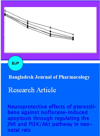
To evaluate the effect of neonatal exposure with isoflurane on potential learning and memory deficits, animals were subjected to Morris water maze test. The rats were trained to explore the swimming pool and to reach on the platform. The time taken to reach the platform was recorded and the duration each animal took to reach up the platform was found to decrease with each training session for all mice, irrespective of pterostilbene administration (Figure 3a).
Cued trials were conducted at postnatal day 30 to evaluate swimming and visual abilities. The rats that were exposed to isoflurane anesthesia took a considerably (p<0.05) longer time to reach the platform as compared to control pups that received no anesthesia. The rats that received pterostilbene were able to reach the platform much quicker as against anesthetic exposed control pups however the rats that received the higher dose of pterostilbene reached the platform at a much lesser time as against mice that received lower doses (Figure 3b).
Figure 3A: Morris water maze test - effects of isoflurane and pterostilbene on memory and learning ability Values are represented as mean ± SD; n=6
Figure 3B: Learning and memory of the rat pups determined by cued, place and probe trials with the Morris water maze Values are represented as mean ± SD, n=6. arepresents statistical significance at p<0.05 compared against control as determined by one-way ANOVA
Further to evaluate the differences in visual judgments and memory after anesthetic exposure on P7, place and probe trials were performed. The trials were conducted to test the ability of the rat pups to learn and remember the location of a new platform (Figure 3b). In place trials, the rats that were supplemented with pterostilbene at 20 and 40 mg/kg showed a marked (p<0.05) improvement in performance. The rats were able to reach the platform in a much lesser time than the anesthesia alone treated rat pups. Pterostilbene at 10 mg dose did present an enhanced performance in rats, however, the difference was negligible as compared against the pronounced positive effects seen in rats that were given higher doses.
Probe trials test the ability of memory retention of the rats following exposure to isoflurane on P7. The anesthesia exposure had a significant impact on memory of the rats that was very evident from the time the animals spent in the target quadrant. Rat pups exposed to isoflurane tend to spend significantly (p<0.05) very less percentage of time in the target quadrant than the control group unexposed to anesthesia (Figure 3b). This suggests that isoflurane caused memory and learning deficits. The rats that were administered with pterostilbene spent more time on the target quadrant looking out for the platform (p<0.05), indicating that pterostilbene markedly improved memory of the rats with 40 mg dose presenting improved results than the lower doses.
Discussion
In the present study, efficiency of pterostilbene on neurotoxicity induced by isoflurane was investigated. While anesthetic-induced neurodegeneration has been observed in many brain regions, we focused on hippocampus, as many previous reports have demonstrated that isoflurane-treated neonatal rats present normal short-term memory, a function predominantly involving the prefrontal cortex, but however have an abnormal response to contextual fear conditioning, indicating severe hippocampal lesions (Sanders et al., 2009).
The raised apoptotic cell counts observed following isoflurane exposure in our study was significantly reduced on pterostilbene treatment, indicating the neuroprotective effects of pterostilbene. Sanders et al. (2009) reported neuroprotective effects of dexmedetomidine against isoflurane-induced neuroapoptosis in the hippocampus.
Cleaved caspase-3 expression was used as marker of apoptosis and cell death. Caspase-3 has been previously validated to be an indicator of apoptosis in anesthesia induced neuroapoptosis (Jevtovic-Todorovic et al., 2003; Rizzi et al., 2008; Istaphanous et al., 2011; Kong et al., 2011). The elevated caspase-3 expressions following isoflurane exposure is indicative of apoptosis. Consistent with the results of TUNEL assay, pterostilbene reduced the levels of caspase expression in a dose dependent way.
The mechanisms involved in inhalational anesthetic-induced neurodegeneration in the developing brain are under extensive investigation. Isoflurane has been demonstrated to act via activation of inositol-1,4,5-trisphosphate (IP3) receptors and also cause change in intracellular calcium homeostasis (Liang et al., 2008; Yang et al., 2008; Zhao et al., 2013). Isoflurane causes intense calcium release from the endoplasmic reticulum than sevoflurane or desflurane (Liang et al., 2008; Yang et al., 2008). [Ca2+]i overload activates the intrinsic mitochondria-dependent apoptotic pathway, which may possibly attribute to the early signs of neuronal injury (Yon et al., 2005). The regulation of mitochondrial membrane integrity and the release of apoptogenic factors from mitochondria are tightly controlled by the proteins of Bcl-2 family (Zhao et al., 2003).
In our study, isoflurane not only increased the expressions of caspase-3 and raised apoptotic cell counts as determined by TUNEL assay; it also markedly decreased the expression of phospho-Akt and phospho-Bad proteins. Obvious raise in the expression of total Bad protein indicates that isoflurane activates function of Bad via both inhibiting Akt activity and increasing gene transcription of Bad.
The antiapoptotic protein Bcl-xL is widely expressed in the central nervous system, which enhances cell survival by maintaining mitochondrial membrane integrity and inhibits cytochrome c release (Zhao et al., 2003). However, isoflurane did not influence much on the expression of Bcl-xL. The elevated levels of Bad observed following isoflurane exposure could have possibly contributed to the decrease in the Bcl-xL/Bad ratio.
Our results demonstrated that pterostilbene effectively reversed isoflurane-induced inhibition of Akt activity and improved the Bcl-xL/Bad ratio, which could contribute to the stabilization of the inner mitochondrial membrane and thus inhibit isoflurane–induced neuroapoptosis.
The JNK signalling pathway is implicated in neuronal apoptosis triggered by several brain injury stimuli, such as ischemia/reperfusion and ethanol (Guan et al., 2006; Han et al., 2008; Fan et al., 2010). The JNK pathways include nuclear and non-nuclear pathways (Han et al., 2008). Activated JNK phosphorylates nuclear substrate, the transcription factor c-Jun, which leads to increase of activator protein-1 transcription activity to modulate transcription of genes related to apoptosis. On the other hand, activated JNK regulates the activation of non-nuclear substrates including Bcl-2 family members (Guan et al., 2006).
In the present study, isoflurane exposure caused marked increase in phospho-JNK levels though the expression levels of total JNK were not much altered. Pterostilbene pretreatment effectively down-regulated the isoflurane-induced increase of phosphorylation of JNK and c-Jun, thus suggesting the involvement of JNK pathway in isoflurane-induced neuroapoptosis. The results of this study are in agreement with previous studies that suggest that JNK signalling promotes apoptosis possibly via transcriptional regulation of Bcl-2 family gene, including Bcl-xL (Jeong et al., 2008; Chu et al., 2009).
Prosurvival pathways, such as Akt pathway have been demonstrated to be inactivated during the apoptotic process (Song et al., 2005; Yin et al., 2011). Our results suggest that pterostilbene treatment effectively maintained the level of activated Akt. This result is in agreement with previous studies that show that there is potential crosstalk between JNK and Akt signalling (Fornoni et al., 2008; Yeste-Velasco et al., 2009). Thus it could be said that pterostilbene was able to offer neuroprotection against isoflurane-induced toxicity via modulating JNK and PI3/Akt pathways.
Several previous studies reported that neonatal exposure to volatile anesthetics led to deficits in learning and memory (Jevtovic-Todorovic et al., 2003; Loepke et al., 2006; Satomoto et al., 2009; Kodama et al., 2011). Working memory refers to a cognitive function that provides concurrent temporary storage and manipulation of the information necessary to perform complex cognitive tasks (Baddeley, 1992). Working memory is thought to be involved in higher executive functioning such as planning and sequential behavior; deficits in working memory are directly related to deficits in behavioural flexibility. The Morris water maze test was chosen to evaluate the cognitive behavior in mice as it is a reliable measure of hippocampus-dependent spatial navigation and reference memory (D'Hooge and De Deyn, 2001).
To assess the spatial working memory, Morris water maze test was performed. Our results indicate that isoflurane caused deficits in memory, consistent with previous reports (Satomoto et al., 2009; Stratmann et al., 2009; Kodama et al., 2011). Pterostilbene treatment was able to bring about marked improvements in the performance of the rats in the Morris water maze tests and also improved working memory of the rats.
Pterostilbene was able to offer effective neuroprotection against isoflurane induced apoptosis and neurodegeneration by modulating JNK and PI3/Akt pathways and as well improved the memory and cognitive performance of rats.
References
Baddeley A. Working memory. Science 1992; 255: 556-59.
Bai T, Dong DS, Pei L. Resveratrol mitigates isoflurane-induced neuroapoptosis by inhibiting the activation of the Akt-regulated mitochondrial apoptotic signaling pathway. Int J Mol Med. 2013; 32: 819-26.
Brambrink AM, Evers AS, Avidan MS, Farber NB, Smith DJ, Zhang X, Dissen GA, Creeley CE, Olney JW. Isoflurane-induced neuroapoptosis in the neonatal rhesus macaque brain. Anesthesiology 2010; 112: 834-41.
Chang J, Rimando A, Pallas M, Camins A, Porquet D, Reeves J, Shukitt-Hale B, Smith MA, Joseph JA, Casadesus G. Low-dose pterostilbene, but not resveratrol, is a potent neuromodulator in aging and Alzheimer's disease. Neurobiol Aging 2012; 33: 2062-71.
Chu R, Upreti M, Ding WX, Yin XM, Chambers TC. Regulation of Bax by c-Jun NH2-terminal kinase and Bcl-xL in vinblastine-induced apoptosis. Biochem Pharmacol. 2009; 78: 241-48.
D'Hooge R, De Deyn PP. Applications of the Morris water maze in the study of learning and memory. Brain Res Brain Res Rev. 2001; 36: 60-90.
DiMaggio C, Sun LS, Li G. Early childhood exposure to anesthesia and risk ofdevelopmental and behavioral disorders in a sibling birth cohort. Anesth Analg. 2011; 113: 1143-51.
Fan J, Xu G, Nagel DJ, Hua Z, Zhang N, Yin G. A model of ischemia and reperfusion increases JNK activity, inhibits the association of BAD and 14-3-3, and induces apoptosis of rabbit spinal neurocytes, Neurosci Lett. 2010; 473: 196-201.
Fornoni A, Pileggi A, Molano RD, Sanabria NY, Tejada T, Gonzalez-Quintana J, Ichii H, Inverardi L, Ricordi C, Pastori RL. Inhibition of c-jun N terminal kinase (JNK) improves functional beta cell mass in human islets and leads to AKT and glycogen synthase kinase-3 (GSK-3) phosphorylation. Diabetologia 2008; 51: 298-308.
Guan QH, Pei DS, Zong YY, Xu TL, Zhang GY. Neuroprotection against ischemic brain injury by a small peptide inhibitor of c-Jun N-terminal kinase (JNK) via nuclear and non-nuclear pathways. Neuroscience 2006; 139: 609-27.
Han JY, Jeong EY, Kim YS, Roh GS, Kim HJ, Kang SS, Cho GJ, Choi WS. C-jun N-terminal kinase regulates the interaction between 14-3-3 and Bad in ethanol-induced cell death. J Neurosci Res. 2008; 86: 3221-29.
Ing C, DiMaggio C, Whitehouse A, Hegarty MK, Brady J, von Ungern Sternberg BS, Davidson A, Wood AJ, Li G, Sun LS. Long-term differences in language and cognitive function after childhood exposure to anesthesia. Pediatrics 2012; 130: e476-85.
Istaphanous GK, Loepke AW. General anesthetics and the developing brain. Curr Opin Anesthesiol. 2009; 22: 368-73.
Istaphanous GK, Howard J, Nan X, Hughes EA, McCann JC, McAuliffe JJ, Danzer SC, Loepke AW. Comparison of the neuroapoptotic properties of equipotent anesthetic concentrations of desflurane, isoflurane or sevoflurane in neonatal mice. Anesthesiology 2011; 114: 578-87.
Jeong HS, Choi HY, Choi TW, Kim BW, Kim JH, Lee ER, Cho SG. Differential regulation of the antiapoptotic action of B-cell lymphoma 2 (Bcl-2) and B-cell lymphoma extra-long (Bcl-xL) by c-Jun N-terminal protein kinase (JNK) 1-involved pathway in neuroglioma cells. Biol Pharm Bull. 2008; 31: 1686-90.
Jevtovic-Todorovic V, Hartman RE, Izumi Y, Benshoff ND, Dikranian K, Zorumski CF, Olney JW, Wozniak DF. Early exposure to common anesthetic agents causes widespread neurodegeneration in the developing rat brain and persistent learning deficits. J Neurosci. 2003; 23: 876-82.
Joseph JA, Fisher DR, Cheng V, Rimando AM, Shukitt-Hale B. Cellular and behavioral effects of stilbene resveratrol analogues: Implications for reducing the deleterious effects of aging. J Agric Food Chem. 2008; 56: 10544-51.
Kodama M, Satoh Y, Otsubo Y, Araki Y, Yonamine R, Masui K, Kazama T. Neonatal desflurane exposure induces more robust neuroapoptosis than do isoflurane and sevoflurane and impairs working memory. Anesthesiology 2011; 115: 979-91.
Kong F, Xu L, He D, Zhang X, Lu H. Effects of gestational isoflurane exposure on postnatal memory and learning in rats. Eur J Pharmacol. 2011; 670: 168-74.
Li Y, Liang G, Wang S, Meng Q, Wang Q, Wei H. Effect of fetal exposure to isoflurane on postnatal memory and learning in rats. Neuropharmacology 2007; 53: 942-50.
Li Y, Liu C, Zhao Y, Hu K, Zhang J, Zeng M, Luo T, Jiang W, Wang H. Sevoflurane induces short-term changes in proteins in the cerebral cortices of developing rats. Acta Anaesthesiol Scand. 2013a; 57: 380-90.
Li Y, Wang F, Liu C, Zeng M, Han X, Luo T, Jiang W, Xu J, Wang H. JNK pathway may be involved in isoflurane-induced apoptosis in the hippocampi of neonatal rats. Neurosci Lett. 2013b; 545: 17-22.
Li Y, Zeng M, Chen W, Liu C, Wang F, Han X, Zuo Z, Peng S. Dexmedetomidine reduces isoflurane-induced neuroapoptosis partly by preserving PI3K/Akt pathway in the hippocampus of neonatal rats. PLOS ONE 2014; 9: e936-39.
Liang G, Wang Q, Li Y, Kang B, Eckenhoff MF, Eckenhoff RG, Wei H. A presenilin-1 mutation renders neurons vulnerable to isoflurane toxicity. Anesth Analg. 2008; 106: 492-500.
Lin HS, Yue BD, Ho PC. Determination of pterostilbene in rat plasma by a simple HPLC-UV method and its application in pre-clinical pharmacokinetic study. Biomed Chromatogr. 2009; 23: 1308-15.
Loepke AW, McCann JC, Kurth C, McAuliffe JJ. The physi¬ologic effects of isoflurane anesthesia in neonatal mice. Anesth Analg. 2006; 102: 75 80.
Luo HR, Hattori H, Hossain MA, Hester L, Huang Y, Lee-Kwon W, Donowitz M, Nagata E, Snyder SH. Akt as a mediator of cell death. Proc Natl Acad Sci USA 2003; 100: 11712-17.
McCormack D, McFadden D. Pterostilbene and cancer: Current review. J Surg Res. 2012; 173: e53-61.
Olney JW, Young C, Wozniak DF, Jevtovic-Todorovic V, Ikonomidou C. Do pediatric drugs cause developing neurons to commit suicide? Trends Pharmacol Sci. 2004; 25: 135-39.
Orliaguet G, Vivien B, Langeron O, Bouhemad B, Coriat P, Riou B. Minimum alveolar concentration of volatile anesthetics in rats during postnatal maturation. Anesthesiology 2001; 95: 34-39.
Paule MG, Li M, Allen RR, Liu F, Zou X, Hotchkiss C, Hanig JP, Patterson TA, Slikker WJ, Wang C. Ketamine anesthesia during the first week of life can cause long-lasting cognitive deficits in rhesus monkeys. Neurotoxicol Teratol. 2011; 33: 220-30.
Remsberg CM, Yanez JA, Ohgami Y, Vega-Villa KR, Rimando AM, Davies NM. Pharmacometrics of pterostilbene: Preclinical pharmacokinetics and metabolism, anticancer, anti-inflammatory, antioxidant and analgesic activity. Phytother Res. 2008; 22: 169-79.
Rizzi S, Carter LB, Ori C, Jevtovic-Todorovic V. Clinical anesthesia causes permanent damage to the fetal guinea pig brain. Brain Pathol. 2008; 18: 198-210.
Roupe KA, Remsberg CM, Yanez JA, Davies NM. Pharmacometrics of stilbenes: Seguing towards the clinic. Curr Clin Pharmacol. 2006; 1: 81-101.
Sanders RD, Xu J, Shu Y, Januszewski A, Halder S, Fidalgo A, Sun P, Hossain M, Ma D, Maze M. Dexmedetomidine attenuates isoflurane-induced neurocognitive impairment in neonatal rats. Anesthesiology 2009; 110: 1077-85.
Satomoto M, Satoh Y, Terui K, Miyao H, Takishima K, Ito M, Imaki J. Neonatal exposure to sevoflurane induces abnormal social behaviors and deficits in fear conditioning in mice. Anesthesiology 2009; 110: 628-37.
Song G, Ouyang G, Bao S. The activation of Akt/PKB signaling pathway and cell survival. J Cell Mol Med. 2005; 9: 59-71.
Sovak M. Grape extract, resveratrol, and its analogs: A review. J Med Food 2001; 4: 93-105.
Stratmann G, Sall JW, May LD, Bell JS, Magnusson KR, Rau V, Visrodia KH, Alvi RS, Ku B, Lee MT, Dai R. Isoflurane differentially affects neurogenesis and long-term neurocognitive function in 60-day-old and 7-day-old rats. Anes thesiology 2009; 110: 834-48.
Wei H, Kang B, Wei W, Liang G, Meng QC, Li Y, Eckenhoff RG. Isoflurane and sevoflurane affect cell survival and BCL-2/BAX ratio differently. Brain Res. 2005; 1037: 139-47.
Yang H, Liang G, Hawkins BJ, Madesh M, Pierwola A, Wei HF. Inhalational anesthetics induce cell damage by disruption of intracellular calcium homeostasis with different potencies. Anesthesiology 2008; 109: 243-50.
Yeste-Velasco M, Folch J, Casadesus G, Smith MA, Pallas M, Camins A. Neuroprotection by c-Jun NH2-terminal kinase inhibitor SP600125 against potassium deprivation-induced apoptosis involves the Akt pathway and inhibition of cell cycle re-entry. Neuroscience 2009; 159: 1135-47.
Yin G, Li LY, Qu M, Luo HB, Wang JZ, Zhou WX. Upregulation of AKT attenuates amyloid-beta-induced cell apoptosis. J Alzheimers Dis. 2011; 25: 337-45.
Yon JH, Daniel-Johnson J, Carter LB, Jevtovic-Todorovic V. Anesthesia induces neuronal cell death in the developing rat brain via the intrinsic and extrinsic apoptotic pathways. Neuroscience 2005; 135: 815-27.
Zhao H, Yenari MA, Cheng D, Sapolsky RM, Steinberg GK. Bcl-2 overexpression protects against neuron loss within the ischemic margin following experimental stroke and inhibits cytochrome C translocation and caspase-3 activity. J Neurochem. 2003; 85: 1026-36.
Zhao YL, Xiang Q, Shi QY, Li SY, Tan L, Wang JT, Jin XG, Luo AL. GABAergic excitotoxicity injury of the immature hippocampal pyramidal neurons’ exposure to isoflurane. Anesth Analg. 2011; 113: 1152-60.
Zhao X, Yang Z, Liang G, Wu Z, Peng Y, Joseph DJ, Inan S, Wei H. Dual effects of isoflurane on proliferation, differentiation, and survival in human neuroprogenitor cells. Anesthesiology 2013; 118: 537-49.