Alpha-Naphthyl isothiocyanate triggering G2/M phase arrest and apoptosis in human brain malignant glioma U87MG cells via mitochondrial pathway
Abstract
Cancer protective effect of cruciferous vegetables is partly attributed to organic isothiocyanates (ITC) with an -N = C = S functional group. Elucidation of the mechanism by which ITCs impart protection against cancer has been the topic of intense research in the past few decades. In this study, we demonstrate that ANIT significantly decreased proliferation and viability of human brain malignant glioma U87MG cells in a dose-dependent manner. The cell cycle analysis showed that ANIT induced significantly G2/M arrest and sub-G1 phase (apoptotic population) in U87MG cells. CDK1 activity assay and Western blot analysis showed that there observed marked reduction in the CDK1/cyclin B activity and protein levels. Pretreatment with specific inhibitors of caspase-3 (Z-DEVE-FMK) and -9(Z-LEHD-FMK) significantly reduced caspase-3 and -9 activity in U87MG cells. Western blot analysis and colorimetric assays also displayed that ANIT caused a time-dependent increase in cytosolic cytochrome C, pro-caspase-9, Apaf-1, AIF, Endo G and the stimulatedcaspase-9 and -3 activity.
Introduction
Glioblastoma is considered to be a death sentence to patients because of little response or resistance to the present therapeutic regimens, leading to short period of survival, usually less than a year between diagnosis and death (Eitel et al., 1999). Therefore, it is necessary to develop new medicine in order to improve the treatment efficacy. Recent studies have found that a large number of edible cruciferous plants, such as broccoli, cabbage and water celery, can reduce the risk of cancer. It has also been confirmed that the isothiocyanates compounds contained in these plants have anti-tumor ability (Pawlik et al., 2012; Singh et al., 2012).
Phenethyl and benzyl isothiocyanates inhibited carcinogenesis induced by carcinogens such as diethyl nitrosamine (DEN), dimethyl benzo(a)anthracene (DMBA) or benzo(a)pyrene (BP) (Wattenberg et al., 1987). The mechanism involved in the inhibition of carcinogenesis by ITCs is most likely due to the blocking of the activation of carcinogens through the inactivation of P450 enzymes and activation of Phase II enzymes (Wattenberg et al., 1987).
We hypothesized that resonance structure of naphthyl moiety would result in more effective anticancer agents based on the observation that in comparison to the phenyl and benzyl derivatives, naphthyl structure provides more electron donation. In addition, no previous study exists addressing whether the ANIT inhibits cell proliferation, promotes cell cycle arrest and induces apoptosis in human brain malignant glioma cells. The aim of the present study is to determine and explore ANIT-triggered G2/M phase arrest and cell death in U87MG cells via a mitochondria-dependent apoptotic signaling.
Materials and Methods
Reagents and chemicals
Alpha-naphthylisothiocyanate (ANIT), [3-(4,5-dimethyl-thiazol-2-yl)-2,5-diphenyltetrazolium bromide] (MTT), propidium iodide (PI), Triton X-100, RNase A, dimethylsulfoxide (DMSO) were purchased from Sigma Chemical Co. (USA). RPMI-1640 medium, fetal bovine serum (FBS), L-glutamine, penicillin-streptomycin and trypsin-EDTA were obtained from Gibco BRL/Invitrogen Corp (USA). Caspase-9 inhibitor (Z-LEHDFMK), caspase-8 inhibitor (Z-IETD-FMK) and caspase-3 inhibitor (Z-DEVD-FMK) were obtained from Invitrogen (R&D Systems, Minneapolis, MN,USA)
Cells, culture method and cell morphology identification
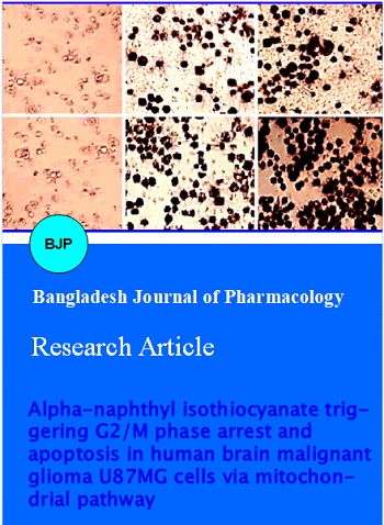
U87MG cell lines (human brain malignant glioma) were purchased from the Food Industry Research and Development Institute (Hsinchu, Taiwan). Cells were plated onto 75 cm2 tissue culture flasks in RPMI-1640 medium supplemented with 10% FBS, 100 U/mL penicillin, 100 ug/mL streptomycin and 2 mM L-glutamine and grown at 37degree C under a humidified 5% CO2 and 95% air at one atmosphere. U87MG cells of about 2 x 10⁵ cells/well in a 12-well plate were treated with different concentrations (0, 10, and 20 uM) ANIT and incubated for 24 and 48 hours. Cells were directly examined and were photographed under contrast-phase microscope (Yang et al., 2009).
MTT assay
U87MG cells of about 1 x 10⁴ cells/well in 96-well plates were treated with to 4, 8, 16, 32, and 64 uM of ANIT and DMSO, 0.1% in media served as a vehicle control. After a 24 hours incubation, 100 uL of 0.5 mg/mL MTT solution was added to each well, and the plate was incubated at 37degree C for 3 hours. An aliquot of 100 uL of 0.04 N HCl in isopropanol was added and the absorbance at 570 nm was measured for each well with the cell survival ratio expressed as % of control.
Cell cycle distribution analysis
The U87MG cells were seeded onto 12-well culture plates at 2 x 10⁵ cells/well then incubated with 10 uM of ANIT for 24 hours. The cells were harvested and washed by centrifugation. For cell cycle and apoptosis determination, cells were fixed by 70% ethanol in -20 degree C overnight and then re-suspended in PBS containing 40 ug/mL PI and 0.1 mg/mL RNase A and 0.1%. Triton X-100 in dark room for 30 min. The cell cycle distribution and apoptotic nuclei were determined by flow cytometry (FACSCaliburTM, USA).
DAPI staining for apoptosis
About 2 x 10⁵ cells/well of U87MG cells in a 12-well plate were treated with 0, and 10 uM of ANIT and 0.1% DMSO as the control. Cells were incubated and grown for 24 hours. Cells were stained by 4'-6-diamidino-2-phenylindole (DAPI) and were photographed under fluorescence microscopy.
CDK1 kinase assay
U87MG cells of about 1 x 10⁷ density were suspended in a buffer containing a final volume of 0.2 mL, 50 mM Tris-HC1(pH 7.5), 1 mM phenylmethylsulfonyl fluoride, 50 pg/mL leupeptin, 2-mercaptoethanol (10 mM), MgCl2 (1 mM), EGTA (2 mM), dithiothreitol (0.5 mM), Brij (350.01%), b-glycerophosphate (25 mM), and NaCl (0.5 M). Cell suspensions were sonicated and centrifuged for 30 min. To determine the CDK1 kinase assay condition using MV Peptide, its absorbance was measured at 492 nm (Yang et al., 2004; Kuo et al., 2009). Assay was carried out according to the protocol of Medical and Biological Laboratories CDK1 kinase assay kit (MBL, Nagoya, Japan).
Western blot
U87MG cells of about 1 x 10⁷ density in a 75-T flask were treated with 10 uM of ANIT for 0, 6, 12, 24 and 48 hours. The examined cells were harvested and washed with cold PBS for detecting the changes of protein levels correlated with G2/M phase-modulated and apoptotic signaling. The total proteins were collected from ANIT-treated U87MG cells before the CDK1, cyclin B and cyclin A were detected and measured. The level of cytosolic fraction proteins such as cytochrome C, Apaf-1, pro-casepase9, AIF, and Endo G were determined and isolated according to the mitochondria/cytosol fractionation kit manufacturer's protocol (BioVision, Inc. Mountain View, CA, USA). U87MG cells after exposure to ANIT were harvested, disrupted and then centrifuged to isolate cytosolic fractions. The total or cytosolic protein (40 μg) from each sample was resolved on 12% sodium dodecyl sulfate polyacrylamide gel electrophoresis (SDS-PAGE) and transferred to nitrocellulose membrane. The blot was soaked in blocking buffer (5% non-fat dry mik/0.05% Tween-20 in 20 mM TBS at pH 7.6) at room temperature for 1 hour and then incubated with anti-CDK1, anti-cyclin B, anti-cyclin A, anti-cytochrome c, anti-Apaf-1, antipro-casepase-9, anti-AIF, and anti-Endo G antibodies (SantaCruz Biotechnology, Inc.) in blocking buffer at 4degree C overnight. A secondary antibody horseradish peroxidase conjugate was added and detected by chemiluminescence and autoradiography using X-ray film. To ensure equal protein loading, each membrane was stripped and reprobed with anti beta-actin antibody.
Assays for caspase-3, -8 and -9 activities
Caspase-3, -8 and -9 activities were assessed according to the manufacturer's instructions (Caspase colorimetric kit, R&D-System Inc.). U87MG cells of about 1 x 10⁷ density in a 75-T flask were pretreated with or without the selective inhibitors (Z-DEVE-FMK forcaspase-3, Z-IETD-FMK for caspase-8 and Z-LEHD-FMK for caspase-8) and then treated with 10 uM ANIT were incubated for 24 hours to detect caspase-3, -8 and -9 activities. Briefly, cells were harvested and lysed in 50 uL lysis buffer containing 2 mM DTT for 10 min. After centrifugation, the supernatants containing 100 ug proteins were incubated with caspase-3, -8 and -9 substrate in reaction buffer. Samples were incubated in a 96-well flat bottom microplate at 37degree C for 1 hour. Levels of released pNA (Z-DEVEpNA, Z-IETD-pNA and Z-LEHD-pNA for cas-pase-3, -8 and -9, respectively) were measured with an ELISA reader (AnthosLabtec Instruments) at an absorbance of 405 nm.
Statistical analysis
Student's t-test was used to analyze the differences between the ANIT-treated and control groups. All data were expressed as mean ± SD from at least three independent experiments. P<0.05, p<0.01 and p<0.001 were indicative of significant difference.
Results
The cell viability of ANIT on a human brain malignant glioma cell line U87MG was investigated by the MTT assay. The results shown in Figure 1A indicated that ANIT significantly reduced the percentage of viable cells when compared to that of control cells. These effects were found to be in a dose-dependent manner.
The IC50 of ANIT was 8.3 ± 0.6 uM after 24 hours treatment. In addition, the examined cells exhibited morphological changes, including rounding and shrinkage after 24 and 48 hours-incubation with 10 and 20 uM of ANIT (Figure 1B). Viable cells were determined by MTT assay. The cell morphological changes were examined and photographed under phase-contrast microscopy (x200) as described in Materials and methods. There was a significant difference (ap<0.001) when compared with the control group by Student's t-test analysis.
Figure 1: Effects of ANIT on cell viability (A) and morphology (B) of U87MG cells. Cells were cultured with 0, 4, 8, 16, 32 and 64 uM of ANIT for 24 hours
Cells were treated with 10 uM of AITC for 24 hours and then were analyzed and determined for cell cycle distribution by PI staining and flow cytometric analysis. AITC induced chromatin condensation (an apoptotic characteristic) in U87MG cells. Cells were incubated with 10 uM of AITC for 24 hours and apoptotic cells were determined by DAPI staining and were photographed by fluorescence microscopy (x200) as described in Materials and Methods. ANIT induced reduction of cell viability could be due to apoptosis (sub-G1 population) mediated by cell cycle arrest which were determined. Representative profiles of cell cycle progression and percentage of each phase are presented in Figure 2A. Significant arrest was seen at G2/M phase at 10 uM of ANIT and it significantly increased apoptotic cells (sub-G1groups) (Figure 2A). Apoptotic chromatistic (nuclear and chromatin condensation) in GBM8401 cells occurred with or without ANIT (10 uM) treatment for 24 hours determined by DAPI staining (Figure 2B).
Figure 2: Effects of ANIT on cell cycle distribution (A) and chromatin condensation (B) in U87MG cells
This experiment was carried out to prove ANIT-induced accumulation of G2/M population in U87MG cells and it was regulated associated proteins. U87MG cells were incubated with 10 uM AITC for 0, 6, 12, 24 and 48 hours, harvested from each sample and then relative protein levels were determined by Western blot analysis. The levels of CDK1, cyclin B and cyclin A were examined, and beta-actin as an internal control using SDS-PAGE and Western blotting as described in Materials and Methods. ap<0.001, was significantly different compared to the control group. The results are shown in Figure 3A and B. U87MG cells after exposure to AITC showed that a significant decrease of CDK1 activity (Figure 3A) and down-regulated the protein levels, including CDK1, cyclin B and cyclin A as shown in Figure 3B.
Figure 3: ANIT alters the levels of CDK1 activity (A), and the CDK1, cyclin (A) and cyclin (B) proteins of G2/M phase arrest in U87MG cells. Cells were incubated with 10 uM AITC for 0, 6, 12, and 24 hours for CDK1 activity
To assess the ANIT-induced apoptotic cell death, the results of Western blot analysis from 10 uM of ANIT co-incubation with U87MG cells for 0, 6, 12, 24 and 48 hours are shown in Figure 4A. The raise in protein levels of cytochrome C, Apaf-1, pro-caspase-9, AIF, and Endo G in cytosolic fractions may contribute to the occurrence of ANIT-triggered mitochondrial apoptosis and this signaling was found to be time dependent. Figure 4B, showed that ANIT induced a significant rise in caspase-3 and -9 activities rather than influence of caspase-8 response. This stimulation of activity of the caspases cascade in U87MG cells was reduced after pre-incubation with specific inhibitors of caspase-3 and -9, respectively. Based on these results, ANIT-induced cell death was correlated with caspase-dependent and -independent mitochondrial apoptotic pathways.
Figure 4: ANIT affects the relative protein levels of apoptosis (A) and caspase-3, -8 and -9 activities (B) in U87MG cells. ap<0.001, significant difference
Figure 5: The proposed model of ANIT-induced G2/M phase arrest and apoptosis in U87MG cells
Discussion
Apoptotic cell death induction is one of the best strategies for cancer treatment (Batista et al., 2009; Sanchez-Munoz et al., 2009). Our results indicated that ANIT induced apoptosis in U87MG cells. This was demonstrated by flow cytometric analysis and DAPI staining was applied to confirm cell apoptosis in examined U87MG cells. The results from the Western blot analysis showed that AITC increased protein levels of cytochrome C, Apaf-1, pro-caspase-9, AIF, and Endo G, suggesting that analyses of protein abundance indicated that AITC induced apoptosis through a mitochondria-dependent pathway. Also, AITC stimulates the loss of mitochondrial membrane potential resulting from the mitochondria dysfunction (data not shown). In this study, we observed the ANIT-induced caspase-9 and -3 activities in U87MG and the specific inhibitors (Z-DEVEFMK for caspase-3 and Z-LEHD-FMK for caspase-9) individually prevented ANIT-induced caspase-3 and -9 activities, respectively in U87MG cells. ANIT-induced G2/M phase arrest and apoptosis in U87MG cells is shown in Figure 5.
Conclusion
ANIT induces morphological changes on U87MG cells, decreases the percentage cell viability, increases G2/M phase arrest, and stimulates the levels of caspase-9, -3, AIF and Endo G through a mitochondria-dependent apoptotic pathway.
References
Batista LF, Kaina B, Meneghini R, Menck CF. How DNA lesions are turned into powerful killing structures: Insights from UV-induced apoptosis. Mutat Res. 2009; 681: 197-208.
Brusewitz G, Cameron BD, Chasseaud LF, Gorler K, Hawkins DR, Koch H, Mennicke WH. The metabolism of benzyl isothiocyanate and its cysteine conjugate. Biochem J. 1977; 162: 99-107.
Drobinica L, Augustine J. Reaction of isothiocyanate with amino acids, peptides and proteins. I. Kinetics of the reaction of aromatic isothiocyanates and glycines. Chem Commun. 1965; 30: 99-104.
Eitel K, Wagenknecht B, Weller M. Inhibition of drug-induced DNA fragmentation, but not cell death, of glioma cells by non-caspase protease inhibitors. Cancer Lett. 1999; 142: 11-16.
Kim SJ, Min HY, Chung HJ, Park EJ, Hong JY, Kang YJ, Shin DH, Jeong LS, Lee SK. Inhibition of cell proliferation through cell cycle arrest and apoptosis by thio-Cl-IB-MECA, a novel A3 adenosine receptor agonist, in human lung cancer cells. Cancer Lett. 2008; 264: 309-15.
Kuo HM, Tsai HC, Lin YL, Yang JS, Huang AC, Yang MD, Hsu SC, Chung MC, Gibson Wood W, Chung JG. Mitochondrial-dependent caspase activation pathway is involved in baicalein-induced apoptosis in human hepatoma J5 cells. Int J Oncol. 2009; 35: 717-24.
Pawlik A, Szczepanski MA, Klimaszewska A, Gackowska L., Zuryn A., Grzanka A. Phenethylisothiocyanate-induced cytoskeletal changes and cell death in lung cancer cells. Food Chem Toxicol. 2012; 50: 3577-94.
Sanchez-Munoz A, Perez-Ruiz E, Mendiola Fernandez C, Alba Conejo E, Gonzalez-Martin A. Current status of anti-angiogenic agents in the treatment of ovarian carcinoma. Clin Transl Oncol. 2009; 11: 589-95.
Schwartz GK, Shah MA. Targeting the cell cycle: A new approach to cancer therapy. J Clin Oncol. 2005; 23: 9408-21.
Singh SV, Kim SH, Sehrawat A, Arlotti JA, Hahm ER, Sakao K, Beumer JH, Jankowitz RC, Chandra-Kuntal K, Lee J. Biomarkers of phenethyl isothiocyanate-mediated mammary cancer chemoprevention in a clinically relevant mouse model. J Natl Cancer Inst. 2012; 104: 1228-39.
Srivastava SK, Singh SV. Cell cycle arrest, apoptosis induction and inhibition of nuclear factor kappa B activation in antiproliferative activity of benzyl isothiocyanate against human pancreatic cancer cells. Carcinogenesis 2004; 25: 1701-09.
Wattenberg LW. Inhibitory effect of benzyl isothiocyanates administered shortly before diethyl nitrosamine or benzo(a)pyrene on pulmonary and fore stomach neoplasia in A/J mice. Carcinogenesis 1987; 12: 1971-73.
Xu K, Thornalley PJ. Studies on the mechanism of the inhibition of human leukemia cell growth by dietary isothiocyanates and their cysteine adducts in vitro. Biochem Pharmacol. 2000; 60: 221-31.
Yang JS, Chen GW, Hsia TC, Ho HC, Ho CC, Lin MW, Lin SS, Yeh RD, Ip SW, Lu HF, Chung JG. Diallyl disulfide induces apoptosis in human colon cancer cell line (COLO 205) through the induction of reactive oxygen species, endoplasmic reticulum stress, caspases casade and mitochondrial-dependent pathways. Food Chem Toxicol. 2009; 47: 171-79.
Yang JS, Hour MJ, Kuo SC, Huang LJ, Lee MR. Selective induction of G2/M arrest and apoptosis in HL-60 by a potent anticancer agent, HMJ-38. Anticancer Res. 2004; 24: 1769-78.