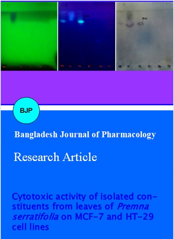
Cytotoxic activity of isolated constituents from leaves of Premna serratifolia on MCF-7 and HT-29 cell lines
Abstract
Premna serratifolia (Syn: Premna integrifolia) is an important medicinal herb known as “Agnimantha†in Ayurveda and traditionally used for anticancer activity. The objective of present study was to isolate the cytotoxic phytoconstituents from the n-hexane soluble fraction of P. serratifolia leaf extract. Unsaponifiable portion of n-hexane soluble fraction was subjected to silica based column chromatography. The major constituents present in all the sub-fractions were identified by TLC and phytochemical tests. Two constituents were isolated and they were purified. Sub-fractions with isolates were tested for cytotoxic effect by BSL bioassay. Two isolates were found to be active and which were tested on cancer cell lines MCF-7 and HT-29 for their cytotoxicity. Among two isolates, one compound has shown significant cytotoxicity. From the results we conclude that the plant isolates showed cytotoxicity against selected human cancer cell lines.
Introduction
Premna serratifolia Linn., commonly known as Arni or Agnimantha, is a large shrub or a small tree common along the Indian peninsular and Andaman coast. Pharmacologically, it has been reported to have activities such as hepatoprotective (Vadivu et al., 2009), immunomodulatory, cardiac stimulant, antibacterial, cytotoxic, antiarthritic, anti-inflammatory (Biradi and Hullatti, 2013), anti-diabetic (Mujumder et al., 2014), antioxidant (Mali et al., 2014), anti-obesity and used in treatment of tumors (Cragg and Newman, 2005).
Plant derived cytotoxic constituents have played an important role in the development of clinically useful anticancer agents (Cragg and Newman, 2005). Various cytotoxic constituents have been isolated from plants and which are used as anticancer agents.
Among all cancer types breast cancer is the most common disease in women worldwide with high mortality rate (18%) and relative risk factors, including dietary factors (Ahmed et al., 2014). Colorectal cancer is the third most leading cause of cancer death in both men and women. About 96% of colorectal cancers are adenocarcinomas, which evolve from glandular tissue (Anonymous, 2013).
However, the efficacy and effect of currently available drugs is very limited and are more toxic on normal cells. By increasing the use of existing knowledge with newer and established screening tests the majority of these cancers and deaths could be prevented.
Although various extracts of P. serratifolia are reported to have cytotoxic activity in the literature (Selvam et al., 2009; Singh, 2011), there are no reports available on isolation based study. As part of ongoing investigation P. serratifolia plant has been selected for bioactivity guided isolation and here cytotoxic activity of two isolated constituents from the n-hexane soluble fraction (unsaponifiable portion) of methanolic extract of P. serratofolia leaves on human cancer cell lines.
Materials and Methods
Collection and authentication of plant material
Leaves of P. serratifolia were collected from the premises of Regional Medical Research Centre, (ICMR), Belgaum during January 2011 and authenticated by Dr. Harsha V. Hegde, Scientist "B" RMRC, Belgaum, India. The voucher specimen is deposited in ICMR Herbarium repository (Accession Number RMRC-554).
Extraction, fractionation and cytotoxicity by BSL assay
The extraction and fractionation of P. serratofolia leaves has been done according modified Cos et al., 2006. Based on the results of previous study (Biradi and Hullatti, 2013), n-hexane soluble fraction was taken up for present study.
Phytochemical investigation
Phytochemical investigation of isolates was carried out according to the standard procedure (Trease and Evans, 2005; Sandjo and Kuete, 2013).
Optimization of TLC system
Different solvents systems were tried for developing a TLC system for identification of constituents in the extracts and the one with good resolution was selected as the mobile phase for the study. Toluene: Ethyl acetate: Glacial acetic acid (7:2:1) was found to be the most suitable solvent system for n-hexane fractions as it gave maximum number of bands.
Isolation of the phytoconstituents
Unsaponifiable portion of n-hexane soluble fraction was subjected to silica based column chromatography. The different fractions were generated using gradient elution technique using petroleum ether, chloroform and acetone with increasing the polarity. Five sub-fractions (SF) generated from unsaponifiable portion (i.e. PS/F1/USM). The chemical nature of all the sub fractions was established by TLC (Plate No. 1-3) and chemical tests (Table I).
Plates 1-3: TLC of sub-fractions at different pre and post derivatization
Processing of the isolated precipitates
The sub fractions PS-01 and PS-02 were purified by washing with cold methanol followed by ethanol. The solid mass was weighed and collected in previously labelled eppendorf tubes and stored in a desiccator.
In vitro cytotoxicity of the isolated compounds on selected cell lines
Brine shrimp lethality (BSL) bioassay
BSL assay was performed for all the five sub fractions to confirm the cytotoxic activity and only the isolates which shown the cytotoxicity were subjected for cell line studies.
Cytotoxicity of the active compounds on MCF-7 and HT-29 cell lines
The active fractions from BSL bioassay were subjected for in vitro cytotoxicity studies using human cancer cell lines MCF-7 (breast carcinoma) and HT-29 (adenocarcinoma).
Cell lines and culture medium (Koba et al., 2009)
HT-29 (adenocarcinoma) and MCF-7 (breast carcinoma) cell lines were procured from National Centre for Cell Sciences (NCCS), Pune, India. Stock cells were cultured in 25 cm2 culture flasks in Dubelcco's Modified Eagle's Minimum Essential Medium (DMEM) supplemented with 10% inactivated fetal bovine serum (FBS), penicillin (100 IU/mL), streptomycin (100 mg/mL) and amphotericin B (5 mg/mL) in an humidified atmosphere of 5% CO2 at 37°C until confluent. The cells were dissociated with TPVG solution (0.2% trypsin, 0.02% EDTA, 0.05% glucose in PBS).
Preparation of test solutions
Stock solution of 1 mg/mL concentration of each test drugs were prepared separately by dissolving in distilled DMSO and made volume with DMEM in 2% FBS and filtered. Serial two fold dilutions were prepared from this for carrying out cytotoxic studies.
Determination of cell viability by MTT assay
The monolayer cell culture was trypsinized and the cell count was adjusted to 1.0 x 10⁵ cells/mL using DMEM containing 10% FBS. To each well of the 96-well microtitre plate, 0.1 mL of the diluted cell suspension (approximately 10,000 cells) was added. After 24 hours, supernatant was flicked off upon formation of partial monolayer. Then washed the monolayer once with medium and 100 mL of different test concentrations (62.5 to 1000 ug/mL) of test drugs were added on to the monolayer in microtitre plates. The plates were then incubated at 37°C for 3 days in 5% CO2 atmosphere and observations were noted every 24 hours interval by microscopic examination. After 72 hours, the drug solutions in the wells were discarded and 50 mL of MTT in PBS was added to each well. The plates were gently shaken and incubated again for 3 hours. The supernatant was removed and 100 mL of propanol was added and the plates were gently shaken to solubilize the formed formazan. The absorbance was measured using a microplate reader at a wavelength of 540 nm. The percentage growth inhibition was calculated using the following formula and concentration of test drug needed to inhibit cell growth by 50% (CTC50) values is generated from the dose-response curves for each cell line.
%Growth inhibition = (Mean OD of individual test group/Mean OD of control group) x 100
Result and Discussion
The study was taken to identify the constituents responsible for cytotoxic activity of P. serratifolia leaf extract. The n-hexane soluble fraction (F1) has shown significant cytotoxicity in the earlier studies (Biradi and Hullatti, 2013). The unsaponifiable portion which was rich in steroids and triterpenoids was subjected for further fractionations using column chromatography. The column chromatography has yielded 5 sub-fractions (PS-01 to PS-05) with gradient elution. Out of these 5 sub-fractions, PS-01 and PS-02 were found single compound fractions as identified by TLC. The chemical tests and TLC has shown that all the 5 sub-fractions are triterpenoids in nature. The BSL bioassay of these sub-fractions has indicated that PS-01 and PS-02 are more cytotoxic than other 3 sub-fractions LC50 values are depicted in Table II. Further after purification these 2 fractions were analysed on MCF-7 and HT-29 cell lines. Result of this study has indicated that the sub-fractions PS-02 has significant cytotoxicity in both the cell lines which are depicted in Figure 1 and 2 (LC50 value 100.0 and 99.9 ug/mL respectively). Various authors have reported cytotoxic activity of number of triterpenoids such as ursolic acid, oleane and sterols from different plants (Da Silva Filho et al., 2009; Awasare et al., 2012). These data indicate that the certain triterpenoids of P. serratifolia may be useful as cytotoxic component that needs further characterization to identify the structure.
Table I: Cytotoxicity screening of the column sub-fractions (BSL assay)
| Fractions | LC50 |
|---|---|
| PS-01 | 54.6a |
| PS-02 | 30.8a |
| PS-03 | 323.3 |
| PS-04 | 143.6 |
| PS-05 | 209.6 |
Table II:TLC profile of the column sub-fractions
| Fractions | Gradient program | Physical nature | Number of spots observed | Rf value |
|---|---|---|---|---|
| PS-01 | Petroleum ether | Yellowish and amorphous | 2 | 0.82,0.62 |
| PS-02 | CHCl3:PE (1:4) | White and amorphous | 1 | 0.65 |
| PS-03 | CHCl3/PE (1:1) | Yellowish and sticky | 3 | 0.69, 0.49, 0.36 |
| PS-04 | CHCl3 | Yellowish and sticky | 2 | 0.69, 0.36 |
| PS-05 | Me2CO | Brownish and sticky | 2 | 0.69,0.33 |
Figure 1: Graphical representation of cytotoxic effect of drugs on MCF-7 cell line
Figure 2: Graphical representation of cytotoxic effect of drugs on HT-29 cell line
From the present study, the triterpenoids of P. serratifolia provide a new insight into the anticancer approach.
Acknowledgments
Authors are thankful to the KLE University for the financial support and the Principal, KLES College of Pharmacy, Belgaum, Karnataka for providing facilities to carry out this research work.
References
Ahmed B, Usman AA, Muhammad TQ, Matloob A. Anticancer potential of phytochemicals against breast cancer: Molecular docking and simulation approach. Bangladesh J Pharmacol. 2014; 9: 545-50.
Anonymous. Agrotechniques of selected medicinal plants. National Medicinal Plants Board, 2008; 2: pp 151-54.
Anonymous. American Cancer Society. Colorectal cancer: Facts & figures, 2011-2013.
Awasare S, Bhujbal S, Nanda R. In vitro cytotoxic activity of novel oleanane type of triterpenoid saponin. Asian J Pharm Clin Res. 2012; 5: 183–88.
Biradi M, Hullatti K. Screening of Indian medicinal plants for cytotoxic activity by brine shrimp lethality (BSL) assay and evaluation of their total phenolic content. Drug Dev Ther. 2014; 5: 139-44.
Cos P, Vlietinck AJ, Berghe DV, Maes L. Anti-infective potential of natural products: How to develop a stronger in vitro proof-of-concept. J Ethnopharmacol. 2006; 106: 290–92.
Cragg GM, Newman DJ. Plant as a source of anticancer agents. J Ethnopharmacol. 2005; 100: 72-79.
Da Silva Filho AA, Resende DO, Fukui MJ, Santos FF, Pauletti PM, Cunha WR, et al. In vitro antileishmanial, antiplasmodial and cytotoxic activities of phenolics and triterpenoids from Baccharis dracunculifolia D.C. (Asteraceae). Fitoterapia. 2009; 80: 478–82.
Evans WC. ‘Trease and Evans’ Pharmaconosy. 15th ed. London: W. B. Saunders Company Ltd; 2005.
Koba K, Sanda K, Guyon C, Raynaud C, Chaumont JP, Nicod L. In vitro cytotoxic activity of Cymbopogon citratus L. and Cymbopogon nardus L. essential oils from to go. Bangladesh J Pharmacol. 2009; 4: 29-34
Majumder R, Akter S, Naim Z, Al-Amin M, Badrul M. Antioxidant and anti-diabetic activities of the methanolic extract of Premna integrifolia bark. Adv Bio Res. 2014; 8: 29-36.
Mali PY. Beneficial effect of extracts of Premna integrifolia root on human leucocytes and erythrocytes against hydrogen peroxide induced oxidative damage. Chron Young Sci. 2014; 5: 53-58.
Sandjo LP, Kuete VSE. Medicinal plant research in Africa. Elsevier Inc.; 2013. Available from: http://dx.doi.org/ 10.1016/B978-0-12-405927-6.00004-7.
Selvam TN, Venkatakrishnan V, Kumar S. Damodar, Elumalai P. Antioxidant and tumor cell suppression potential of Premna serratifolia L. leaf. Toxicol Int. 2012; 19: 31–34.
Singh RC. Antimicrobial effect of Callus and Natural plant extracts of Premna serratifolia L. Int J Pharm Biomed Res. 2011; 2: 17-20.
Vadivu R, Jerad S, Girinath K, Kannan PB, Vimala R, Kumar NM. Evaluation of hepatoprotective and in vitro cytotoxic activity of leaves of Premna serratifolia Linn. J Sci Res. 2009; 1: 145-52.

