Antiviral mode of action of Lactobacillus plantarum YML009 on Influenza virus H1N1
Abstract
The development of resistance against antiviral agents has augmented a threat to public health sector. Consequently there is an expanding demand for the development of unconventional antiviral agents that could efficiently replace the existent in-use drugs. The probiotic strains of lactic acid bacteria (LAB) have earned the status of being efficient, economical and safe nutraceutical in health care armamentarium. A total of 2272 LAB strains were screened against H1N1 virus. The isolate YML009 displayed a pronounced antiviral activity. Sequencing and biochemical assays identified the isolate as Lactoballius plantarum which, exhibited resistance to the damage caused by the acidic conditions such as gastric juice and 5% bile salt. The anti-H1N1 activity of the strain was confirmed by hemagglutination assay and was found to display enhanced efficacy in comparison to the commercially available antiviral drug. This is a primary report on anti-influenza activity of a bacterium L. plantarum YML009.
Introduction
Influenza, commonly called as flu is a contagious respiratory disease induced by flu viruses. It is a severe form of infection which results over 220,000 hospitalizations and approximately 36,000 annual deaths in the United States every year (Thompson et al., 2004). Considering the restricted efficacy of the current vaccination program (Hancock et al., 2009) and limited use of antiviral drugs (Beigel and Bray, 2008; Long et al., 2000), there is an alarming demand for the development of unconventional form of measure against influenza virus.
Probiotics also called as friendly bacteria are live microorganisms, which are similar to the microflora present in the gut. When consumed in adequate amounts probiotics confer health benefits on the host. The ability of lactic acid bacteria (LAB) to thrive in extreme conditions such as gastric acid in the stomach and bile acid in the intestines (Nomura et al., 2006; Dunne et al., 2001). Fermented foods and dairy products have been known contain a variety of probiotic LAB strains (Hanniffy et al., 2004; Drouault and Corthier, 2001; Parvez et al., 2006). It was demonstrated that LAB probiotic strains were able to protect against infectious diseases (Corr et al., 2007; Youn et al., 2012), and display anti-allergic effects on immune diseases in mice (Fujiwara et al., 2004; Nagai et al., 2011), chicken (Seo et al., 2012) and humans (Nagao et al., 2000; Ishida et al., 2005). However, it remains unknown whether treatments with LAB can confer improved protection.
Materials and Methods
Isolation and identification of lactobacillus plantarum YML009
LAB were isolated by homogenizing different types of Kimchi samples and then filtering the homogenate through a sieve (100 mesh). An appropriate volume of the filtered homogenate was diluted by decimal dilution and spread onto MRS agar (Difco laboratories, USA), followed by incubation at 37°C for 24 hours.
Colonies of LAB were then isolated by sequential subcultivation. The isolated strains were stored at -80°C subjected to screening against H1N1.
Kimchi isolate showing highest activity against H1N1 was sequenced by 16S rRNA gene sequencing. The sequence was compared for homology using BLAST and multiple aligned with 16S rRNA gene sequences of the different strains for similarity using ClustalW program of the MEGA 5 software. A phylogenic tree was constructed by the neighbor-joining method using the same software.
Characterization of L. plantarum YML009
The strain was characterized biochemically using the API 50 CH strip and API CHL medium systems according to the manufacturer's instructions (API bioMerieux, USA). Briefly, a fresh grown colony of L. plantarum YML009 was harvested and resuspended in sterile water to achieve a cell density of 108 CFU/mL. A 2 mL aliquot of the cell suspension was inoculated into 10 mL APL 50 CHL medium and mixed gently by inversion. Further, 120 µL of this suspension was inoculated into API 50 CH strips that were overlaid with mineral oil and were incubated for 48 hours before reading color changes.
Resistance against artificial gastric juice and bile
For artificial gastric juice the method of Kobayashi et al. (1974) was followed. In brief, L. plantarum YML009 was incubated in MRS broth with pH 2.5, and 1N HCl was then added to a final concentration of 1%. The bacterium was incubated for 24 hours at 37°C. Survival rate was measured by decimal dilution method on MRS agar at 37°C for 24 hours.
Artificial bile juice was prepared as described in the Difco (Detroit, USA) manual. Briefly, MRS broth was prepared by adding pancreatin (Wako Fine Chemicals, japan) at a final concentration of 5%. The pH was adjusted to pH 6.8 using sterile 5% bile acid (oxagall, Sigma-Aldrich, USA). Resistance against bile acid was tested by the modified method of Oh et al. (2000).
Hemolytic phenomenon test
For hemolytic activity test, L. plantarum YML009 was cultured in MRS broth and then streaked on tryptone soya agar (Oxoid) with an addition of 5% sheep blood. The plates were examined for hemolytic reaction after incubating under CO2 environment at 37°C for 48 hours. Hemolytic activity was confirmed by the presence of a clear zone around bacterial colony. Strains that produce green-hued zones around the spots (α-hemolysis) or do not produce any effect on the blood plates (γ- hemolysis) are considered non-hemolytic, and the strains displaying blood lysis zones around the spots were classified as hemolytic (beta-hemolysis) (Petros et al., 2009).
Antibiotic susceptibility
The antibiotic susceptibility of an isolate YML009 was carried out using microdilution method. Prior to the assay, YML009 strain was pre-cultured at 37°C for 24 hours, and subjected to LAB susceptibility test medium (LSM) formulation consisting of a mixture of IST broth (90%) and MRS broth (10%) adjusted to pH 6.7 as previously described (ES ISO, 2012). After 24 hours of incubation, inoculum was prepared, a colony was suspended in a sterile plastic culture tube containing 2 mL sterile saline until a density corresponding to a McFarland (McF) standard of 1 or a spectrophotometric equivalent (3 x 10⁸ CFU/mL) was obtained. The inoculated saline suspension was diluted 1:500 to obtain a final concentration of 3 x 10⁵ using LSM broth (ES ISO, 2012). According to the European Food Safety Authority (EFSA) guidelines the following antibiotics were tested at a concentration ranges (mg/L) given in parenthesis: Gentamicin (0.5 to 256 mg/L; Tokyo chemical industry co; Ltd, Tokyo, Japan), kanamycin (2 to 1024 mg/L; Biopure reagent, Seoul Korea), streptomycin (0.5 to 256 mg/L; Tokyo chemical industry co; Ltd, Tokyo, Japan), tetracycline (0.1 to 64 mg/L; Tokyo Chemical Industry Co; Ltd, Tokyo, Japan), erythromycin (0.02 to 8 mg/L; Tokyo Chemical Industry co; Ltd, Tokyo, Japan), clindamycin (0.03 to 16 mg/L; Sigma-Aldrich, St. Louis, MO, USA), chloramphenicol (0.1 to 64 mg/L; Sigma-Aldrich, St. Louis, MO, USA), and ampicillin (0.03 to 16 mg/l; Biopure reagent, Seoul Korea). Further, 50 µL of 3 x 10⁵ CFU/mL inoculum was added to each well (already contained antibiotics) of the microdilution plate within 30 min after the preparation of the standardized inoculum. The well without antibiotic served as positive control, but contains mixture of test strain and the medium containing solvent which was used to dissolve the antibiotics at the highest concentration. While well without test strain and the antibiotic, but with the medium served as negative control. The susceptibility panel was incubated anaerobically at 37°C for 24 to 48 hours after inoculation; growth in susceptibility panel was evaluated visually by comparing the pellet at the bottom of each well comparing with the positive and negative controls. Available MIC break points, recommended by EFSA (EFSA, 2012), were considered for all antibiotics. Irrespective of the bactericidal or bacteriostatic mechanism of the tested agent, the MIC was defined as the lowest antimicrobial concentration for which at least 80% visual reduction in growth was reported (EFSA., 2012). The test was conducted in triplicate.
Preparation of anti-influenza component of L. plantarum YML009
plantarum YML009 was grown in MRS medium at 37°C for 24 hours. The culture was centrifuged at 12,000 rpm for 15 min. The supernatant was collected and filter sterilized by cellulose acetate filter (0.2 µm, Milipore Co., USA). Simultaneously, another batch of culture medium was sonicated for 30 min and heat-killed at 121°C for 15 min. The culture was centrifuged and cell-mass was used to check the activity. The three samples, cell-free supernatant (CS), heat-killed and sonicated culture medium (HK), cell-mass (CM) were subjected to in vitro antiviral activity screening against H1N1.
Antiviral test on MDCK cells
Influenza virus H1N1 (A/Korea/01/2009) was obtained from Korea Centers for Disease Control and Prevention, Korea. H1N1 was propagated in the MDCK cell for 72 hours at 37°C, under 4% CO2 environment. The virus was harvested from infected MDCK cell by centrifugation at 1,500 rpm for 5 min. The H1N1 titer was determined as 106.5 median embryo infection dose (EID50)/0.1 mL as described by Reed and Muench (1938). Further, MDCK cells were cultured in Dulbecco's Modified Eagle's Medium (DMEM) with 10% (v/v) fetal bovine serum (FBS), 1% (v/v) 100 U/mol penicillin, and 100 µg/mL streptomycin solution. The filter sterilized cell-free supernatant of L. plantarum YML009 was serially 2-fold diluted in DMEM with 2% (v/v) FBS, 1% (v/v) 100U/mol penicillin, and 100 µg/mL streptomycin solution. The H1N1 virus was treated with 2-fold diluted bacterial cell-free supernatant at 37°C, under 5% CO2 for 1 hour. The mixture was then injected into MDCK cells and incubated in DMEM with 2% (v/v) FBS, 1% (v/v) 100 U/mol penicillin, and 100 µg/mL streptomycin solution at 37°C in a humidified chamber under 5% CO2 for 2 days. The plates were observed for cytopathic effect (CPE) after 2 days and the absence of CPE was considered as presence of antiviral activity (Seo et al., 2012).
Antiviral testing with SPF embryonated eggs and hemagglutination assay
The H1N1 was harvested from infected MDCK cells by centrifugation at 1,500 rpm for 5 min. The virus titer was determined as 106.5 EID50/0.1 mL as described by Reed and Muench (1938). The cell-free supernatant (CS), heat killed and sonicated culture medium (HK), cell-mass (CM) of L. plantarum YML009 were used for hemagglutination assay. The mixture of bacterial supernatant and IFV H1N1 (105.5 EID50/0.1 mL) was injected into allantoic cavity of 11 days old SPF embryonated eggs and inoculated for 5 days at 37°C on 70% humidity. Inoculated SPF embryonated eggs were chilled at 4°C for overnight. Allontoic fluid was harvested and harvested samples were transferred in new tubes for hemagglutination test. 2-fold dilution of treated samples were made in 50 µL with phosphate buffered saline in V-form shape 96-well microplate. Serially diluted samples were reacted with equal volume of 1% chicken red blood cell at room temperature to allow hemagglutination reaction (Seo et al., 2012).
Results
Isolation of lactic acid bacteria from Kimchi: After preliminary screening of antiviral activity of 2272 bacteria strains isolated from different kimchi, YML009 showed highest against H1N1. Based on the molecular systematic analysis with 16S rDNA gene sequences, YML009 showed 99.9% similarity with different L. plantarum sp. The sequence was submitted in GenBank with nucleotide accession number JN408516.1 (Figure 1). Thus, the strain YML009 was finally confirmed as L. plantarum.
Figure 1: Neighbor-joining phylogenetic tree showing the position of strain YML009 among the different Lactobacillus strains based on 16s rDNA sequences
Physiological test
The carbohydrate fermenting profile of L. plantarum was determined using API 50 CH strips in conjunction with API 50CHL medium. The analysis of API 50 CHL strip showed that strain L. plantarum YML009 was most closely related to L. plantarumas shown in Table I. The strain showed typical carbohydrate utilization according to API web software that used D-xylose, D-galactose, D-glucose, D-fructose, D-mannose, L-rhamnose, D-mannitol, D-sorbitol, N-ace-tylglucosamine, amygdalin, arbutin, esculin, salicin, D-cellobiose, D-maltose, D-lactose, D-melibiose, D-sac-charose, D-trehalose, D-melezitose, D-raffinose, gentio-biose, D-turanose, D-tagatose, and D-arabitol. Fermentation was indicated by a color and, the color changes observed were from violet to yellow in the strip capsule (complete fermentation).
Table I: Carbohydrate interpretation of L. plantarum YML009 using API 50 CHL
| Active ingredient | Result | Active ingredient | Result |
|---|---|---|---|
| Glycerol | -1 | Salicin | + |
| Erythritol | - | D-cellobiose | + |
| D-arabinose | - | D-maltose | + |
| L-arabinose | - | D-lactose (bovine origin) | + |
| D-ribose | - | D-melibiose | + |
| D-xylose | +2 | D-saccharose | + |
| L-xylose | - | D-trehalose | + |
| D-adonitol | - | Lnulin | - |
| Methyl-β- D-xylopyranoside |
- | D-melezitose | + |
| D-galactose | + | D-raffinose | + |
| D-glucose | + | Amidon (starch) | - |
| D-fructose | + | Glycogen | - |
| D-mannose | + | Xylitol | - |
| L-sorbose | - | Gentiobiose | + |
| L-rhamnose | + | D-turanose | + |
| Dulcitol | - | D-lyxose | - |
| Inositol | - | D-tagatose | + |
| D-mannitol | + | D-fuccose | - |
| D-sorbitol | + | L-fuccose | - |
| Methyl-α-D-man- nopytanoside |
- | D-arabitol | + |
| Methyl-alpha-D-glucopyranoside | - | L-Arabitol | - |
| N-acetylglucosamine | + | Potassium gluconate | - |
| Amygdalin | + | Potassium 2-ketogluconate | - |
| Arbutin | + | Potassium 5-ketogluconate | - |
| Esculin | + |
Resistance against artificial gastric juice and bile juice
The results corresponding to the loss in cell viability after the exposure to gastric juice, and growth in the presence of bile acid (Figure 2). L. plantarum YML009 showed survival of 6.9 x 10⁸ after exposing to artificial gastric juice and 1.5 x 10⁸ CFU/mL with bile juice (Kobayashi et al., 1974; Oh et al., 2000). These charac teristics of probiotics were exemplified by the probiotics bacteria that colonize the intestine, hence, could be administered orally. Therefore, L. plantarum YML009 proved to be quite resistant even in presence of 5% of the bile acid and artificial gastric juice.
Figure 2: Lactobacillus plantarum YML009 tolerance in artificial gastric acid (A) and bile juice (B)
Hemolytic phenomenon test
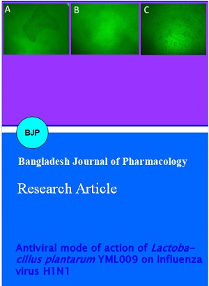
After streaking the L. plantarum YML009 on tryptone soy agar with 5% sheep blood with incubation at 37°C for 48 hours, no beta-hemolysis (clear zones around colonies) was observed on the plate agar. The L. plantarum YML009 produced green-hued zones around the colony (Figure 3A). Streptococcus agalactiae KCCM 40417 was used as control for beta-hemolysis (Figure 3B). Whereas, L. paracasei KCTC 3510 was used as control for α-hemolysis (Figure 3C). Therefore, L. plantarum YML009 showed α–hemolytic phenomenon and caused no hemolysis.
Figure 3: Hemolytic phenomenon test. (A) Test strain: Lactobacillus plantarum YML009. (B) Control: Streptococcus agalactiae KCCM 40417. (C) Control: Lactobacillus paracasei KCTC 3510
Antibiotic susceptibility
The MIC values obtained for L. plantarum YML009 are shown in Table II. YML0009 was sensitive to all the recommend antibiotics tested. The MIC values obtained after 24 and 48 hours of incubation were as follows; gentamicin (0.5 mg/L), kanamycin (4 mg/L), streptomycin (2 mg/L), tetracycline (8 mg/L), erythromycin (0.1 mg/L) clindamycin (0.1 mg/L), chloramphenicol (4 mg/L), and ampicillin (0.3 mg/L). These results suggest that L. plantarum YML009 is safe. Since, it does not show any resistance to the antibiotics, YML009 could be considered as probiotic bacterium
Table II: Antibiotic susceptibility test of L. plantarum YML009
| Antibiotics | 1a | 2 | 3 | 4 | 5 | 6 | 7 | 8 | 9 | 10 | 11 | 12b |
|---|---|---|---|---|---|---|---|---|---|---|---|---|
| Gentamicin | P | 0.500 | 1 | 2 | 4 | 8 | 16 | 32 | 64 | 128 | 256 | N |
| Kanamycin | P | 2 | 4 | 8 | 16 | 32 | 64 | 128 | 256 | 512 | 1024 | N |
| Streptomycin n.r. | P | 0.500 | 1 | 2 | 4 | 8 | 16 | 32 | 64 | 128 | 256 | N |
| Tetracycline | P | 0.125 | 0.250 | 0.500 | 1 | 2 | 4 | 8 | 16 | 32 | 64 | N |
| Erythromycin | P | 0.016 | 0.032 | 0.064 | 0.125 | 0.250 | 0.500 | 1 | 2 | 4 | 8 | N |
| Clindamycin | P | 0.032 | 0.063 | 0.125 | 0.250 | 0.500 | 1 | 2 | 4 | 8 | 16 | N |
| Chloramphenicol | P | 0.125 | 0.250 | 0.500 | 1 | 2 | 4 | 8 | 16 | 32 | 64 | N |
| Ampicillin | P | 0.032 | 0.063 | 0.125 | 0.250 | 0.500 | 1 | 2 | 4 | 8 | 16 | N |
Blue marked values are <MIC (mg/L); Green marked values are MIC (mg/L); aPositive control well without antibiotics, but with the test strain containing solvent which is used to dissolve the antibiotics at the highest concentration; bNegative control well without test strain and the antibiotics, but with the medium
In vitro antiviral activity of L. plantarum YML009 using MDCK cells
Here we tested L. plantarum YML009 for its potential antiviral activity against influenza virus H1N1 on MDCK cells. We observed that H1N1 induced cytopathic effect (CPE) in MDCK cells when used alone (Figure 4A). However, the same CPE was not observed in MDCK cells treated with cell-free supernatant of L. plantarum YML009 even after 72 hours of the viral injection (Figure 4B). Further, microscopic observation results showed that MDCK cells treated with H1N1 and YML009 showed similar morphology as control MDCK cells (without any treatment) as shown in Figure 4C. Hence, it proved L. plantarum YML009 could be considered as a potent anti-influenza bacterium able to control CPE in MDCK cells. In addition, 10x cell-free supernatant of YML009 showed more fascinating results. Serial dilutions of 21, 22, 23 of 10x cell-free supernatant were also able to control the CPE in MDCK cells (Table III).
Table III: Antiviral activity of L. plantarum against H1N1
| Two-fold dilution | |||||
|---|---|---|---|---|---|
| 20 | 21 | 22 | 23 | 24 | |
| CS(a) | + | - | - | - | - |
| CS(X10) | + | + | + | + | - |
Figure 4: Cytopathogenic effects H1N1 virus infection in MDCK cells. (A) Cytopathogenic effects in MDCK cells treated with H1N1. (B) MDCK cells treated with Lactobacillus plantarum YML009 and H1N1. (C) Control cells without any treatment. The photos were taken under microscope at a magnification of 40
In vitro antiviral activity of L. plantarum YML009 using SPF embryonated eggs and hemagglutination assay
Embry-noated SPF eggs were inoculated with viral dose (105.5 EID50/0.1 mL) and similarly were treated with cell-free supernatant (CS), heat-killed supernatant (HK) and cell-mass (CM) of L. plantarum YML009. Tamiflu (T) was taken as a positive control. Our results showed that the survival rate of embryonated eggs were highest with CS followed by CM, whereas HK and T showed same level of inhibition (Table IV).
Table IV: Antiviral activity of L. plantarum YML009 using embryonated SPF eggs
| Group | Dilution rate | Survival/total |
|---|---|---|
| H1N1 virus + CSa | 1:104 | 2/2 |
| 1:105 | 0/2 | |
| H1N1 virus + HKb | 1:105 | 1/2 |
| 1:106 | 1/2 | |
| H1N1 virus + CMc | 1:104 | 2/2 |
| 1:105 | 0/2 | |
| H1N1 virus + Td | 5 mg/100 µL | 1/2 |
| 3 mg/100 µL | 1/2 |
The hemaggluttination (HA) assay is a tool used to screen cell culture or amnioallantoic fluid harvested from embryonating eggs for hemagglutinating agents, such as influenza virus. Since, live and inactivated viruses are detected by the HA test, we performed the same test to confirm the antiviral effects of L. plantarum YML009. Our results showed that cell-free supernatant of L. plantarum YML009 completely revoked viral infection. In addition to cell-free supernatant of YML009, heat killed supernatant (HS), and cell-mass (CM) were also able to inhibit viral proliferation and showed antiviral activity. Similar test was conducted with tamiflu (containing oseltamivir phosphate) as a positive control (Table V).
Table V: Hemagglutinating activity of L. plantarum YML009 on embryonating SPF eggs infected with H1N1
| Treatment | ||||
|---|---|---|---|---|
| A | B | C | D | |
| Hemagglutinating activity | 1 : 4 | 1 : 0 | 1 : 0 | 1 : 4 |
Discussion
In recent years, interest in LAB has increased due to their probiotic and antimicrobial properties suggesting them to be promising alternatives to chemical drugs (Rather et al., 2013; Park et al., 2013). In this study, a total of 2272 LAB strains isolated from different kimchi samples were screened for their antiviral activity against H1N1 influenza virus. A highly potent strain named as L. plantarum YML009 was isolated from kimchi showed strong antiviral effects against H1N1 influenza. The biochemical characterization using API 50 CHL showed that strain used 25 carbohydrates which include: D-xylose, D-galactose, D-glucose, D-fructose, D-mannose, L-rhamnose, D-mannitol, D-sorbitol, N-acetylglucosamine, amygdalin, arbutin, esculin, salicin, D-cellobiose, D-maltose, D-lactose, D-melibiose, D-saccharose, D-trehalose, D-melezitose, D raffinose, gentiobiose, D-turanose, D-tagatose, and D-arabitol. Almost similar results of fermentation test were reported by Rather et al. (2013).
plantarum YML009 showed great level of resistance against artificial gastric and bile juice. An initial cell culture (2.67 x 10⁹ CFU/mL) of YML009 was exposed to artificial bile and after incubation, a higher level of survival (1.5 x 10⁸ CFU/mL) was observed. Similarly, a cell culture of YML009 (6.9 x 10⁸ CFU/mL) was exposed to artificial gastric juice and a survival of 6.9 x 10⁸ CFU/mL was observed. This suggested L. plantarum YML009 as a potent strain and can withstand against the internal environment of stomach.
Further, L. plantarum YML009 was tested for hemolysis test. It was found that YML009 showed alpha hemolysis which suggested L. plantarum YML009 to be considered as a safe candidate without effecting red blood cells.
There are many reports published regarding the antibiotic resistance of LAB which has become a major concern. L. plantarum YML009 was tested for antibiotic susceptibility against eight antibiotics as suggest my EFSA. Our results showed that YML009 was very sensitive against all the antibiotics tested. This further confirmed that L. plantarum YML009 could be as a novel bacterium to use as a probiotic against influenza virus.
In vitro antiviral activity assays with MDCK cell line showed that L. plantarum YML009 has very strong antiviral activity. A 10-fold concentrated cell-free supernatant and its dilutions were also effective against H1N1. Using embryonated SPF eggs, antiviral activity was seen up to 104 dilution of CS (cell-free supernatant) and CM (cell-mass), and up to 105 dilution of HK (heat-killed and sonication culture medium).
The antiviral efficacy of L. plantarum YML009 was compared with oseltamivir (tamiflu). It was shown that YML009 proved to be more effective than tamiflu.
Conclusion
L. plantarum YML009 may be a novel probiotic candidate as anti-influenza prevention and infection.
References
Beigel J, Bray M. Current and future antiviral therapy of severe seasonal and avian influenza. Antiviral Res. 2008; 78: 91-102.
Corr SC, Gahan CG, Hill C. Impact of selected Lactobacillus and Bifidobacterium species on Listeria monocytogenes infection and the mucosal immune response. FEMS Immunol Med Microbiol. 2007; 50: 380-88.
Drouault S, Corthier G. Health effects of lactic acid bacteria ingested in fermented milk. Vet Res. 2001; 32: 101-17.
Dunne C, O’Mahony L, Murphy L, Thornton G, Morrissey D, O'Halloran S, Feeney M, Flynn S, Fitzgerald G, Daly C, Kiely B, O'Sullivan GC, Shanahan F, Collins JK. In vitro selection criteria for probiotic bacteria of human origin: Correlation with in vivo findings. Am J Clin Nutr. 2001; 73: S386-92.
EFSA Panel on Additives and Products or Substances used in Animal Feed (FEEDAP). Guidance on the assessment of bacterial susceptibility to antimicrobials of human and veterinary importance. EFSA J. 2012; 10: 2740.
ES ISO. Milk and milk products: Determination of the minimal inhibitory concentration (MIC) of antibiotics applicable to bifidobacteria and non-enterococal lactic acid bacteria. ESA. 2012; 10932.
Fujiwara D, Inoue S, Wakabayashi H, Fujii T. The anti-allergic effects of lactic acid bacteria are strain dependent and mediated by effects on both Th1/Th2 cytokine expression and balance. Int Arch Allergy Immunol. 2004; 135: 205-15.
Hancock K, Veguilla V, Lu X, Zhong W, Butler EN, et al. Cross-reactive antibody responses to the 2009 pandemic H1N1 influenza virus. N Engl J Med. 2009; 361: 1945–52.
Hanniffy S, Wiedermann U, Repa A, Mercenier A, Daniel C, Fioramonti J, Tlaskolova H, Kozakova H, Israelsen H, Madsen S, Vrang A, Hols P, Delcour J, Bron P, Kleerebezem M, Wells J. Potential and opportunities for use of recombinant lactic acid bacteria in human health. Adv Appl Microbiol. 2004; 56: 1-64.
Ishida Y, Nakamura F, Kanzato H, Sawada D, Hirata H, Nishimura A, Kajimoto O, Fujiwara S. Clinical effects of Lactobacillus acidophilus strain L-92 on perennial allergic rhinitis: A double-blind, placebo-controlled study. J Dairy Sci. 2005; 88: 527-33.
Kobayashi Y, Toyama K, Terashima T. Biological characteristics of Lactobacillus. II. Tolerance of a multiple antibiotic resistant strain, Lactobacillus casei PSR 3002, to artificial digestive fluids. Japanese J Bacteriol. 1974; 27: 691-97.
Long JK, Mossad SB, Goldman MP. Antiviral agents for treating influenza. Cleve Clin J Med. 2000; 67: 92-95.
Nagai T, Makino S, Ikegami S, Itoh H, Yamada H. Effects of oral administration of yogurt fermented with Lactobacillus delbrueckii ssp. Bulgaricus OLL1073R-1 and its exopolysaccharides against influenza virus infection in mice. Int Immunopharmacol. 2011; 11: 2246-50.
Nagao F, Nakayama M, Muto T, Okumura K. Effects of a fermented milk drink containing Lactobacillus casei strain Shirota on the immune system in healthy human subjects. Biosci Biotechnol Biochem. 2000; 64: 2706-08.
Nomura M, Kobayashi M, Narita T, Kimoto-Nira H, Okamoto T. Phenotypic and molecular characterization of Lactococcus lactis from milk and plants. J Appl Microbiol. 2006; 101: 396-405.
Oh S, Kim SH, Worobo RW. Characterization and purification of a bacteriocin produced by a potential probiotic culture, Lactobacillus acidophilus 30SC. J Dairy Sci. 2000; 83: 2747-54.
Park MK, NGO V, Kwon YM, Lee YT, Yoo S, et al. Lactobacillus plantarum DK119 as a probiotic confers protection against influenza virus by modulating innate immunity. PLoS ONE. 2013; 8: e75368.
Parvez S, Malik KA, Ah Kang S, Kim HY. Probiotics and their fermented food products are beneficial for health. J Appl Microbiol. 2006; 100: 1171-85.
Petros AM, Konstantinos CM, Dimitris P, Silvia C, Jana F, Mette D C, Effie T. Functional properties of novel protective lactic acid bacteria and application in raw chicken meat against Listeria monocytogenes and Salmonella enteritidis. J Food Microbiol. 2009; 130: 219-26.
Rather IA, Seo BJ, Rejish VJK, Choi UH, Choi KH, Lim JH, Park YH. Isolation and characterization of a proteinaceous antifungal compound from Lactobacillus plantarum YML007 and its application as a food preservative. Lett Appl Microbiol. 2013; 57: 69-76.
Reed LJ, Muench H. A simple method of estimating fifty percent endpoints. Am J Hyg. 1938; 27: 493-97.
Seo BJ, Rather IA, Kumar VJR, Choi UH, Moon MR, Lim JH, Park YH. Evaluation of Leuconostoc mesenteroides YML003 as a probiotic against low-pathogenic avian influenza (H9N2) virus in chickens. J Appl Microbiol. 2012; 113: 163-71.
Thompson WW, Shay DK, Weintraub E, Brammer L, Bridges CB, Cox NJ, Fukuda K. Influenza-associated hospitalizations in the United States. JAMA. 2004; 292: 1333-40.
Youn HN, Lee DH, Lee YN, Park JK, Yuk SS, et al. Intranasal administration of live Lactobacillus species facilitates protection against influenza virus infection in mice. Antiviral Res. 2012; 93: 138-43.