Tanshinone IIA inactivates Akt and induces caspase-dependent death in cervical cancer cells via the mitochondrial pathway
Abstract
In human cervical cancer cells pro-apoptotic effect of tanshinone IIA isolated from the ethanol extract of Scutellaria barbata was investigated. Tanshinone IIA treatment resulted in apoptosis and mitochondrial membrane potential loss in the cervical cancer cells. The viability of SW756 and C4-1 cells was reduced in a concentration dependent manner on treatment with tanshinone IIA for 36 hours. Flow cytometric analysis in SW756 cells showed marked increase in accumulation of sub-G1-phase cell population. Tanshinone IIA treatment also caused significant increase in DNA fragmentation in these cells. DAPI staining revealed significant increase in nuclear condensation and apoptotic body formation on tanshinone IIA treatment in SW756 cells However, the effect of tanshinone IIA was reversed by caspase inhibitor, z-VAD-fmk. In tanshinone IIA treated cells Akt phosphorylation was markedly reduced and this decrease was inhibited by LY294002 (phosphatidylinositol-3'-kinase inhibitor). Tanshinone IIA treated apoptotic cells exhibited decrease in expression of Mcl-1. Thus tanshinone IIA induces apoptosis in cervical cancer cells through mitochondrial pathway.
Introduction
Cervical cancer is a commonly observed cancer in the females throughout the world with around 290,000 deaths every year (Saslow et al., 2007). Contraction of human papillomavirus (HPV) is the major risk inducing factor found in all the cervical cancer patients (Steben and Duarte, 2007). Cervical cancer if detected at the early stage using regular Pap tests makes treatment favorable compared to late stage cancer (Long, 2007; Brinkman et al., 2005).
Currently used cervical cancer treatment strategies include radiation and chemotherapy along with the herbal remedies (Leitao and Chi, 2002; Yang, et al., 2014). Apoptosis is induced by the activation of enzymes known as caspases which cleave essential proteins producing changes in the morphological characteristics of the cells (Cohen, 1993; White, 1996; Williams and Smith, 1993; Wyllie et al., 1980). Apoptosis induces release of some proteins like cytochrome c from mitochondria into the cytosol inducing pro-apoptotic functions. In cytoplasm, cytochrome c binds to the adaptor protein, Apaf-1 causing its oligomerization and caspase activation (Danial and Korsmeyer, 2004).
One of the antiapoptotic Bcl-2 homologues is Mcl-1 which binds to subset of BH3 domain-only proteins (Chen et al., 2005; Kuwana et al., 2005). Mcl-1 is also reported to inhibit apoptosis after UV irradiation (Nijhawan et al., 2003). In the present study, we investigated the potential therapeutic effects of tanshinone IIA in human cervical cancer. It was observed that tanshi-none IIA induces death in cervical cancer cells through the mitochondrial apoptotic pathway. The pathway is mediated by inactivating Akt and decreasing Mcl-1 expression.
The plants belonging to Scutellaria genus have a long traditional importance of possessing potential anticancer activity. Some of the flavonoids isolated from the plant extract of this genus include apigenin, baicalein, baicalin, chrysin, scutellarein, and wogonin. All of these molecules exhibit antitumor activity. It is reported that the leaf extracts of Scutellaria angulosa, Scutellaria integrifolia, Scutellaria ocmulgee and Scutellaria scandens show potent anticancer activity (Parajuli et al., 2009). Recently the ethanol extract of Scutellaria barbata was shown to significantly reduce the growth of A549 cells with an IC50 value of 0.2 mg/mL (Yin et al., 2004). Another, constituent tanshinone IIA exhibits potential anticancer activity against breast cancers. It exhibits its effect by inducing apoptosis, inhibiting cell proliferation, transcriptional regulation, angiogenesis, invasive potential and metastatic potential of cancer cells (Wang et al., 2005). Tanshinone IIA has been shown to exhibit anticancer effects both in vitro and in vivo (Lee et al., 2008). Other species of Scutellaria like S. baicalensis, S. litwinowii etc. also act as potent anticancer agents in various in vitro as well as in vivo models (Ye et al., 2002; Katiyar et al., 1997).
Materials and Methods
Cell lines and culture: SW756 and C4-1 human cervical cell lines were obtained from American Type Culture Collection (Manassas, VA, USA). The cells were cultured in DMEM medium (Gibco BRL, Gaithersburg, MD, USA) containing 10% fetal bovine serum (FBS) at 37°C in a 5% CO2 humidified atmosphere.
Reagents and drug: Tanshinone IIA and z-VAD-fmk were purchased from Sigma-Aldrich (St. Louis, MO, USA). Polyclonal antipolyclonal anti-poly (ADP-ribose) polymerase (PARP), anti-Bc1-XL and anti-actin antibodies were purchased from Qiagen (Valencia, CA, USA). Anti-phospho Akt and anti-caspases from Cell Signaling Technology (San Diego, CA, USA). Monoclonal antibodies against caspase-8 and Bcl-2 were purchased from Qiagen (Valencia, CA, USA).
MTT assay: Cervical carcinoma cell lines, SW756 and C4-1 were incubated in growth media containing tanshinone IIA for 36 hours. After incubation growth media was removed and cells were rinsed three times in sterile Dulbecco's phosphate-buffered saline (DPBS). The 3-(4,5-dimethylthiazol-2-yl)-2,5-diphenyltetrazolium bromide (MTT) was added to plates and then incubated for 2 hours. The formazan crystals were solubilized by adding dimethyl sulfoxide and Spectra MaxPlus 384 microplate reader was used to measure the absorbance.
Reverse transcription-polymerase chain reaction (RT-PCR): 2.5 x 10⁵ cells were seeded into each well of a 6 cm dish and cultured for 72 hours. Tanshinone IIA was added to each well and incubation was continued for 2 hours. NucleoSpin® RNA II kit (Macherey-Nagel, Duren, Germany) was used for the isolation of total RNA from the cells according to the manufacturer's instructions. The complementary DNA was synthesized using AMV reverse transcriptase (Promega) and random primers (Takara Bio Inc., Shiga, Japan).
Immunoblot analysis: Cervical cells, 2.5 x 10⁶ were seeded into each well of a 6 cm dish and incubated for 72 hours. To each well tanshinone IIA was added and incubated for 12 hours. Electrophoresis using sodium dodecyl sulfate-polyacrylamide gel for cell lysates was followed by transfer to a nitrocellulose membrane (Millipore, Bedford, MA, USA). Incubation of the membrane was performed with primary antibody for 2 hours. We used horseradish peroxidase-conjugated anti-rabbit as the secondary antibody. The enhanced chemiluminescence (Amersham, Arlington Heights, IL, USA) was used for the immunoreactive protein visualization.
DNA fragmentation analysis and 4',6'-diamidino-2-phenylindole (DAPI) staining: Tanshinone IIA treated cells were lysed in lysis buffer (10 mM Tris (pH 7.4), 150 mM NaCl, 5 mM EDTA, and 0.5% Triton X-100) for 30 min. Centrifugation of the lysates at 12,000 x g was performed for 30 min to get clear lysis mass. Extraction and separation of the fragmented DNA was achieved on 1.5% agarose gel containing 0.1 ug/mL EtBr. After tanshinone IIA treatment for 24 hours cells were fixed with paraformaldehyde for 1 hour, washed with PBS and then incubated with 300 nM DAPI (Microprobe, CA, USA) for 30 min. Fluorescence microscopy was used for the examination of cells.
Flow cytometric analysis of propidium iodide (PI) and JC-1 staining: The SW756 cells (2.5 x 10⁵) suspended in PBS were supplemented with 100 μL ethanol and incubated at 4°C for 1 hour. The cells were washed with PBS, resuspended in 200 uL of sodium citrate buffer and RNase then incubated for 1 hour. The cellular DNA was stained and incubated with PI for 45 min. FACScan flow cytometer was employed for fluorescence activated cell sorting (FACS) to measure relative DNA content. SW756 cells treated with tanshinone IIA were subjected to apoptosis measurement by flow cytometry after PI staining.
Transfection of HeLa cells with Akt, Mcl-1 and Bcl-2 expression vectors: Lipofectamine Plus reagent (Invitrogen) was used to transfect SW756 cells with plasmids (Akt, Mcl-1, or Bcl-2) according to manual protocol. In 6-well culture plates, 2.5 x 10⁶ cells were cultured in DMEM supplemented and incubated for 36 hours. Cells washed with PBS were incubated for 4 hours followed by addition of lipofectamine reagent along with pcDNA 3.1 or Akt, Mcl-1, or Bcl-2 expressing vector. Replacement of the medium was followed by incubation for 36 hours then transfection in primary cell culture medium.
Analysis of cytochrome c release: Tanshinone IIA treated cells after harvesting and washing with ice-cold PBS were lysed ice-cold lysis buffer (25 mM HEPES, pH 7.5; 250 mM sucrose; 1 mM EDTA; 1 mM EGTA; 1 mM DTT; 10 mM KCl; 1.5 mM MgCl2; 1 mM PMSF; protease inhibitor cocktail). Cell lysates were centrifuged at 12,000 rpm to obtain supernatants and pellets. The supernatant was analyzed by immunoblot using an anti-cytochrome c antibody.
Statistical analysis: The data presented are the mean of S.D. All the experiments were performed in triplicates. Student t test or analysis of variance was used for statistical analysis. The differences were considered statistically significant at p<0.05.
Results
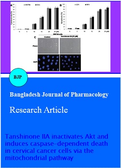
Induction of apoptosis by tanshinone IIA in cervical cancer cells: We used MTT assay to investigate the effect of tanshinone IIA on the viability of SW756 and C4-1 human cervical cells. The viability of SW756 and C4-1 cells was reduced in a concentration dependent manner on treatment with tanshinone IIA for 36 hours (Figure 1A). SW756 cells showed marked increase in accumulation of sub-G1-phase cells in a concentration- and time-dependent manner following tanshinone IIA treatment using flow cytometric analysis (Figure 1B). Tanshinone IIA treatment also caused significant increase in DNA fragmentation in these cells. DAPI staining revealed significant increase in nuclear condensation and apoptotic body formation on tanshinone IIA treatment in SW756 cells (Figure 1C). Thus tanshinone IIA induces cytotoxic effect in cervical cancer cells through apoptosis.
Figure 1: Tanshinone IIA induced cytotoxic effect in cervical cancer cells. (A) Viability of SW756 and C4-1 cells following 12 or 36 hours treatment with the indicated concentration of tanshinone IIA using MTT assays. (B) The percentage of SW756 apoptotic cells following treatment with the indicated concentrations of SW756 for 12 and 36 hours. For apoptosis assessment DNA content was examined after propidium iodide staining. (C) DAPI staining showing death of SW756 cells after exposure to 20 μM tanshinone IIA for 12 hours. The values presented are the mean ± SD of three experiments
Effect of tanshinone IIA on caspase processing: Tanshinone IIA treated SW756 and C4-1 cells showed cleaved pro-caspase-8 and pro-caspase-9 after 12 h. Similar results were observed for caspase-3. Tanshinone IIA treated SW756 and C4-1 cells also showed increased cleavage of PARP by immunoblot analysis (Figure 2A). However, exposure of tanshinone IIA treated SW756 cells to caspase inhibitor (z-VAD-fmk) inhibited cell death (Figure 2B). These findings suggest that tanshinone IIA-induced apoptosis is associated with the activation of caspases.
Figure 2: Effect of tanshinone IIA on caspase activation in cervical cancer cells. (A) Immunoblot analysis of caspase-8, caspase-9, caspase-3 and PARP in SW756 and C4-1 cell lysates. β-actin served as a loading control. (B) Apoptosis in SW756 cells incubated with 25 μM z-VAD-fmk or solvent for 1 h before treatment with 20 μM tanshinone IIA for 1 hour. Apoptosis was assessed by the DNA content after propidium iodide staining. The values presented are the mean ± S. D. from triplicate experiments
Effect of tanshinone IIA on Bcl-2 family proteins in cervical cancer cells: SW756 cells on treatment with tanshinone IIA did not show any change in the expression of Bcl-2, Bcl-xL and Bax proteins (Figure 3A). However, the expression of Mcl-1 was significantly decreased by tanshinone IIA after 24 hours (Figure 3B). RT-PCR results showed decrease in the levels of Mcl-1 mRNA on tanshinone IIA treatment. Therefore, tanshinone IIA-induced Mcl-1 expression down-regulation is controlled at the transcriptional level.
Figure 3: Effect of tanshinone IIA on Bcl-2 family proteins in cervical cancer cells. Immunoblot analysis of (A) Bcl-2, Bcl-xL and Bax and (B) Mcl-1 in lysates of SW756 cells incubated with tanshinone IIA for 36 hours. β-actin served as a loading control
Effect of tanshinone IIA on Akt phosphorylation in SW756 cells: In tanshinone IIA treated cells a significant decrease in the phosphorylated Akt level was observed (Figure 4). However, tanshinone IIA treated cells when exposed to LY294002, an inhibitor of the Akt activating kinase suppressed that tanshinone IIA-mediated apoptosis (Figure 4).
Figure 4: Effect of tanshinone IIA on PI3K/Akt signaling in SW756 cells. (A) Immunoblot analysis of phospho-Akt and Akt in SW756 cells exposed to tanshinone IIA for 36 hours. (B) Apoptosis in SW756 cells treated for 36 hours with tanshinone IIA with or without 25 uM LY294002
Tanshinone IIA-induced activation of the mitochondrial cell death pathway in SW756 cells: Transfection of SW756 cells with plasmid encoding dominant-negative caspase-9 (caspase-9 dN) and treatment with tanshinone IIA for 36 hours showed that caspase-9 dN cells were resistant to tanshinone IIA (Figure 5A). This suggests that caspase-9 is essential for tanshinone IIA-induced cell death and that tanshinone IIA induces apoptosis via the mitochondrial pathway. Measurement of cytochrome c release and Δm in SW756 cells revealed significant increase in cytochrome c in the cytosol of cells treated with tanshinone IIA (Figure 5B).
Figure 5: Induction of mitochondrial apoptotic events by tanshinone IIA in SW756 cells. (A) Immunoblot analysis of caspase-9 in SW756 cells that were transfected with pcDNA (vector) or caspase-9 dN and treated with or without tanshinone IIA for 24 hours. (B) Immunoblot analysis of cytochrome c in cells treated with tanshinone IIA for 24 hours
Discussion
Our study clearly revealed that tanshinone IIA induced apoptosis in SW756 and C4-1cervical cancer cells. The results showed that tanshinone IIA-induced cervical cancer cell death depends on inhibition of Akt activation and activation of caspase-9. It was also observed that an antiapoptotic factor, Mcl-1 that exhibits crucial role in countering the apoptosis pathway was inhibited by tanshinone IIA. In various cancers, one of the main signaling pathways to inhibit apoptosis is PI3K/Akt pathway (Song et al., 2005). Usually in cancer cells expression of PI3K/Akt is found to be significantly higher and is believed to be responsible providing resistance to cancer cells against chemotherapy (McCubrey et al., 2006). Our study showed that in SW756 cells contained substantial levels of phosphorylated Akt. Therefore, sustained Akt activity in these cells may facilitate their growth and/or survival. However, we found that tanshinone IIA had an inhibitory effect on Akt in SW756 cells. Because Akt is a pro-survival protein, the inhibition of Akt may be involved in tanshinone IIA mediated growth suppression of SW756 cells. A variety of cellular proteins participate in the induction of apoptosis. Among these are the caspases, which are essential for the execution of cell death induced by apoptotic stimuli (Cohen, 1997). We have demonstrated that tanshinone IIA exposure leads to the processing of caspase-8, -9 and -3 in cervical cancer cells. An important finding was that tanshinone IIA induced caspase-3 processing and PARP cleavage in parallel with the induction of apoptosis. Furthermore, the inhibition of caspase-3 by the broad-spectrum caspase inhibitor z-VAD-fmk blocked cell death by tanshinone IIA, suggesting that tanshinone IIA-induced cell death is caspase-dependent. The finding that transfection of caspase-9 dN imparted resistance to tanshinone IIA indicates that the activation of caspase-9 and subsequent activation of caspase-3 activation (i.e., the mitochondrial pathway of caspase activation) are critical to the action of tanshinone IIA. The release of mitochondrial intermembrane space proteins to the cytosol is a key event during apoptosis (Fulda and Debatin, 2004a; Fulda and Debatin, 2004b). Our results clearly showed that tanshinone IIA induced cytochrome c release into the cytosol and decreased Δm, supporting the idea that tanshinone IIA activates the mitochondrial apoptotic pathway.
Conclusion
Tanshinone IIA induces apoptosis in cervical cancer cells, likely by inducing multiple cellular events such as caspase activation, Mcl-1 inactivation, inhibition of Akt signaling, and loss of Δm. The findings presented here suggest that tanshinone IIA is a potential anticancer drug for cervical cancer.
References
Brinkman JA, Caffrey AS, Muderspach LI, Roman LD, Kast WM. Inhibitory effect of ginsenoside Rg5 and its metabolite ginsenoside Rh3 in an oxazolone induced mouse chronic dermatitis model. Eur J Gynaecol Oncol. 2005; 26: 129-42.
Chen L, Willis SN, Wei A, Smith BJ, Fletcher JI, Hinds MG, Colman PM, Day CL, Adams JM, Huang DC. Differential targeting of prosurvival Bcl-2 proteins by their BH3-only ligands allows complementary apoptotic function. Mol Cell. 2005; 17: 393-403.
Cohen GM. Caspases: The executioners of apoptosis. Biochem J. 1997; 326: 1-16.
Cohen J J. Apoptosis. Immunol Today. 1993; 14: 126-30.
Danial NN, Korsmeyer SJ. Cell death: Critical control points. Cell 2004; 116: 205-19.
Fulda S, Debatin KM. Apoptosis signaling in tumor therapy. Ann NY Acad Sci. 2004a; 1028: 150-56.
Fulda S, Debatin KM. Targeting apoptosis pathways in cancer therapy. Curr Cancer Drug Targets. 2004b; 4: 569-76.
Katiyar SK, Korman NJ, Mukhtar H, Agarwal R. Protective effects of silymarin against photocarcinogenesis in a mouse skin model. J Nat Cancer Inst. 1997; 89: 556.
Kuwana T, Bouchier-Hayes L, Chipuk JE, Bonzon C, Sullivan BA, Green DR, Newmeyer DD. BH3 domains of BH3-only proteins differentially regulate Bax-mediated mitochondrial membrane permeabilization both directly and indirectly. Mol Cell. 2005; 17: 525-35.
Lee CY, Sher HF, Chen HW, Liu CC, Chen CS, Lin CS, Yang PC, Tsay HS, Chen JJW. Anticancer effects of tanshinone I in human non-small cell lung cancer. Mol Cancer Ther. 2008; 7: 3527.
Leitao MM Jr, Chi DS. Recurrent cervical cancer. Curr Treat Options Oncol. 2002; 3: 105-11.
Long HJ 3rd. Management of metastatic cervical cancer: Review of the literature. J Clin Oncol. 2007; 25: 2966 74.
McCubrey JA, Steelman LS, Abrams SL, Lee JT, Chang F, Bertrand FE, Navolanic PM, Terrian DM, Franklin RA, D'Assoro AB, Salisbury JL, Mazzarino MC, Stivala F, Libra M. Roles of the RAF/MEK/ERK and PI3K/PTEN/AKT pathways in malignant transformation and drug resistance. Adv Enzyme Regul. 2006; 46: 249-79.
Nijhawan D, Fang M, Traer E, Zhong Q, Gao W, Du F, Wang X. Elimination of Mcl-1 is required for the initiation of apoptosis following ultraviolet irradiation. Genes Dev. 2003; 17: 1475-86.
Parajuli P, Joshee N, Rimando AM, Mittal S, Yadav SK. In vitro antitumor mechanisms of various Scutellaria extracts and constituent flavonoids. Planta Medica. 2009; 75: 41.
Saslow D, Castle PE, Cox JT, et al. American Cancer Society Guideline for human papillomavirus (HPV) vaccine use to prevent cervical cancer and its precursors. CA Cancer J Clin. 2007; 57: 7 28.
Song G, Ouyang G, Bao S. The activation of Akt/PKB signaling pathway and cell survival. J Cell Mol Med. 2005; 9: 59-71.
Steben M, Duarte Franco E. Human papillomavirus infection: Epidemiology and pathophysiology. Gynecol Oncol. 2007; 107: S2 S5.
Wang X, Wei Y, Yuan S, Liu G, Lu Y, Zhang J, Wang W. Potential anticancer activity of tanshinone II A against human breast cancer. Int J Cancer. 2005; 116: 799.
White E. Life, death, and the pursuit of apoptosis. Genes Dev. 1996; 10: 1-15.
Williams GT, Smith CA. Molecular regulation of apoptosis: Genetic controls on cell death. Cell 1993; 74: 777-79.
Wyllie AH, Kerr JF, Currie AR. Cell death: The significance of apoptosis. Int Rev Cytol 1980; 68: 251-306.
Yang J, Li J, Sun M, Chen K. Studies of traditional Chinese medicine monomer on HeLa cell of cervical cancer. Pak J Pharm Sci 2014; 27: 1063-1068.
Ye F, Xui L, Yi J, Zhang W, Zhang DY. Anticancer activity of Scutellaria baicalensis and its potential mechanism. J Altern Complement Med. 2002; 8: 567.
Yin X, Zhou J, Jie C, Xing D, Zhang Y. Anticancer activity and mechanism of Scutellaria barbata extract on human lung cancer cell line A 549. Life Sci. 2004; 75: 2233.