MHC class I-presented tumor antigen appraisable for T-cell responses against ovarian cancer
Abstract
The purpose of this study is to assess whether MHC class I-presented tumor antigen is appraisable for T-cell responses against ovarian cancer. In ovarian cancer cell, human leukocyte antigen A2 (HLA-A2) associated with peptides was used to promote the activation of naive T cells so as to activate antigen-specific T cells. 7 or 4 patients were observed grade 1 or 2 injection site reactions, respectively. 5, 2 or 1 patients were observed grade 1, 2 or 3 pain reactions, respectively. 4 or 1 patients were observed grade 1 or 2 induration reactions. Total number mean value of patients experiencing response to the particular peptide was 7.73, and total number mean value of peptides to which the patients responded was 7.45. MHC class I-presented tumor antigen is appraisable for T-cell responses against ovarian cancer in China.
Introduction
Ovarian cancer is one of the three malignant tumors of female reproductive system, and the ovarian cancer mortality ranks first in gynecological malignancies (Meng et al., 2011). The reason may be that the position of the anatomical parts is relatively centered in the onset of ovarian cancer, and the early abdominal symptoms are not easy to be distinguished and differentiated from those of neighboring gastrointestinal diseases, especially the common symptoms such as bloating, constipation and abdominal pain (Kang et al., 2014). About 70% of ovarian cancer patients have come to an advanced stage before diagnosis with complications (Sanchez-Munoz et al., 2009). Currently, the specificity of ovarian cancer-related examinations is low, lacking in simple methods of operation, unfavorable for the early diagnosis, and there are not enough researches on the occurrence of ovarian cancer, without accepted theory that can clearly explain its pathogenesis, which limit the progress of ovarian cancer diagnosis and treatment as well as prevention techniques to a great extent (Liu et al., 2010).
Major Histocompatibility Complex I (MHC-I) participates in the processing and handling of endogenous antigens (viral antigens, tumor antigens, etc.), of which the role is to transform the protein antigen into antigen peptides, present them to antigen-specific T lymphocytes, and implement antigen-antibody reaction, protecting the body from infection outside (Cao et al., 2015; Lisik et al., 2009). Many viruses can infect the skin and mucosa, and interfere with MHC-I antigen presentation process to evade the host immune response. Studies have shown that MHC-I protein molecules are down-regulated in the esophagus cancer, stomach cancer, colorectal cancer, lung cancer, cervical cancer and other carcinomas, reducing cytotoxic T lymphocyte-mediated immune response in the body (Catamo et al., 2014; Chen et al., 2005; Goodridge et al., 2013; Haimiti et al., 2014; Paulson et al., 2014). We therefore evaluated whether MHC Class I-presented tumor antigens was appraisable for T-Cell responses against ovarian cancer.
Materials and Methods
Subjects: Heparinized blood from healthy HLA-A2 + donors was purchased from selected patients. All patients were obtained following written informed consent approved by the Second Hospital of Tianjin Medical University Medical Center Institutional Review Board (No. 20140034).
Primary cells from human tissues: Kidney and liver tissue samples were obtained from HLA-A2 + human donors. Briefly, all samples were minced and digested with enzyme. Then, cell suspensions were generated per standard methods and supplemented in Roswell Park Memorial Institute-1640 (RPMI-1640, Invitrogen Company, USA) with 2 concentration of antibiotics and antimycotics at 37°C for 6 hours. Cell suspensions were pelleted and washed several times with PBS and RPMI-1640 supplemented with 10% fetal calf serum (FBS, Invitrogen Company, Australia).
Synthetic peptides: Synthetic peptides were supplied by GenScript Corporation and were reconstituted in RPMI-1640. P1, P2, P3, P4, P5, P6, P7, P8, P9, P13, P14, and P15 were formulated by mixing equal amounts of each the component peptides. Low, medium and high binding affinity based on the time for dissociation data published elsewhere (Ramakrishna et al., 2003) and used a t1/2 of 1,000 min as the cutoff separating low/medium from high affinity.
In vitro generation of peptide-specific T cells: Peripheral blood mononuclear cells (PBMC) were obtained from HLA-A2 + human donors and ovarian cancer patients. PBMC were purified by using lymphocyte separation medium (Sangon Biotech, China) by using differential centrifugation according to standard methods. All cells (1 x 10⁶ cells/well) were seeded in 6-well plates and cultured 2 mL RPMI-1640 with 10% FBS, 300 mg/mL L-glutamine, nonessential amino acids, sodium pyruvate, penicillin, and streptomycin at a temperature of 37°C in a humidified atmosphere of 5% CO2 for 1 day. Meanwhile, non-adherent cells were removed and saved. Non-adherent cells were added back in 5 mL complete medium supplemented with 50 ng/mL interleukin-4 (IL-4), 5 ng/mL IL-7, 5 mg/mL keyhole limpet hemocyanin (Invitrogen Company, USA), 25 ng/mL GM-CSF.
Plastic adherent cells were pulsed in complete medium with 50 mg/mL synthetic peptides and 1.5 mg/mL human b2-microglobulin (Invitrogen Company, USA) and cultured in complete medium at a temperature of 37 °C in a humidified atmosphere of 5 % CO2. 2 ml medium was removed and replaced with fresh complete medium supplemented with 10 U/mL IL-2 for 2 days. IL-2 treatment and restimulation were repeated thrice at the indicated time intervals prior and were use to expand T cells in ELISpot assays (Beyotime, Nanjing, China).
Statistical analysis: All data was expressed as the means ± standard deviation (SD) and performed with SPSS 17.0 software. For statistical comparisons of two groups of samples, the Mann-Whitney U-test was used. P<0.05 was considered to be significant.
Results
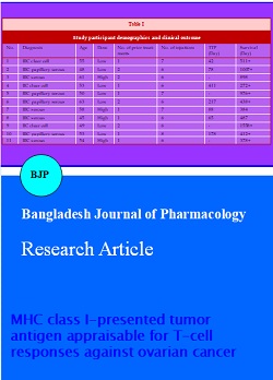
Basic situation of patient: Eleven patients were selected and their basic situation was expressed as Table I, and the average age was 53.55 years. Meanwhile, 4 patients were received with the high dose (1 mg of each peptide) and 7 patients were received with the low dose (100 mg of each peptide). All patients were received cytoreductive surgery and then had 1 to 2 prior chemotherapy regimens.
Clinical activity: Survival average follow-up time of all patients was 665.5 days, 3 patients have died and 6 patients have relapsed (Table I, Figure 1). The median survival has not been reached on either study.
Table I: Study participant demographics and clinical outcome
| No. | Diagnosis | Age | Dose | No. of prior treatments | No. of injections | TTP (Day) |
Survival (Day) |
|---|---|---|---|---|---|---|---|
| 1 | IIIC clear cell | 55 | Low | 1 | 7 | 42 | 511+ |
| 2 | IIIC papillary serous | 48 | Low | 2 | 6 | 78 | 1007+ |
| 3 | IIIC serous | 61 | High | 2 | 6 | - | 898 |
| 4 | IIC clear cell | 53 | Low | 1 | 6 | 411 | 272+ |
| 5 | IIIC papillary serous | 50 | Low | 1 | 7 | - | 976+ |
| 6 | IIIC papillary serous | 63 | Low | 2 | 6 | 217 | 438+ |
| 7 | IIIC serous | 58 | High | 1 | 7 | 88 | 384 |
| 8 | IIIC serous | 45 | High | 1 | 6 | 65 | 487 |
| 9 | IIC clear cell | 49 | Low | 2 | 6 | - | 1558+ |
| 10 | IIIC papillary serous | 53 | Low | 1 | 8 | 178 | 412+ |
| 11 | IIIC serous | 54 | High | 1 | 6 | - | 378+ |
Figure 1: Clinical activity
Adverse event: Table II showed that 7 patients were observed Grade 1 injection site reactions, and 4 patients were observed Grade 2 injection site reactions. 5, 2 or 1 patients were observed Grade 1, 2 or 3 pain reactions, respectively. 4 or 1 patients were observed Grade 1 or 2 induration reactions. Flu-like syndrome, pruritis, fever, chills, fatigue, myalgia, hypotension, weight gain, anorexia, nausea, incontinence or urinary, and headache, etc. were appeared Grade 1 or 2. Meanwhile, confusion, depression, anxiety, and agitation were appeared Grade 3 or 4, and the quantity of these adverse events was very few (Table II).
Table II: Adverse events
| Grade | ||||
|---|---|---|---|---|
| 1 | 2 | 3 | 4 | |
| Injection site reaction | 7 | 4 | ||
| Pain | 5 | 2 | 1 | |
| Induration | 4 | 1 | ||
| Flu-like syndrome | 2 | 1 | ||
| Pruritis | 1 | 1 | ||
| Fever | 2 | |||
| Chills | 2 | |||
| Fatigue | 4 | 2 | ||
| Myalgia | 1 | |||
| Hypotension | 1 | 1 | ||
| Weight gain | 1 | 1 | ||
| Anorexia | 1 | |||
| Nausea | 1 | 1 | ||
| Incontinence, urinary | 1 | |||
| Headache | 1 | 1 | ||
| Confusion | 1 | 1 | 1 | |
| Depression | 1 | 1 | ||
| Anxiety | 1 | |||
| Agitation | 1 | 1 | ||
| Elevated creatinine | 3 | 1 | ||
| Elevated AST/LFTs | 2 | 1 | 1 | |
| Decreased leukocytes | 3 | 1 | 1 | |
| Hypercalcemia | 1 | 1 | ||
| Decreased hemoglobin | 3 | 2 | ||
| Decreased platelets | 1 | |||
| Hypoglycemia | 1 | 1 | ||
| Hypokalemia | 1 | |||
Peptide-specific T-cell responses: Table III showed that total number mean value of patients experiencing response to the particular peptide were 7.73, and total number mean value of peptides to which the patient responded were 7.5. There into, 2 patients were observed that total number of peptides to which the patient responded was zero (Table III).
Table III: Peptide-specific T-cell responses
| No | P1 | P2 | P3 | P4 | P5 | P6 | P7 | P8 | P9 | P13 | P14 | P15 | Amountb |
|---|---|---|---|---|---|---|---|---|---|---|---|---|---|
| 1 | + | + | + | + | + | + | + | + | + | - | + | + | 11 |
| 2 | + | + | + | - | + | - | + | + | + | + | - | + | 9 |
| 3 | + | + | - | - | - | - | + | + | + | + | + | + | 8 |
| 4 | + | + | + | + | + | + | + | + | + | + | + | + | 12 |
| 5 | - | - | - | - | - | - | - | - | - | - | - | - | 0 |
| 6 | + | + | + | + | + | + | + | + | + | - | + | + | 9 |
| 7 | - | + | - | - | - | + | + | + | + | + | + | + | 7 |
| 8 | + | + | + | + | + | + | + | + | + | + | + | + | 12 |
| 9 | - | - | + | + | - | - | - | - | - | - | + | + | 4 |
| 10 | - | - | - | - | - | - | - | - | - | - | - | - | 0 |
| 11 | + | + | - | - | + | + | + | + | + | + | + | + | 10 |
| Amounta | 7 | 8 | 6 | 5 | 6 | 6 | 8 | 8 | 8 | 6 | 8 | 9 |
Discussion
According to a survey from 2008 by International Cancer Research Center, the average incidence of ovarian cancer worldwide is 6.6 cases per 100,000 population, with a 1.9-fold higher incidence in developed versus developing countries. In China, the incidence is 6.0/100,000, close to the world average (Liu et al., 2015). The average mortality rate for ovarian cancer is 3.8/100,000 worldwide, and 2.3/100,000 for China, ranking 166 in the world, at a lower level. A survey from 2009 shows that ovarian cancer accounts for 3.1% of female malignant tumors and for 2.5% of (female) mortality. The morbidity and mortality from ovarian cancer are higher in urban area than in rural area and
the current study, eleven ovarian cancer patients were selected, with the average age of 53.6 years. Four patients received 1 mg of each peptide and seven patients received 100 mg of each peptide. Average follow-up time of these patients was 665.6 days. However, three patients have died and six patients have relapsed. The median survival has not been reached on either study.
MHC is a group of protein molecules or antigen genomes in encoding cell surface; MHC of mouse is H-2, while it is known as human leukocyte antigen (HLA) in humans. HLA-â… type is mainly responsible for processing and presentation of endogenous and exogenous antigens (such as tumor antigens) (Chen et al., 2015). HLA-â…¡ type, namely HLA-DR, HLA-DP and HLA-DQ, mainly take part in processing and presentation of endogenous antigens (Chen et al., 2015). The decrease or absence of MHC expression density in tumor cell surface can make the tumor unable to render or show only weak antigenicity, which is an important reason for the evasion of tumor cells from the host immune surveillance, the proliferation and metastasis. Studies have shown that for certain tumors, the loss of one single antigen sometimes cannot cause immune evasion of tumor, but as long as the abnormality appears at some point of MHC molecules, it can make the tumor evade the immune destruction of the body for ever (Weston and Connor, 2014). We found that 7 or 4 patients had Grade 1 or 2 injection site reactions, respectively. 5, 2 or 1 patients had Grade 1, 2 or 3 pain reactions, respectively. 4 or 1 patients had Grade 1 or 2 in duration reactions. Some other minor (Grade 1-2) adverse reactions were observed as well, such as flu-like syndrome, pruritus, fever, chills, fatigue, myalgia, hypotension, weight gain, anorexia, nausea, urinary incontinence and headache. However, few patients experienced confusion, depression, anxiety, and agitation, which scored Grade 3 or 4.
HLA-DR expression is on the surface of antigen-presenting cells. The antigen-presenting cells with phagocytic function play a key role in the activation of CTL immune response (Lueg et al., 2015). Antigen is hydrolyzed into peptide fragments by the action of lysosomes in APC, and then combined with MHC-I molecules to form a stable MHC-I antigen complex which is transported to the cell surface, recognized by the CD4+T cells and activate CD4+T cells (Reynolds et al., 2014). In this study, the average number of patients experiencing a response to a particular peptide was 7.7 per peptide, and the average number of peptides to which a particular patient responded was 7.5 per patient. However, two patients did not respond to any peptide.
Conclusion
This research can be potentially used to enhance dendritic cell uptake of the peptides, which might lead to injection reactions. Potential deficiency of this research is that the immune responses remain a problem. We nonetheless believe that MHC class I presented tumor antigens should be assessed for T cell-based therapy against ovarian cancer.
References
Cao YH, Fan JW, Li AX, Liu HF, Li LR, Zhang CL, Zeng L, Sun ZZ. Identification of MHC I class genes in two Platyrrhini species. Am J Primatol. 2015, 10.1002/ajp.22372.
Catamo E, Zupin L, Crovella S, Celsi F, Segat L. Non-classical MHC-I human leukocyte antigen (HLA-G) in hepatotropic viral infections and in hepatocellular carcinoma. Hum Immunol. 2014; 75: 1225-31.
Chen D, Gaborieau V, Zhao Y, Chabrier A, Wang H, Waterboer T, Zaridze D, Lissowska J, Rudnai P, Fabianova E, Bencko V, Janout V, Foretova L, Mates IN, Szeszenia-Dabrowska N, Boffetta P, Pawlita M, Lathrop M, Gyllensten U, Brennan P, McKay JD. A systematic investigation of the contribution of genetic variation within the MHC region to HPV seropositivity. Hum Mol Genet. 2015, 10.1093/hmg/ddv015.
Chen K, Wei H, Ling S, Yi C. Expression and significance of transforming growth factor-beta1 in epithelial ovarian cancer and its extracellular matrix. Oncol Lett. 2014; 8: 2171-74.
Chen LC, Lan H, Sun L, Deng YL, Tang KY, Wan QH. Genomic organization of the crested ibis MHC provides new insight into ancestral avian MHC structure. Sci Rep. 2015; 5: 7963.
Chen W, Cai MY, Wei DP, Wang X. Pivotal molecules of MHC I pathway in human primary hepatocellular carcinoma. World J Gastroenterol. 2005; 11: 3297-99.
Goodridge JP, Lee N, Burian A, Pyo CW, Tykodi SS, Warren EH, Yee C, Riddell SR, Geraghty DE. HLA-F and MHC-I open conformers cooperate in a MHC-I antigen cross-presentation pathway. J Immunol. 2013; 191: 1567-77.
Haimiti A, Hailiman Y, Gulina A, Du J, Hao Z, Rong XL, Zainuer A, Qin W, Lalai S. Reduced expression of members of the MHC-I antigen processing machinery in ethnic Uighur women with cervical cancer in the Xinjiang region of China. Curr Oncol. 2014; 21: e67-74.
Kang K, Nho CW, Kim ND, Song DG, Park YG, Kim M, Pan CH, Shin D, Oh SH, Oh HS. Daurinol, a catalytic inhibitor of topoisomerase II alpha, suppresses SNU-840 ovarian cancer cell proliferation through cell cycle arrest in S phase. Int J Oncol. 2014; 45: 558-66.
Lisik W, Tejpal N, Gong Y, Skelton TS, Ganachari M, Bremer EG, Kloc M, Ghobrial RM. Down regulation of genes involved in T cell polarity and motility during the induction of heart allograft tolerance by allochimeric MHC I. PLoS One. 2009; 4: e8020.
Liu J, Zhang H, Jia L, Sun H. Effects of Treg cells and IDO on human epithelial ovarian cancer cells under hypoxic conditions. Mol Med Rep. 2015; 11: 1708-14.
Liu JJ, Lin B, Hao YY, Li FF, Liu DW, Qi Y, Zhu LC, Zhang SL, Iwamori M. Lewis(y) antigen stimulates the growth of ovarian cancer cells via regulation of the epidermal growth factor receptor pathway. Oncol Rep. 2010; 23: 833-41.
Lueg G, Gross CC, Lohmann H, Johnen A, Kemmling A, Deppe M, Groger J, Minnerup J, Wiendl H, Meuth SG, Duning T. Clinical relevance of specific T-cell activation in the blood and cerebrospinal fluid of patients with mild Alzheimer's disease. Neurobiol Aging. 2015; 36: 81-89.
Mao HL, Pang Y, Zhang X, Yang F, Zheng J, Wang Y, Liu P. Smac peptide potentiates TRAIL- or paclitaxel-mediated ovarian cancer cell death in vitro and in vivo. Oncol Rep. 2013; 29: 515-22.
Meng CF, Su B, Li W. DNA demethylation is superior to histone acetylation for reactivating cancer-associated genes in ovarian cancer cells. Mol Med Rep. 2011; 4: 1273-78.
Paulson KG, Tegeder A, Willmes C, Iyer JG, Afanasiev OK, Schrama D, Koba S, Thibodeau R, Nagase K, Simonson WT, Seo A, Koelle DM, Madeleine M, Bhatia S, Nakajima H, Sano S, Hardwick JS, Disis ML, Cleary MA, Becker JC, Nghiem P. Down-regulation of MHC-I expression is prevalent but reversible in Merkel cell carcinoma. Cancer Immunol Res. 2014; 2: 1071-79.
Ramakrishna V, Ross MM, Petersson M, Gatlin CC, Lyons CE, Miller CL, Myers HE, McDaniel M, Karns LR, Kiessling R, Parmiani G, Flyer DC. Naturally occurring peptides associated with HLA-A2 in ovarian cancer cell lines identified by mass spectrometry are targets of HLA-A2-restricted cytotoxic T cells. Int Immunol. 2003; 15: 751-63.
Reynolds CJ, Jones C, Blohmke CJ, Darton TC, Goudet A, Sergeant R, Maillere B, Pollard AJ, Altmann DM, Boyton RJ. The serodominant secreted effector protein of Salmonella, SseB, is a strong CD4 antigen containing an immunodominant epitope presented by diverse HLA class II alleles. Immunology 2014; 143: 438-46.
Sanchez-Munoz A, Jurado JM, Perez-Ruiz E, Alba E. Second complete remission induced by cyclophosphamide plus bevacizumab in two patients with heavily pre-treated ovarian cancer. Clin Transl Oncol. 2009; 11: 329-31.
Weston C, Connor J. Evidence for the influence of the iron regulatory MHC class I molecule HFE on tumor progression in experimental models and clinical populations. Transl Oncogenomics. 2014; 6: 1-12.
Yang Z, Liu YI, Wei X, Zhou X, Gong C, Zhang T, Jin P, Xu S, Ma D, Gao Q. Cotargeting EGFR and autophagy impairs ovarian cancer cell survival during detachment from the ECM. Curr Cancer Drug Targets. 2015; 15: 215-26.