Effects of paeonol on the expression of NF-kB pathways in human umbilical veins endothelial cells induced by homocysteine
Abstract
The aim of this study was to investigate the effects of paeonol on the expression of NF-kB pathway induced by homocysteine. After Human umbilical vein endothelial cells exposed to homocysteine for 24 hours, paeonol (0.15-0.6 mmol/L) improved the cell viability (p<0.05). NF-kB p65 mRNA expression was reduced largely (p<0.05) and IkB-alpha protein expression increased significantly (p<0.01). The staining of NF-kB p65 in nucleus was not as much as those in homocysteine injured model group (p<0.01). Therefore, paeonol can inhibit IkB-alpha protein degradation and suppress NF-kB transferred into nuclear in order to inhibit the activation of NF-kB.
Introduction
Homocysteine is a non-protein-forming sulfur amino acid and it is an intermediary generated in the metabolism of essential amino acid methionine. Nowadays experimental and clinical studies provide convincing evidence that homocysteine is a risk factor for atherosclerosis (Held et al., 2008). It plays an important role in the pathogenesis of atherosclerosis and cardiovascular diseases. Hyperhomocysteinemia induces endothelial injury, which leads to atherosclerosis in the end.
Paeonol (2'-hydroxy-4'-methoxyacetophenone) is isolated from the root bark of Paeonia moutan. It has been shown to have a wide range of pharmacological activities, such as antiatherosclerotic, antiplatelet and antioxidative activity (Ye et al., 2009).
Up to now, some reports have indicated that paeonol has beneficial effects on endothelial cells and it can decrease the progression of atherosclerosis in vivo (Zhao et al., 2010). However, there is no experimental research on cellular models injuried by homocysteine that shows the protective effect of paeonol.
In this article, we developed human umbilical endothelial cellular models exposed to homocysteine. We observed the potentially beneficial effects of paeonol, and we explored the possible molecular mechanisms of paeonol in the view of NF-kB pathways. Paeonol may be used to prevent as well as remedy to atherosclerosis in future, and our study will provide valuable experimental and theoretical basis.
Materials and Methods
Reagents
Paeonol injection was obtained from Ningbo Tianzhen company. DMEM medium was purchased from Gibco. Collagenase I, trypsin, homocysteine were purchased from Sigma. bFGF were purchased from pepro Tech. Trizol was purchased from Invitrogen and RT-PCR kit from TaKaRa Biotechnology. All other chemicals used were of the purest grade commercially available.
Cell culture
Primary endothelial cells were obtained from freshly human umbilical cord veins by an adaptation of the method (Jaffe et al., 1973). The vein was perfused with about 50 mL of PBS to wash out the blood and allowed to drain. Collagenase I (0.1%), trypsin (0.25%) and EDTA (0.02%) were the infused into the umbilical vein, and incubated at 37°C for 13 min. The effluent was collected, and was centrifuged at 1,000 rpm for 10 min. Supernatant was removed, and the cells were inoculated in a 25 cm2 culture flask. The flask was incubated in an atmosphere of 5% CO2 at 37°C in the culture incubator. The culture medium was DMEM supplemented with 20% fetal bovine serum and 20 ug/L bFGF. The cells were fed one time a day with a complete change of fresh culture medium. Primary cells were harvested with 0.125% trypsin- 0.01% EDTA digestion when the cells were confluent at a degree of 80%. The cultured cells were identified by morphological observation and immunohistochemistry detecting factor VIII.
Experimental grouping
We divided the human umbilical vein endothelial cells (HUVECs) into 5 groups randomly: normal control group, homocysteine injured model group (10 mmol/L homocysteine), the low-dose group of paeonol (10 mmol/L homocysteine + 0.15 mmol/L paeonol), the middle-dose group of paeonol (10 mmol/L homocysteine + 0.3 mmol/L paeonol), the high-dose group of paeonol (10 mmol/L homocysteine + 0.6 mmol/L paeonol). Normal control group cells are maintained in DMEM supplemented with 10%(v/v) fetal calf serum. Normal control group cells are maintained in DMEM supplemented with 20% fetal bovine serum.
MTT assay for cell viability detection
Cells were cultured in 96-well plates at a density of 1 x 10⁵ cells with 200 μL medium for 24 hours. After the cells were grown to 80% volume of the culture flasks, we removed the medium, and only added DMEM medium so as to make cells synchronize. We treated the cells according to the above, and each group contained six wells. At the same time we set blank control, which consisted of culture medium without cells. After 24 hours, MTT solution (5 mg/mL) was added to the plates. The cells were incubated at 37°C for another 4 hours. The medium was then carefully removed, so as not to disturb the formazan crystals formed. The resulting formazan crystals were solubilized by the addition of 150 uL DMSO to each well. The absorbance was detected at 490 nm.
RT-PCR was used to measure the expression of NF-kB p65 mRNA in HUVECs in each group
The second generation HUVECs were seeded in 6-well plate, and we added 1 mL Trizol (total RNA isolation) reagent to each well. Total RNA was extracted according to the manufacturer's protocol, with some modification. To obtain cDNA, 0.5 ug of isolated RNA from each group was transcribed using a TaKaRa RNA PCR Kit (AMV) Ver. 3.0 kit. NF-kB p65 PCR was performed with the following parameters for 30 cycles: pre-denaturation at 94°C for 2 min, denaturation at 94°C for 30 sec, annealing at 57°C for 30 sec, extension at 72°C for 1 min. And the final single step was extension at 72°C for 10 min. The annealing tempereration of beta-actin was 58°C. The primer sequences used for NF-kB p65 were 5'-GCACTTACGGATTCTGGTGG-3', and 5'-CTCAAACG CTGGTGTTAGGC-3'. The size of the PCR NF-kB p65 product was 426 bp. The primer sequences used for beta-actin were 5'-AGCGGGAAATCGTGCGTGAC-3', and 5'-ACATCTGCTGGAAGGTGGAC-3'. The size of the PCR beta-actin product was 453 bp. The RNA of the b-actin gene was used in all reactions as internal controls for the efficiencies of the cDNA synthesis and the PCR amplification. 5 uL PCR products were applied on 2% agarose gels, stained with ethidium bromide and visualized under a UV light. The relative intensity of NF-kB p65 mRNA expression = NF-kB p65 mRNA scan values/beta-actin scan value.
Western blotting for detecting IkB-alpha, ICAM-1 protein expression in HUVECs in each group
The second generation HUVECs were seeded in 25 cm2 culture flask. After HUVECs were treated by the following experimental grouping, total proteins were extracted. Total protein concentrations were determined with the BCA method. Total proteins (30 ug) were separated by SDS-PAGE and transferred to the polyvinyl difluoride membrane for two hours. Blots were then incubated in 5% non-fat milk in Tris-buffered saline-tween buffer to block the nonspecific binding overnight, and primary antibodies (1:200) were added and incubated for 3 hours, followed by 1 hour incubation with the respective secondary antibodies(1:5000). Immunoreactive proteins were detected with the enhanced chemiluminescenced Western blotting detection system. Membranes were incubated in ECL-plus chemiluminescence reagent, and films were scanned with a laser scanning densitometer. The relative density of the protein expression was scanned by densitometry and quantified by Quantity One 4.0 software.
Immunohistochemistry was used to observe the change of NF-kB p65 nuclear translocation in HUVECs in each group.
The second generation HUVECs were seeded in small Petri dishes equipped with slides. Cells were treated with drugs according to the experimental grouping, and the cells were 80% confluent at the time of treatment. Slides were fixed in acetone for 10 min, and to suppress endogenous peroxidase, slides were treated with 3% peroxide for 10 min. Solution A was applied to the cover slips, and the cover slips were incubated at 37°C for 30 min. Subsequently, cover slips were incubated with primary antibody NF-kB p65 at a dilution of 1:50 in a humidity chamber at 4°C overnight. Then we added solution B, and the slides were incubated at 37°C for 30 min. Then we added solution C, and the slides were incubated at 37°C for 30 min. In a final step, the slides were stained using the DAB method and they were counterstained with hematoxylin. The slides were dehydrated and were immediately cover slipped with mounting medium. Staining intensity was evaluated using the semi-quantitative analysis, which was done under Mivnt microscopic image analysis system. The results are expressed as lesion area positively brown stained. We selected two stained slices from each group, and we observed three fields of vision. For signal analysis the x400 objective was used. Positive expression products were brown in cytoplasm. The images were collected, density scan were measured, and measuring the average gray value in each field of view images. We set the negative control group. We used PBS instead of primary antibody in negative control group, and other steps were the same as other groups.
Statistical analysis
All statistical analyses were performed using the SPSS 11.5 software. Results were expressed as mean ± SEM. Statistical comparisons were performed by non-paired Student's t test and one-way ANOVA followed by the Newman-Keuls test. Probability values p<0.05 were considered significant.
Results
Effect of paeonol and homocysteine on HUVECs cell viability
The cell viability in homocysteine injured model group (10 mmol/L homocysteine decreased obviously compared with normal control group (p<0.01). The cell viability in the low-dose, the middle-dose and the high-dose group of paeonol was remarkably higher than that of homocysteine injured model group (p<0.05). This suggests that 10 mmol/L homocysteine can cause cellular damage. However, different concentrations of paeonol can inhibit the damage and increase the cell viability (Figure 1).
Figure 1: A value of HUVECs in each group by MTT
MTT assay and cell proliferation rates of HUVECs. HUVECs were treated with 0, 0, 0.15, 0.3, 0.6 mmol/L Pae for 48 hours. After these periods, cell viability was measured by MTT assay. The proliferation rate correlates with the absorbance value; Compared with normal control group, △p<0.01; Compared with homocysteine injured model group, *p<0.01;Normal: normal control group; Model: Homocysteine injured model group; L-Pae: low-dose group of paeonol; M-Pae: middle-dose group of paeonol; H-Pae: high-dose group of paeonol
Effects of paeonol and homocysteine on NF-kB p65 mRNA expression
Compared with nomal control group, NF-kB p65 mRNA expression in homocysteine injured model group increased significantly (p<0.01). NF-kB p65 mRNA expression in the low-dose, the middle-dose and the high-dose group of paeonol was remarkably lower than that of homocysteine model group (p<0.05, p<0.01) and they were in a dose-dependent manner (p<0.05) (Figure 2).
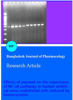
Figure 2: NF-kB p65 mRNA expression of HUVECs in each group by RT-PCR
Total RNA was extracted in HUVECs treated with different doses of paeonol for 48 hours and RT-PCR analysis was performed using specific primers for NF-kB p65 and beta-actin. △p<0.01 versus normal control group; ☆p<0.05, *p<0.01 versus homocysteine injured model group; (Upper) 1: normal control group; 2: homocysteine injured model group; 3: low-dose group of paeonol; 4: middle-dose group of paeonol; 5: high-dose group of paeonol; (Lower) M: Marker; Normal: normal control group; Model: Homocysteine injured model group; L-Pae: low-dose group of paeonol; M-Pae: middle-dose group of paeonol; H-Pae: high-dose group of paeonol
Effects of paeonol and homocysteine on IkB-alpha protein expression
As shown in Figure 2, compared with normal control group, IkB-alpha protein expression in homocysteine injured model group decreased significantly (p<0.01). IkB-alpha protein expression in the low-dose, the middle-dose and the high-dose group of paeonol markedly increased than that of homocysteine model group (p<0.01) (Figure 3).
Figure 3: IκB-α protein expression of HUVECs in each group by western blotting
Total proteins were extracted in HUVECs treated with different doses of paeonol for 48 hours. Protein levels were detected by western blot analysis. The blots were quantified by using densitometry, and represented by the ratio of beta-actin. Compared with normal control group, △p<0.01; Compared with homocysteine injured model group; *p<0.01; 1: normal control group; 2: homocysteine injured model group; 3: low-dose group of paeonol; 4: middle-dose group of paeonol; 5: high-dose group of paeonol; Normal: normal control group; Model: homocysteine injured model group; L-Pae: low-dose group of paeonol; M-Pae: middle-dose group of paeonol; H-Pae: high-dose group of paeonol
Effects of paeonol and homocysteine on ICAM-1 protein expression
Compared with nomal control group, ICAM-1 protein expression in homocysteine injured model group increased significantly (p<0.01). ICAM-1 protein expression in the low-dose, the middle-dose and the high dose group of paeonol was remarkably lower than that of homocysteine injured model group (p<0.05, p<0.01) (Figure 4).
Figure 4: ICAM-1 protein expression of HUVECs in each group by western blotting
Total proteins were extracted in HUVECs treated with different doses of paeonol for 48 hours. Protein levels were detected by western blot analysis. The blots were quantified by using densitometry, and represented by the ratio of beta-actin. Compared with normal control group, △p<0.01; Compared with homocysteine injured model group; *p<0.01; 1: normal control group; 2: homocysteine injured model group; 3: low-dose group of paeonol; 4: middle-dose group of paeonol; 5: high-dose group of paeonol; Normal: normal control group; Model: homocysteine injured model group; L-Pae: low-dose group of paeonol; M-Pae: middle-dose group of paeonol; H-Pae: high-dose group of paeonol
Effects of paeonol and homocysteine on the change of NF-kB p65 nuclear translocation by immunohistochemistry in HUVECs
NF-kB p65 expression products were brown. In the normal control group, there was almost no staining in the cells. The brown staining was located in nucleus and cytoplasm in homocysteine injured model group. Compared with the normal control group, the difference was significant in homocysteine injured model group (p<0.01). In paeonol low-dose group, there was much positivity in the nucleus, and the positivity in the nucleus in paeonol middle-dose group decreased significantly. Furthermore, there were almost no buffy positivity in the nucleus in paeonol high-dose group. Compared with homocysteine injured model group, the NF-kB p65 positivity in the nucleus was significantly reduced in Pae dose groups (p<0.01) (Figure 5).
Figure 5: Expression of NF-kB p65 in HUVECs by immunohistochemistry assay x400
NF-kB p65 expression products were brown. Compared with normal control group, △p<0.01; Compared with homocysteine injured model group, *p<0.01; A: negative control group; B: normal control group; C: homocysteine injured model group; D: low-dose group of paeonol; E: middle-dose group of paeonol; F; high-dose group of paeonol; Normal: normal control group; Model: homocysteine injured model group; L-Pae: low-dose group of paeonol; M-Pae: middle-dose group of paeonol; H-Pae: high-dose group of paeonol
Discussion
Homocysteine is a thiol containing amino acid derived when the essential amino acid methionine is metabolized to cysteine. The normal level of plasma total homocysteine concentrations is about 15 umol/L. The total homocysteine concentration (>12-50 umol/L) was considered as mild hyperhomocysteinemia. The total homocysteine concentration (>50-100 umol/L) was considered as moderately elevated hyperhomocysteinemia. The total homocysteine concentration (>100 umol/L) was considered as hyperhomocysteinemia (Zhou and Austin, 2009). Homocysteine has been proposed as a risk factor for atherosclerosis. How do hyperhomocysteine contribute to atherosclerosis remain unclear, however studies have demonstrated that endothelial cell dysfunction is one of the initiating events in the process of atherosclerosis. The inflammatory nature of atherosclerosis was previously described by Carl von Rokitanskyn and by Rudolf Virchow. The transcription nuclear factor-kB (NF-kB) plays a key role in inflammation (Frostegard, 2010).
NF-kB is a dimeric transcription factor formed by the Rel family of proteins. It is composed of p50 and p65 members (Sandur et al., 2007). Under most basal conditions, NF-kB complexes are maintained in an inactive form primarily through interactions with the inhibitor of kB (IkB) family of proteins in the cytoplasm. The activation of NF-kB involves the phosphorylation, ubiquitination, and degradation of IkB-alpha and phosphorylation of p65, which leads to the release of the NF-kB complexes from their inhibitory interaction. The released complexes then accumulate in the nucleus, where they bind to target DNA sequences and regulate the expression of a number of genes such as adhesion molecules, growth factors (Donato et al., 2008). NF-kB complexes activate the target genes, and then up-regulate the expression of adhesion molecules such as ICAM-1 and chemotactic proteins in blood vessel endothelium. Monocytes adhere to the endothelial cell surface due to the exaggerated production, and monocytes transmigrate through the endothelial monolayer into the intima, and monocytes become macrophages, foam cells in the end, which resulting in the pathogenesis of atherosclerosis (Wang and Li, 2009).
Paeonol is a traditional Chinese herb and it has been shown to have many pharmacological activities, such as antiatherosclerotic, antithrombotic and antiarrhythmic activity. The mechanism of its antiatherosclerotic may be protective effect on vascular endothelial cells (Wang and Wang, 2002). It was reported that feeding quails on paeonol had positive effect on experimental arteriosclerosis (Zhao and Li, 2005).
In this study, We used pathology model of vascular endothelial cells induced by 10 mmol/L homocysteine for antiatherosclerotic research. We have shown that the cell viability decreased obviously in homocysteine injured model group, and NF-kB p65 mRNA expression increased significantly, and the expression of IkB-alpha was decreased, and a great deal of NF-kB p65 translocated to the nucleus, furthermore, the expression of ICAM-1 Protein was increased largely. However, after treatment with different concentrations of paeonol, the cell viability was improved, NF-kB p65 mRNA expression was inhibited largely, IkB-alpha protein degradation was suppressed significantly, staining of NF-kB p65 in the nucleus was decreased significantly, a lot of NF-kB p65 translocated to cytoplasm, and the level of ICAM-1 protein was reduced.
Homocysteine can contribute to IkB-alpha protein degradation and activate NF-kB so that it can up-regulate NF-kB target genes such as adhesion molecules and chemotactic proteins. So, our research infers homocysteine can promote the initiation and progression of atherosclerosis. After all, paeonol can inhibit IkB-alpha protein degradation and suppress NF-kB transferred into nuclear in order to inhibit the activation of NF-kB. Thus, paeonol can reduce NF-kB target genes such as adhesion molecules and chemotactic proteins overexpression. It is possible that paeonol eventually slow down and reverse the formation and development of atherosclerosis.
Conclusion
Paeonol inhibits the expression of NF-kB mRNA in endothelial cells injured by homocysteine, and then paeonol suppresses the activation of NF-kB. Paeonol down-regulates the expression of ICAM-1 protein.
References
Donato AJ, Black AD, Jablonski KL, Gano LB, Seals DR. Aging is associated with greater nuclear NF κB reduced Iκ Bα , and increased expression of proinflammatory cytokines in vascular endothelial cells of healthy humans. Aging Cell. 2008; 7: 805-12.
Frostegard J. Rheumatic diseases insights into inflammation and atherosclerosis. Arterioscler Thromb Vasc Biol. 2010; 30: 892-93.
Held C, Sumner G, Sheridan P, McQueen M, Smith S, Dagenais G, Yusuf S, Lonn E. Correlations between plasma homocysteine and folate concentrations and carotid atherosclerosis in high-risk individuals: Baseline data from the Homocysteine and Atherosclerosis Reduction Trial (HART). Vascular Med. 2008; 13: 245-53.
Jaffe EA, Nachman RL, Becker CG, Minick CR. Culture of human endothelial cells derived from umbilical veins. Identification by morphologic and immunologic criteria. J Clin Invest. 1973; 52: 2745-56.
Sandur SK, Ahn KS, Ichikawa H, Sethi G; Shishodia, S, Newman, RA, Aggarwal, BB. Zyflamend, a polyherbal preparation, inhibits invasion, suppresses osteoclastogenesis, and potentiates apoptosis through down-regulation of NF-κB activation and NF-κB-regulated gene products. Nutr Cancer. 2007; 57: 78-87.
Wang Q, Wang H. Natural medicines and their monomers with antiatherosclerosis effects. World Sci Technol. 2002; 4: 52-58.
Wang X, Li WJ. A study on the correlation of NF-κB with atherosclerosis and interference effect of Chinese. Med J Liaoning Univ Traditional Chinese Med. 2009; 11: 55-56.
Ye JM, Deng T, Zhang JB. Influence of paeonol on expression of COX-2 and p27 in HT-29 cells. World J Gastroenterol. 2009; 15: 4410-14.
Zhao L, Li Q. The research on pharmacological action of paeonol in the cardiovascular system. Anhui Med Pharmaceut J. 2005, 9: 725-26.
Zhao X, Xu XJ, Li B, Yang J. Protective effect of paeonol on experimental hyperlipidemia in rats. J Chinese Med Materials. 2010; 33: 1312-14.
Zhou J, Austin RC. Contributions of hyperhomocysteinemia to atherosclerosis: Causal relationship and potential mechanisms. Biofactors 2009; 35: 120-29.