Cyanobacterial phycoerythrin purified from marine Lyngbya sp. induces apoptosis in lung carcinoma cells
Abstract
Phycoerythrins from various cyanobacterial sources have been extensively studied globally for their therapeutic potentials. This study shows the apoptotic potential of cyanobacterial phycoerythrin isolated, purified and characterized from marine Lyngbya sp. A09DM on human lung carcinoma cells (A549 cell). Results indicated that purified cyanobacterial phycoerythrin accounted for a dose-dependent decrement in cell viability, mitochondrial membrane potential and an increment in lactate dehydrogenase release. Higher dose of cyanobacterial phycoerythrin caused loss of cell viability and nuclear condensation/fragmentation. Flow cytometry accounted for more number of cells in G0/G1 phase and an increase in total number of early apoptotic cells (39.5%). Overall, this study is the first to report on apoptotic property of cyanobacterial phycoerythrin from marine Lyngbya sp. A09DM against A549 human lung carcinoma cells and requires a detailed scrutiny to establish its anti-cancer potential.
Introduction
Lung cancer accounts for about 17% of the newly reported cases of cancer and 23% of the total cancer deaths that occur annually primarily due to nicotine intake and various occupational hazards (Report on carcinogenesis, 2011). Extensive use of chemotherapy for alleviating symptoms and for treating lung cancer also involves undesired physiological effects. In recent times, metal complexes (Singh et al., 2012; Vyas et al., 2013; Vyas et al., 2014; Ramadevi et al., 2015; Thakore et al., 2015), herbal extracts (Sankhari et al., 2012; Patel et al., 2012), structured nano sized particles (Valodkar et al., 2011; Valodkar et al., 2012) or novel proteins from bacterial or fungal origins (Huang et al., 2002) have been extensively researched as a therapeutic option.
Phycobiliproteins are light-harvesting accessory pigments with immunomodulatory, anti-inflammatory, antioxidant and nutritional properties (Belay, 2002; Rodriguez-Sanchez et al., 2012). Cyanobacterial phycoerythrin (absorption- 520 to 580 nm and emission peak at 575 ± 10 nm) is used as a dietary supplement (Romay et al., 2003) that also inhibits growth of tumor cells (Huang et al., 2002; Pan et al., 2013; Madamwar et al., 2015). However, cytotoxic property of proteins from a marine cyanobacteria Lyngbya sp. against carcinoma cells has so far not been reported.
This study investigates the impact of cyanobacterial phycoerythrin on A549 lung carcinoma cells wherein, alterations in cell morphology, cell cycle and possible induction of apoptosis has been scrutinized in detail.
Materials and Methods
Chemicals
Sephadex G-150 powdered matrix (bead-diameter 20-300 um and fractionation range 5-300 kDa) was purchased from GE Healthcare UK Limited (UK), 2, 4, 6-tripyridyl-s-triazine and ferric chloride (hexahydrate) from Sigma, USA; protein molecular mass standard from Bangalore Genei (India); sodium dodecyl sulfate, electrophoresis grade acrylamide and bisacrylamide from Merck (Germany). Phosphate buffer saline, Dulbeco's Modified Eagle Medium, trypsin phosphate versene glucose solution, trypsin and methylthiazolyl-diphenyltetrazolium bromide (MTT) were purchased from HiMedia Laboratories Pvt. Ltd. (India). Fetal bovine serum was purchased from Biosera (UK) and dimethyl sulfoxide was purchased from the Sisco Research Laboratories Pvt. Ltd. (India).
Isolation and growth condition
The enrichment, isolation and purification of the cyanobacterial cell culture were carried out according to Shah et al. (2001). Samples were isolated from the rocky shores of Bet Dwarka, Okha and estuarine mouth of river Daman Ganga and further cultivated in ASN III medium (Waterbury and Stanier, 1981) with 12:12 light:dark (L:D) cycles at 27 ± 2ºC and illumination of 36 W white fluorescent lamps at a flux density of 130 umol photons.m-2.sec-1. Identification by 16S-rRNA gene sequencing showed that the isolated strain was Lyngbya sp. A09DM (accession number: HM446280).
Extraction
Marine Lyngbya sp. A09DM culture was harvested within 30 days of inoculation (Soni et al., 2006). Cyanobacterial cell mass was washed with 1 M Tris Cl buffer (pH 8.1) containing 3 mM sodium azide. One volume of washed cell mass was resuspended in five volumes of the same buffer and subjected to repeat freeze-thaw cycles of -30ºC and 4ºC shocks for the release of phycobiliprotein. The cell debris was removed by centrifugation at 17,000 xg (Kubota 6500, Bunkyo-Ku, Tokyo, Japan) for 40 min and the supernatant was termed as crude extract.
Purification
Steps of purification were carried out as described by Parmar et al., (2011).
Ammonium sulfate precipitation
The crude extract was subjected to two step ammonium sulfate precipitation (20% and 70%) followed by centrifugation at 15,000 xg for 40 min at 4ºC. The supernatant were discarded and the pellet was resuspended in 10 mM Tris Cl buffer (pH 8.1). The extracts, thus, obtained were termed as 70% ammonium sulfate cut that was further subjected to dialysis for 24 hours against 100 times volume of MilliQ water containing 3 mM sodium azide.
Gel permeation chromatography
Dialysed 70% ammonium sulfate cut was applied on a sephadex G-150 column (45 cm x 1.5 cm, bed height 35 cm) pre-equilibrated and eluted with 10 mM Tris Cl buffer (pH 8.1). The flow rate was maintained at 60 mL/hour using peristaltic pump (Model P1; Pharmacia, Sweden). Bright pink color elutes were collected as 1 mL fractions.
Biochemical estimations and characterization of protein
The protein contents were determined by the method of Lowry et al., (1951) with bovine serum albumin as the standard. Phycoerythrin from all the three steps– crude, dialyzed 70% ammonium sulfate cut and gel permeation chromatography elutes were characterized by UV-Visible spectrophotometer (Analytik Jena AG Specord® 210, Germany). The amount of cyanobacterial phycoerythrin, cyanobacterial phycocyanin, and cyanobacterial allophycocyanin was calculated according to equations of Bennett and Bogorad (1973). The ferric reducing ability of plasma assay was performed according to Benzie and Strain (1996). A fresh cocktail solution was prepared by mixing solution A (300 µM acetate buffer- pH 3.6), solution B (10 mM TPTZ 2, 4, 6-tripyridyl-s-triazine prepared in 40 mM hydrochloric acid) and solution C (20 mM ferric chloride hexahydrate) at a ratio of 10:1:1. Later, 30 µL sample and 90 µL distilled water was added to 900 µL of cocktail solution and the mixture was incubated at 37°C for 4 min. Absorbance was read at 593 nm and was recorded against a reagent blank. The purified phycoerythrin was also characterized in terms of the molecular masses of the subunits. Non-denaturing and denaturing polyacrylamide gel electrophoresis was carried out using 15% polyacrylamide slab gels (Singh et al., 2009) and the same was visualized by silver staining (Garfin, 1990). For calibration, marker proteins ranging from 6 kDa to 43 kDa (aprotinin- 6 kDa, lysozyme- 14.3 kDa, soyabean trypsin inhibitor- 20.1 kDa, carbonic anhydrase- 29 kDa and ovalbumin- 43 kDa) were used. Bilin chromophore was detected using zinc-acetate staining as per Brekelman and Lagarias (1986), and the bilin fluorescence was observed under UV light using AlphaEase FC Imaging System (Alpha Innotech Corp., USA).
Cell line and culture
Human lung carcinoma (A549) cells were obtained from National Centre for Cell Science, Pune, India and were seeded (1 x 10⁵ cells/T25 flask) and cultured in Dulbeco's Modified Eagle Medium containing 10% fetal bovine serum and 1% antibiotic-antimycotic solution at 37°C with 5% in water jacketed CO2 incubator (Thermo scientific, forma series II 3111, USA). Cells were sub-cultured every third day by trypsinization with trypsin phosphate versene glucose solution. Reagents used herein were filtered through 0.22 um filter (Millipore Biomedical Aids Pvt. Ltd, Pune) prior to their use for the experiment. A549 cells were maintained for a period of 24 hours in absence or presence of phycoerythrin at a cell density of 5.0 x 10³ cells/well in 96 well plate for MTT and LDH assay and 1 x 10⁵ cells/well in 6 well plate for other assays mentioned herein.
Cell viability assay
A549 cells (7 x 10³ cells/well) were maintained in 96-well culture plates (Tarson India Pvt. Ltd.) for 24 hours with or without phycoerythrin (10, 25, 50, 100, 200 ug/mL). Later, 10 uL of 3-(4,5-dimethylthiazol-2-yl)-2,5-diphenyltetrazolium bromide (MTT, 5 mg/mL) was added to the wells and plates were incubated at 37°C for 4 hours. Subsequently, the culture media were discarded and wells were washed with phosphate buffered saline. The resultant formazan formed was dissolved in 150 uL of DMSO and absorbance was read at 540 nm in BIOTEK ELX800 Universal Microplate Reader (USA).
LDH release assay
Cellular integrity of A549 cells was measured with LDH release assay, wherein cells were maintained in 96 well plates with or without phycoerythrin for 24 hours as mentioned above. Later, supernatant from each well was collected and LDH was assayed with commercially available kit (Reckon diagnostics Ltd., India) on Merck microlab L 300 semi-autoanalyzer (India). Based upon the readings obtained, percentage cytotoxicity was calculated.
Intracellular oxidative stress
Intracelluar oxidative stress due to reactive oxygen species generation in A549 cells was studied using 2,7-dichlorodihydrofluoroscein diacetate (CM-H2-DCFDA) staining. Cells were treated with cyanobacterial phycoerythrin for 18 hours as per the method mentioned above. Later, the cells were incubated with 7.5 μM DCFDA at 37°C for 30 min and cells were photographed in Leica DMRB fluorescent microscope (Germany) using Canon Power Shot camera.
Mitochondrial membrane potential
The changes in mitochondrial membrane potential were measured using the fluorescent cationic dye rhodamine 123 (RHO 123). A549 cells were incubated for 24 hours with or without cyanobacterial phycoerythrin as mentioned above. Later, cells were incubated with 1 uM RHO 123 for 10 min at 37°C and the fluorescence was determined at an excitation and emission wavelengths of 485 and 530 nm, respectively using spectroflurometer (Jasco FP-6300, Japan) and expressed as fluorescence intensity units.
Assessment of cyanobacterial phycoerythrin-induced cell cycle analysis and apoptosis
Assessment of nuclear morphology was done using DAPI staining. A549 cells (5 x 10⁴ cells/well) were plated into 6-well plate. After 80% confluence was achieved, the cells were treated with or without different concentrations of cyanobacterial phycoerythrin at 37°C for 24 hours. Single-cell suspensions of treated cells were washed with phosphate buffered saline and fixed with 70% ethanol for 20 min at room temperature. Cells were re-washed with phosphate buffered saline and stained with DAPI (0.6 ug/mL in phosphate buffered saline) for 5 min. Nuclear morphology (condensed/fragmented nuclei) was examined under Leica DMRB fluorescence microscope (Germany).
Apoptotic cells were studied using AO/EB staining. A549 cells (1 x 10⁵ cells/well) were maintained in 6 well plates for 24 hours with or without cyanobacterial phycoerythrin as described earlier. At the end of experiment period, cells were collected using TPVG solution. One microliter of dye mixture (1 mg/mL AO and 1 mg/mL EB in phosphate buffered saline) was mixed with 9 uL of cell suspension (0.5 x 10⁶ cells/mL) on a clean microscope slide and examined and photographed under Leica DMRB fluorescence microscope (Germany). A minimum of 300 cells were counted in every sample to calculate percentage cell death.
Cells (1 x 10⁶ cells/well) were cultured with or without phycoerythrin as mentioned earlier for 24 hours. After incubation, the cells were washed once in ice-cold phosphate buffered saline and subjected to cell cycle analysis. Briefly, 1 x 10⁵ cells were fixed in 4.5 mL of 70% (v/v) cold ethanol for 30 min, centrifuged at 400 xg for 5 min. Supernatant was removed and cells were washed with 5 mL of phosphate buffered saline. Cells were then re-suspended in 0.5 mL of phosphate buffered saline and 0.5 mL of DNA extraction buffer (Mix 192 mL of 0.2 M Na2HPO4 with 8 mL of 0.1% Triton X-100 v/v) was added. The pH was adjusted to 7.8. Cells were incubated at room temperature for 5 min and then centrifuged at 400 xg for 5 min. Supernatant was discarded and cells were re-suspended in 1 mL of DNA staining solution (200 mg of PI in 10 mL of phosphate buffered saline + 2 mg of DNase free RNase). Cells are then incubated for at least 30 min at room temperature in the dark and the cell cycle distribution was then analyzed on a flow cytometer (BD FACS Aria III, USA) using FlowJo (Oregon, USA).
Annexin V/propidium iodide double staining assay was used to quantify apoptosis, according to the manufacturer's protocol (Alexa Fluor 488 annexin V/dead cell apoptosis kit, Invitrogen). After incubation, cells were harvested using TPVG solution and washed with ice-cold phosphate buffered saline and suspended in 100 uL of 1x binding buffer (10 mM HEPES, 140 mM NaCl, and 2.5 mM CaCl2, pH 7.4). To this mixture, 5 uL of annexin V- Alexa Fluor 488 conjugate and 1 uL of propidium iodide solution were added to each cell suspension and incubated for 15 min at room temperature in the dark. Samples were analyzed on flow cytometer (BD FACSAria III, USA) using FlowJo (Oregon, USA). Double staining of cells with Alexa Fluor 488 annexin-V and propidium iodide enables the discrimination of live cells (Alexa Fluor 488-PI-), early apoptotic (Alexa Fluor 488+PI-), late apoptotic (Alexa Fluor 488+PI+) or necrotic cells (Alexa Fluor 488-PI+).
Statistical analysis
Data was analyzed for statistical significance using one way analysis of variance (ANOVA) followed by Bonferroni's multiple comparison test and results were expressed as mean ± SEM using Graph Pad Prism version 3.0 for Windows, Graph Pad Software, San Diego, California, USA.
Results
Precipitation of cyanobacterial phycoerythrin could not be achieved in first step because the supernatant of 20% ammonium sulfate cut in 70% ammonium sulfate was not efficient for its extraction. Hence, in the second step, the contents were dialyzed overnight and precipitate of cyanobacterial phycoerythrin with a purity ratio of 1.66 was obtained. In the final step, fractionation of the resultant cyanobacterial phycoerythrin was done with sephadex G-150 to achieve a purity ratio of 3.67 and a final yield of 65.2%. The purity of cyanobacterial phycoerythrin was monitored at each step by UV-Visible spectroscopy and gel electrophoresis. The purity ratio was monitored at each step by overlay spectra using UV-Visible spectroscopy. The absorbance maxima (λmax) for phycoerythrin was recorded at 562 nm. The purity ratios of phycoerythrin to total protein (A562/A280), phycoerythrin to phycocyanin (A562/A620), and phycoerythrin to allophycocyanin (A562/A650) were 3.57, 4.09 and 11.41, respectively (Table I).
Table I: Antiemetic effect of crude extract of J. gossypifolia on copper sulfate and Brasica juice-induced emetic chicks
| Treated groups | Copper sulfate-induced | Brasica-induced | ||
|---|---|---|---|---|
| Treated groups | Mean number of retches | %Inhibition | Mean number of retches | %Inhibition |
| Control (10 mL/kg) | 78 ± 1.1 | ____ | 77 ± 1.1 | ____ |
| Chlorpromazine (150 mg/kg) | 44 ± 0.3 | 43.6a | 42 ± 0.5 | 45.5a |
| Ethanolic extract of J. gossypifolia (75 mg/kg) | 36 ± 1.3 | 53.8a | 39 ± 1.2 | 49.4a |
| Ethanolic extract of J. gossypifolia (150 mg/kg) | 20 ± 0.8 | 74.4b | 18 ± 0.8 | 76.6b |
| (ap<0.05 and bp<0.005 vs control showing significant and most significant values by using student's t-test) | ||||
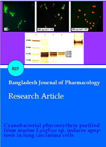
Non-denaturing gel electrophoresis of purified cyanobacterial phycoerythrin showed two bands corresponding to phycoerythrin-I and phycoerythrin-II, suggesting homogeneity of purified cyanobacterial phycoerythrin (Figure 1a). Denaturing gel electrophoresis of cyanobacterial phycoerythrin showed the presence of four bands having molecular masses of 23, 20, 18.5 and 18 kDa (Figure 1b). These bands were obtained in the last step of purification and corresponded to alpha and beta subunits of each of phycoerythrin-I and phycoerythrin-II. The presence of bilin chromophore group attached to the phycoerythrin subunit was confirmed by zinc-acetate staining that showed characteristic orange fluorescence under UV light. No fluorescence was observed in molecular mass standard due to the absence of bilin-linked peptides. These results also confirmed attachment of bilin chromophore groups to the subunits of phycoerythrin (Figure 1c).
Figure 1: Concentration-response curves, showing the effect of J. gossypifolia in the absence and presence of atropine (0.1 µM) on spontaneously contracting rabbit jejunum preparation. The spasmogenic responses are expressed as percent of acetylcholine maximum (values are expressed as mean ± SEM, n=3)
The antioxidant capacity of purified cyanobacterial phycoerythrin obtained herein was 48.45 µm AEAC as per the results obtained in FRAP assay. Cytotoxic potential of cyanobacterial phycoerythrin against lung carcinoma cell line is shown in (Figure 2a). Cells treated with cyanobacterial phycoerythrin showed a dose dependent inhibitory effect. Also, there was a significant dose dependent increment in LDH release following cyanobacterial phycoerythrin treatment as compared to untreated control cells (Figure 2b). Overall, a maximum cell growth inhibition (>80%) and LDH release was recorded at 200 μg/mL dose of cyanobacterial phycoerythrin.
Figure 2: Bar diagram showing the effect of J. gossypifolia in comparison to the acetylcholine maximum response in rabbit jejunum preparation
The fluorimetric analysis of cyanobacterial phycoerythrin treated samples recorded a dose-dependent decrement in MMP as compared to untreated group. The lowest MMP was recorded in 200 ug/mL dose of phycoerythrin (Figure 2c).
The apoptosis induced by phycobiliprotein in A549 cells was confirmed by AO/EtBr staining. The cells of untreated control group showed green fluorescence indicating viability whereas, these cells were progressively less in number in a dose-dependent manner in phycoerythrin-treated group. More number of yellow colored cells was present in 100 ug/mL phycoerythrin-treated group. The same indicates the early apoptotic stage. However, 200 ug/mL phycoerythrin-treated group showed more number of reddish or orange stained cells that symbolize late apoptotic phase (Figure 3a). DAPI staining phycoerythrin-treated cells showed higher number of cells with condensed nuclei in phycoerythrin-treated groups as compared to control group (Figure 3b). The cell cycle analysis (Figure 4a) showed 93.1 % in G0/G1 and 4.5% of cells in sub G0/G1 phase in control group whereas, 15% in G0/G1 and 83.3% of cells in sub G0/G1 phase in phycoerythrin-treated group. Flow cytometric assay with annexin V/propidium iodide double staining of control and phycoerythrin-treated cells showed that apoptosis rate was increased after phycoerythrin-treatment as they recorded 39.5% annexin V positive (early apoptotic) and 0.51% annexin V and propidium iodide positive cells (late apoptotic) compared to 0.9% and 0.6% in control group (Figure 4b).
Figure 3: Concentration-dependent inhibitory effect of J. gossypifolia and verapamil against K+(80 mM)-induced contractions in isolated rabbit jejunum preparations (values are expressed as mean ± SEM, n=3)
Figure 4: Concentration-response curves of Ca2+ in the absence and presence of different concentrations of (A) J. gossypifolia and (B) verapamil in isolated rabbit jejunum preparations (values are expressed as mean ± SEM, n=3)
Discussion
This study elaborates the method of purification of phycoerythrin from a novel isolate Lyngbya sp. A09DM and its subsequent cytotoxicity against A549 lung carcinoma cells. Culturing of Lyngbya sp. under white light can be considered as an efficient source for obtaining cyanobacterial phycoerythrin with a high purity ratio of 3.67. Also, the two-step ammonium sulfate precipitation method is equally efficient for purification of cyanobacterial phycoerythrin and these results are in agreement with Rito-Palomares et al., (2001). Cytotoxicity (MTT) assay of cells treated with cyanobacterial phycoerythrin indicated a dose-dependent impact on cell viability wherein, higher dose (200 ug/mL) was most potent and accounted for maximum cell growth inhibition (>80%). Transmembrane potential of mitochondria is an important parameter to assess mitochondrial function and overall health of cell (Patel et al., 2013; Gajaria et al., 2015). In the present study, a dose-dependent decrement in mitochondrial membrane potential was recorded following cyanobacterial phycoerythrin treatment suggesting possible mitochondrial dysfunction. These results provide supportive evidence on cyanobacterial phycoerythrin-induced cytotoxicity recorded in the MTT assay. Any damage to the plasma membrane of an animal cell causes leaching out of LDH from cytoplasm into the media and the same is measured as an indication of the extant of cell damage (Devkar et al., 2012). Hence, biochemical estimation of LDH is a reliable and popularly used parameter to assess cell damage. In our study, high levels of LDH are in support of the results obtained in cytotoxicity (MTT) assay.
Experiments with natural anticancer agents have reported that heightened intracellular oxidative stress also accounts for cell death by causing damage to the plasma membrane (Gajaria et al., 2015). But in our study, the control and treated cells showed comparable levels of weak fluorescence suggesting that the cyanobacterial phycoerythrin failed to initiate oxidative stress in A549 cells. At this stage, it was assumed that, cyanobacterial phycoerythrin induced cell death was possibly due to the trigger of apoptotic pathway. Inhibition of a tumor due to the arrest of cell division or cytotoxicity and subsequent induction of apoptotic cell death are key features for treating any type of cancer. The down-stream pathway of apoptotic cascade is reportedly triggered in presence of cytochrome c that leaches out from the transitional pores of mitochondrial membrane (Jiang and Wang, 2004). This event results due to an altered mitochondrial membrane potential as observed in our study. Also, presence of more number of EB positive cells (red/orange) observed herein, further corroborate the findings that cyanobacterial phycoerythrin is instrumental in inducing cytotoxicity. As compared to the lower dose (100 ug/mL), the higher dose (200 ug/mL) showed qualitative evidence of higher extent of cell damage in form nuclear fragmentation with possible indications of an early apoptosis (Figure 4a). Chromatin condensation, nuclear fragmentation, nuclear shrinkage and formation of apoptotic bodies are characteristic features of apoptosis (Elmore, 2007). Hence, higher dose was chosen for further cell cycle analysis. Flow cytometric analysis revealed that, cyanobacterial phycoerythrin-treated cells showed cell arrest in sub G0/G1 phase (83.3%) as compared to the control. Further confirmation of cyanobacterial phycoerythrin-induced apoptosis was obtained with annexin V-propidium iodide staining wherein, a large number of early apoptotic (39.5%) and only few late apoptotic (0.51%) cells were observed. These observations provide compelling evidence on cyanobacterial phycoerythrin induced apoptosis of A549 cells.
Conclusion
Cyanobacterial phycoerythrin induced altered mito chondrial membrane potential triggers apoptosis in human lung carcinoma cells. This study is the first to report on apoptotic potential of cyanobacterial phycoerythrin from marine Lyngbya sp. A09DM and the findings warrant a detailed scrutiny on its efficacy as an anticancer agent.
Acknowledgement
RVD acknowledges MSU-DBT-ILSPARE for infrastructural and technical support.
References
Belay A. The potential application of spirulina (arthrospira) as a nutritional and therapeutic supplement in health management. J Am Nutraceutl Assoc. 2002; 5: 26-48.
Bennet A, Bogorad L. Complementary chromatic adaptation in a filamentous blue-green algae. J Cell Biol. 1973; 58: 419–35.
Benzie IFF, Strain JJ. The ferric reducing ability of plasma (FRAP) as a measure of ‘‘antioxidant power’’: The FRAP assay. Anal Biochem. 1996; 239: 70–76.
Brekelman TR, Lagariaz JC. Visualization of bilin-linked peptides and proteins in polyacrylamide gels. Anal Biochem. 1986; 156: 194–201.
Devkar RV, Pandya AV, Shah NH. Protective role of Brassica olerecea and Eugenia jambolana extracts against Hâ‚‚Oâ‚‚ induced cytotoxicity in H9C2 cells. Food Function. 2012; 3: 37-43.
Elmore S. Apoptosis: A review of programmed cell death. Toxicol Pathol. 2007; 35: 495–516.
Gajaria TK, Patel DK, Devkar RV, Ramachandran AV. Flavonoid rich extract of Murraya Koenigii alleviates in-vitro LDL oxidation and oxidized LDL induced apoptosis in raw 264.7 Murine macrophage cells. J Food Sci Technol. 2015; 52: 3367-75.
Garfin D. One-dimensional gel electrophoresis. In: Guide to protein purification. Deutscher MP, Abelson JN, Simon MI, (eds). California, Academic Press, 1990, pp 425-41.
Huang B, Wang GC, Zeng CK, Li ZG. Experimental research of R-phycoerythrin subunits on cancer treatment: A new photosensitizer in PDT. Cancer Biother Radio. 2002; 17: 35-42.
Jiang X, Wang X. Cytochrome C-mediated apoptosis. Annu Rev Biochem. 2004; 73: 87-106.
Lowry HO, Rosenbrough NJ, Farr AL, Randall RJ. Protein measurement with the folin phenol reagent. J Biol Chem. 1951; 193: 265–75.
Madamwar D, Patel DK, Desai SN, Upadhyay KK, Devkar RV. Apoptotic potential of c-phycoerythrin from phormidium sp. A27DM and halomicronema sp. A32DM on human lung carcinoma cells. EXCLI J. 2015; 14: 527-39.
Pan Q, Chen M, Li J, Yan Wu, Zhen C, Liang B. Antitumor function and mechanism of phycoerythrin from Porphyra haitanensis. Biol Res. 2013; 46: 87-95.
Parmar A, Singh NK, Kaushal A, Sonawala S, Madamwar D. Purification, characterization and comparison of phycoerythrins from three different marine cyanobacterial cultures. Bioresource Technol. 2011; 102: 1795–802.
Patel D, Desai SN, Gajaria TK, Devkar R, Ramachandran AV. Coriandrum sativum L. seed extract mitigates lipotoxicity in raw 264.7 cells and prevents atherogenic changes in rats. EXCLI J. 2013;12: 313-34.
Patel DK, Desai SN, Gandhi HP, Devkar RV, Ramachandran AV. Cardio protective effect of Coriandrum sativum L. on isoproterenol induced myocardial necrosis in rats. Food Chem Toxicol. 2012; 50: 3120-25.
Ramadevi P, Singh R, Jana SS, Devkar R, Chakraborty D. Ruthenium complexes of ferrocene mannich bases: DNA/BSA interactions and cytotoxicity against A549 cell line. J Photoch Photobio A. 2015; 305: 1-10.
Report on carcinogens. US Department of Health and Human Services, Public Health Service. National Toxicology Program, 2011, 12 iii-499.
Rito-Palomares M, Nunez L, Amador D. Practical application of aqueous two-phase systems for the development of a prototype process for c-phycocyanin recovery from Spirulina maxima. J Chem Technol Biot. 2001; 76: 1273-80.
RodrÃguez-Sánchez R, Ortiz-Butrón R, Blas-Valdivia V, Hernández-GarcÃa, A, Cano-Europa E. Phycobiliproteins or C-phycocyanin of Arthrospira (spirulina) maxima protect against HgCl2-caused oxidative stress and renal damage. Food Chem. 2012; 135: 2359–65.
Romay CH, González R, Ledón N, Remirez D, Rimbau V. C-phycocyanin: A biliprotein with antioxidant, anti-inflammatory and neuroprotective effects. Curr Protein Pept Sc. 2003; 4: 207-16
Sankhari JM, Thounaojam MC, Jadeja RN, Devkar RV, Ramachandran AV. Anthocyanin-rich red cabbage (Brassica oleracea L.) extract attenuates cardiac and hepatic oxidative stress in rats fed an atherogenic diet. J Sci Food Agric. 2012; 92: 1688-93.
Schmidt M, Bastians H. Mitotic drug targets and the development of novel anti-mitotic anticancer drugs. Drug Resist Update. 2007; 10: 162–81.
Shah V, Garg N, Madamwar D. Record of the marine cyanobacterium from the rocky shores of Bet-Dwarka and Okha, India. Acta Bot Malacit. 2001; 26: 188–93.
Singh NK, Parmar A, Madamwar D. Optimization of medium components for increased production of C-phycocyanin from Phormidium ceylanicum and its purification by single step process. Bioresource Technol. 2009; 100: 1663–69.
Singh R, Jadeja RN, Thounaojam MC, Devkar RV, Chakraborty D. Synthesis characterization DNA binding and cytotoxicity studies of moxifloxacinato complexes. Transit Metal Chem. 2012; 37: 541-51.
Soni B, Kalavadia B, Trivedi U, Madamwar D. Extraction, purification and characterization of phycocyanin from Oscillatoria quadripunctulata: Isolated from the rocky shores of Bet-Dwarka, Gujarat, India. Process Biochem. 2006; 41: 2017–23.
Thakore SI, Nagar PS, Jadeja RN, Thounaojam M, Devkar RV, Rathore PS. Sapota fruit latex mediated synthesis of Ag, Cu mono and bimetallic nanoparticles and their in vitro toxicity studies. Arab J Chem. 2015.
Valodkar M, Jadeja RN, Thounaojam MC, Devkar RV, Thakore S. In vitro toxicity study of plant latex capped silver nanoparticles in human lung carcinoma cells. Mater Sci Eng: C. 2011; 31: 1723-28.
Valodkar M, Rathore PS, Jadeja RN, Devkar RV, Thakore S. Cytotoxicity evaluation and antimicrobial studies of starch capped water soluble copper nanoparticles. J Hazard Mater. 2012; 201: 244-49.
Vyas KM, Jadeja RN, Patel D, Devkar RV, Gupta VK. Effect of ligand substitution in pyrazolone based binary and ternary Cu(II) complexes on DNA binding, protein binding and anticancer activity on A549 lung carcinoma cell lines. Polyhedron 2014; 80: 20-23.
Vyas KM, Jadeja RN, Patel D, Devkar RV, Gupta VK. A new pyrazolone based ternary Cu (II) complex: Synthesis, characterization, crystal structure, DNA binding, protein binding and anticancer activity towards A549 human lung carcinoma cells with a minimum cytotoxicity to non-cancerous cells. Polyhedron 2013; 65: 262-74.
Waterbury JB, Stanier RY. Isolation and growth of cyanobacteria from marine and hypersaline environments. In: The Prokaryotes. Starr MP Stolp H, Trüper HG, Balows A, Schlegel HG (eds). Berlin, Springer-Verlag, 1981, pp 221-23.