Evaluation of anticonvulsant, antimicrobial and hemolytic activity of Aitchisonia rosea
Abstract
The purpose of this study was to evaluate the anticonvulsant, antimicrobial and hemolytic effect of Aitchisonia rosea. The anticonvulsant effect was studied at doses 400 and 800 mg/kg against pentylenetetrazole, strychnine and picrotoxin-induced seizures in albino mice. The antimicrobial assay was conducted by disc diffusion method and minimum inhibitory concentration. Hemolytic effect was analyzed by reported method. Phenolic compounds present in the n-butanol fraction of the plant were estimated by HPLC. The plant showed maximum response against drug-induced convulsions and provided protection to animals at both doses. It also showed maximum zone of inhibition and highly significant MIC against all bacterial and fungal strains. The plant protected the RBCs from hemolysis. The highest amount of phenolics found was caffeic acid (7.5 ± 0.04).
Introduction
Aitchisonia rosea Hemsl. ex Aitch belongs to plant family Rubiaceae. It is an important medicinal plant mostly distributed in hilly areas of Pakistan (Baluchistan) and Afghanistan. It is also found in Iran. The genus Aitchisonia has only one specie i.e. Aitchisonia rosea (Ali and Nasir, 1989). This plant has been collected by Aitchison in 1882 (Mozaffarian, 2006).
In some areas of Baluchistan, this plant is used for the cure of some infectious skin diseases. Two glycosides of iridoid nature, Aitchisonides A and B were isolated and identified from the n-butanol fraction of A. rosea. Deacetylasperulosidic acid and nepetanudoside B were also separated along with these two glycosides. These compounds were isolated from this plant for the first time (Noor et al., 2009a). Anthraquinone derivatives named Rosenones A and B were first time isolated and identified from ethyl acetate fraction of A. rosea by using liquid column chromatography monitored by prepared thin layer chromatography. Another compound 1,3,6-trihydroxy-2-methylanthraquinone was separated from this plant (Noor et al., 2009b).
It is revealed from literature survey that pharmacological studies on A. rosea have not been documented till to date. In this paper, we for the first time reported the pharmacological, biological and safety profile of this plant which might be utilized to standardize the plant for therapeutic purposes. The plant was also estimated for the presence of phenolic constituents by HPLC that might be responsible for these pharmacological activities.
Materials and Methods
Collection and preparation of plant material
A. rosea was collected from the hilly areas of Quetta, Balochistan, Pakistan. The specimen was further identified and authenticated by Taxonomists, Prof. Rasool Bakhsh Tareen, Department of Botany, University of Baluchistan, Quetta and Prof. Zaheer Ahmad Khan, Department of Botany, GC University, Lahore, Pakistan. Voucher specimen number 1911 was deposited in the Sultan Ayoub Herbarium, GC University Lahore for further reference. Stems and branches of the plant were spread on laboratory tables and dried under the shade for seven days at room temperature. The plant was pulverized prior to use by using an electric mill. The powdered plant material was extracted thrice with methanol by dipping for seven days. The methanol extract was concentrated to dryness under reduced pressure using a rotary evaporator (Heidolph, model Laborata 4000, Germany). The methanol extract was further fractioned using a successive solvent extraction method with different polarity based solvents such as n-hexane, chloroform, ethyl acetate and n-butanol. After fractionation samples were concentrated to dryness under reduced pressure and stored in a refrigerator at 4°C, until used for analysis (Brain and Turner, 1975).
Preparation of test microorganism
The pathogenic bacterial strains namely Bacillus subtilis (ATCC 6633), Pseudomonas aeruginosa (ATCC 27853) and Staphylococcus aureus (ATCC 12228); and fungal strains namely Candida albicans (ATCC 18804) and Penicillium notatum (ATCC 11709) were obtained from Musaji Adam and Sons, Rawalpindi, Pakistan and used for antibacterial and antifungal activity respectively. All the microbial strains were transferred to nutrient agar slants then shifted to nutrient broth and stored at 4°C until used.
Experimental animals
Healthy adult male/female Swiss albino mice (22-30 g) were purchased from the local market and used for experimental purposes. The animals were housed in metabolic animal cages and maintained at controlled room temperature of 25 ± 10°C. All the animals had free access to food and water ad libitum.
Phytochemical screening
Different chemical tests were conducted, using reported methods, to check out the presence of various phytoconstituents from methanol extract and its fractions (Edeoga et al., 2005; Kokate et al., 2007; United State Pharmacopoeia, 1980).
Anticonvulsant activity
The anticonvulsant effect of plant crude extract and its various fractions was performed according to the method with some modifications as described by Swinyard et al. (1989). Swiss albino mice of either sex were divided into 12 groups of 6 mice each. All the animals were fasted for 24 hours before the start of the experiment. The plant methanol extract and its fractions were given at a dose of 400 and 800 mg/kg. Standard drug diazepam (4 mg/kg) and vehicle control (80 mg/kg) were administered 30 min prior to convulsions inducing drugs i.e. pentylenetetrazole (PTZ), strychnine and picrotoxin. After administration of plant extract and fractions, following parameters were observed: a) latency (onset of clonus), b) duration of tonic convulsions, c) status of animal after 30 min, d) status of animal after 24 hours and e) percentage protection
Antimicrobial activity
The antimicrobial assay for plant extract and fractions against different bacterial and fungal strains was conducted by disc diffusion method and minimum inhibitory concentration (MIC) (Ergene et al., 2006).
Disc diffusion method
Filter paper discs prepared from Whatman No. 1 having a diameter of 6 mm were loaded with 2 mg of dried extract per disc. Discs were dried and sterilized (Nishanta et al., 2002). The 0.1 mL of culture (3 x 10⁸ cells/mL) according to McFarland standard was spread over muller hinton agar medium for antibacterial assay and sabouraud dextrose liquid medium for antifungal assay. Filter paper discs were placed over the surface of the media with flamed forceps pressed gently to ensure contact. The plates were placed for 2 hours in the refrigerator to ensure prediffusion of the extract into the surface of the agar. The plates were incubated at 37°C for 24 hours and at 28°C for 48 hours against bacterial and fungal strains respectively. Commercial antibiotic ciprofloxacin and antifungal fluconazole were used as positive control. After the incubation period, the antimicrobial activity was determined by measuring the zones of inhibition in mm, i.e. diameter of the paper disc plus the diameter of the zone (Ergene et al., 2006; Nishanta et al., 2002).
MIC
Broth microdilution method was used for the determination of MIC by making some modifications in the procedure as adopted by Sarker et al. (2007). For this purpose, sterilized 96-microtitre well plates were used. A stock solution of methanolic extract and all the fractions was prepared up to 20 mg/mL. From the stock solution, a volume of 50 µL of extract and all fractions was added into the first well of the plate. A volume of 50 µL of nutrient broth was added in the first well of the plate. After mixing, 50 µL of mixture from the first well was transferred to the next well and mixed with 50 µL of broth. Similarly, this serial dilution procedure was repeated up to the last well and then 50 µL was discarded. The tip of micropipette was discarded after single use. Then, 10 µL of resazurin indicator solution (270 mg in 40 mL sterilized distilled water) was added to each well. Finally 10 µL of inoculum (microorganism suspension) was added to all wells except the last well, which was taken as negative control (extract and medium). Positive control for bacterial growth was also included without extract and medium. The plates were incubated at 37°C for 24 hours for bacterial and at 28°C for 48 hours for fungal strains. After incubation the change in color was observed and well numbers were noted. A color change from purple to pink was considered as positive. The lowest concentration at which color changes was taken as MIC value.
Hemolytic activity
The cytotoxicity of the plant crude extract and its subsequent fractions was analyzed by the hemolytic method as described by Sharma et al. (2001) and Aslam et al. (2011) with minor modifications. A solution of plant extract and each fraction was prepared at a concentration of 1 mg/mL in dimethyl sulfoxide solution. A volume of 3 mL of heparinized human blood (freshly obtained) was poured into 15 mL polystyrene tube and centrifuged for 5 min. The supernatant obtained was poured off and RBCs were washed with 5 mL of chilled (4°C) sterile isotonic phosphate-buffered saline (PBS) solution thrice and its pH is adjusted to 7.4. Then cells were counted on hemacytometer. The erythrocyte count was maintained to 7.068 x 10⁸ cells/mL for each test. An aliquot of 20 μL of plant extract and fractions was taken in 2.0 mL eppendorff's tubes aseptically and 180 uL of diluted RBCs suspension was added to them. 0.1% Triton X-100 was used as positive control and PBS as negative control. The samples were incubated at 37°C for 35 min. After incubation the tubes were immediately placed on ice for 5 min and then centrifuged for 5 min. 100 uL of supernatant obtained after centrifugation was diluted with 900 uL of chilled PBS solution in eppendorff's tubes. The tubes were then maintained on water ice. After this, 200 uL mixture from each tube was transferred in 96-will microtitre plates. Three replicates were taken, including one positive and one negative. Absorbance was measured at 576 nm using BioTek, μ CuantTM instrument (BioTeck, USA). The % lysis of RBCs was calculated by the following formula
% Lysis of RBCs = (Absorbance of sample/Absorbance of triton X-100) x 100
HPLC analysis
Phenolic compounds present in the n-butanol fraction of the plant were estimated by HPLC using a procedure with some modification as mentioned by Sultana et al., (2007). Sample for HPLC analysis was prepared by mixing small amounts of plant ethyl acetate fraction in 5 mL of distilled water. Then 12 mL of methanol was added to it. After shaking 6 mL of distilled water was added and left for 5 min. Finally 10 mL of 15 M HCl was added to it and kept in oven at 90°C for 2 hours. The solution was filtered using syringe filter and analyzed for the presence of phenolics.
The analysis of phenolics was performed by using HPLC (model LC-10AT, Shimadzu, Japan) equipped with two LC-10 AS pumps, SCL-10A system control unit, Rheodyne injector, CTO-10A column oven, SPD-10AV, UV-VIS detector and data acquisition class CSW32 software. The 20 µL volume of the filtered sample was injected into an analytical column (Shim-Pak CLC-ODS, 25 cm x 4.6 mm; 5 µm particle sizes). The mobile phase used was gradient; a) water: acetic acid 94: 6; b) acetonitrile 100%. The detection was carried out at 280 nm. The identification was made by external standard methods by comparing with the known standard used in the same conditions.
Statistical analysis
Results were presented as mean ± S.E.M. Statistical significance between the groups was analyzed by means of an analysis of variance followed by Dunnett's multiple comparison tests. P value less than 0.05 was considered as significant.
Results
Phytochemical screening
The qualitative phytochemical analysis showed the occurrence of different chemical constituents like alkaloids, glycosides, tannins, saponins, terpenoids, fats, phenols and flavonoids in methanol extract and its various fractions.
Anticonvulsant activity
The results of drug-induced convulsions are depicted in Figure 1. The test extract of A. rosea and all the fractions with the standard drug PTZ delayed the onset of time and increased the duration of convulsion expect the chloroform fraction at the dose of 400 mg/kg when compared to the solvent control group. The methanolic extract most significantly reduced the onset time and prolonged the duration time in a dose-dependant manner and provides maximum protection to mice.
The crude methanol extract and all the fractions of plant with standard drug strychnine delayed the onset of time and increased the duration of convulsion except the ethyl acetate fraction at the dose of 400 mg/kg and n-hexane fraction at the dose of 400 and 800 mg/kg when compared to the solvent control group. Chloroform-treated group significantly decreased the duration of convulsion at the dose of 400 and 800 mg/kg. However, onset time was not delayed at both doses.
The results of picrotoxin-induced convulsions showed that the extract and all the fractions delayed the onset of time and decreased the duration of convulsion except n-hexane and chloroform fraction at the dose of 400 mg/kg when compared to the control group (Figure 1).
Figure 1: Anticonvulsant effect of A. rosea extract and fractions against PTZ (A)- , strychnine (B)- and picrotoxin (C)-induced convulsions; Mean ± SEM; n=6; *p<0.05, **p<0.01 when compared with control
Antimicrobial activity
All the fractions and methanolic extract of A. Rose was evaluated for their antibacterial activity by disc diffusion method against P. aeruginosa, S. aureus and B. subtilis and their results were compared against standard antibacterial agent ciprofloxacin. An inhibition zone of 14 mm was considered as significant. The methanolic extract, ethyl acetate and n-butanol fractions of the plant showed maximum zone of inhibition against all bacterial strains especially S. aureus. Chloroform and n-hexane fractions, however, did not show any promising results (Table I).
All the fractions of the plant showed antifungal effects when compared with the standard antifungal fluconazole, however the results were not significant with n-hexane and chloroform fractions. The ethyl acetate, n-butanol fractions and methanolic extract significantly produced antifungal effects; however the results were not as promising as antibacterial effects (Table I).
MIC value was also determined against bacterial and fungal strains. The methanolic extract and ethyl acetate fraction showed highly significant MIC against all bacterial and fungal strains. However, n-hexane fraction did not show any significant results in the MIC assay (Table I).
Table I: Antimicrobial activity of methanol extract and fractions of A. rosea
| Diameter of Inhibition Zone (mm) | |||||||
|---|---|---|---|---|---|---|---|
| Tested Microoganisms | Methanol | n-butanol | Ethyl acetate | Chloroform | n-hexane | Ciprofloxacin | Fluconazole |
| P. aeruginosa | 18 ± 2a | 16 ± 1.3a | 17 ± 1.5a | 12 ± 2.0a | 11 ± 2a | 36 ± 2 | |
| S. aureus | 23 ± 2.5a | 21 ± 2.5a | 22 ± 2.5a | 17 ± 2a | 14 ± 2a | 35 ± 1 | |
| B. subtilis | 19 ± 1.3a | 15 ± 10a | 16 ± 1.5a | 11 ± 1.5a | 8 ± 1.5a | 30 ± 2.5 | |
| P. notatum | 11 ± 1.5 | 11 ± 1.5a | 12 ± 2.3a | 5 ± 2.1 | 3 ± 1.5 | 19 ± 1.5 | |
| C. albicans | 14 ± 1.5a | 10 ± 2.0 | 12 ± 2.0a | 7 ± 1.5 | 3 ± 1.5 | 20 ± 1.5 | |
| Minimum Inhibitory Concentration (MIC) value, mg/mL | |||||||
| P. aeruginosa | 3.0 | 3.4 | 3.1 | N.A | 3.7 | 0.07 | |
| S. aureus | 3.0 | N.A | 3.3 | 3.4 | 3.1 | 0.02 | |
| B. subtilis | 3.0 | 3.6 | 3.1 | 3.6 | N.A | 0.06 | |
| P. notatum | 3.1 | N.A | 3.1 | N.A | 3.0 | 0.10 | |
| C. albicans | 3.3 | 3.1 | 3.2 | N.A | N.A | 0.01 | |
| Mean ± S.E.M (n=3) ap˂0.05; N.A= no activity | |||||||
Hemolytic activity
To evaluate the cytotoxicity of the selected medicinal plant extract and its various organic fractions, the hemolytic activity method was used against human red blood cells. Triton X-100 was used as standard. The highest hemolytic effect was shown by methanol extract (9.4%) followed by the n-butanol fraction (5.7%), chloroform (4.0%), and n-hexane fraction (3.0%). Ethyl acetate fraction showed the least hemolytic effect. However, the data obtained was in a safe range and the plant extracts and fractions may be safe for human use (Table II).
Table II: %Hemolysis caused by A. rosea extract and its fractions
| Extract and fractions | %Hemolysis ± SEM |
|---|---|
| Methanol | 9.4 ± 0.04 |
| n-Butanol | 5.7 ± 0.05 |
| Ethyl acetate | 2.7 ± 0.04 |
| Chloroform | 4.0 ± 0.02 |
| n-Hexane | 3.0 ± 0.02 |
| Triton X-100 | 99.7 ± 0.60 |
HPLC analysis
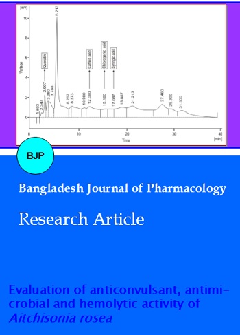
The n-butanol fraction of A. rosea showed significant results for anticonvulsant and antimicrobial activities and also exhibited maximum protection of DNA from damaging, so it was put forward for HPLC analysis to evaluate the amount of phenolics in this particular fraction. The amount (ppm) of phenolics evaluated in the ethyl acetate fraction of the plant was quercitin (2.33 ± 0.03), caffeic acid (7.54 ± 0.04), chlorogenic acid (6.02 ± 0.02) and syringic acid (1.72 ± 0.06) as shown in Figure 2.
Figure 2: HPLC profile of phenolics in n-butanol fraction of A. rosea
Discussion
In the current study pharmacological activities, safety and HPLC profile of A. rosea was evaluated. Among many experimental models used for induction of convulsions, pentylenetetrazole (PTZ) has been most frequently employed for preliminary screening of anticonvulsant effect. PTZ produces petit mal type of epilepsy and convulsions in experimental animals by counteracting the action of inhibitory neurotransmitter GABA and thus producing hyper excitation and excessive neuronal firing, which leads to the development of epilepsy (Ha et al., 2004). PTZ also produces convulsions by destabilizing neuronal membranes (Curtis et al., 1971; Gnyther and Curtis, 1986). The results of the present study have clearly shown potent anticonvulsant activity of plant methanol extract and its n-butanol and ethyl acetate fractions. The tested fractions have been found to exert anticonvulsant effect due to their potent ability to increase the time of onset of convulsions and to decrease the time of duration of convulsions caused by administration of PTZ. The strong anticonvulsant action of A. rosea against PTZ-induced convulsions might be due to their action as a GABA agonist (Bum et al., 2001).
In addition, strychnine, another very potent convulsant has been shown to act on the spinal cord and produce an inhibitory effect on the inhibitory neurotransmitter glycine, which produces hyper excitability in CNS and ultimately convulsions (Adeyemi et al., 2010). Strychnine counteracts the action of excitatory neurotransmitters like AMPA and NMDA and it also produces inhibitory effects on calcium-induced excitotoxicity in neurons. These factors are pro-epileptogenic and precipitate seizures. So, the suppression of the inhibitory neurotransmitter, glycine leads to tonic-clonic seizures in experimental animals (Rogawski, 2011). In our study, methanol extract, n-butanol and ethyl acetate fractions showed anticonvulsant activity against strychnine-induced convulsions in a dose-dependent manner. However, the effect was relatively more potent against PTZ-induced convulsions (Figure 1). The anticonvulsant effect of plant against strychnine-induced convulsions has indicated their ability to interfere with glycine transmission in the spinal cord.
Furthermore, picrotoxin, a non-competitive antagonist at GABA receptors, has been reported to block the GABA-activated chloride ionophore (Vogel and Vogel, 1997). Methanol extract, n-butanol and ethyl acetate fraction have also demonstrated anticonvulsant activity against picrotoxin induced convulsions. This also indicated that the plant extract and its various fractions exert their anticonvulsant effect due to modification of function of GABA receptor-mediated chloride channels.
The results of different studies have provided evidence that some medicinal plants do possess antimicrobial activities and may act as a potential source of new antibacterial agents even against some antibiotic-resistant strains (Kone et al., 2004). In the present study conducted by using the disc diffusion method, it has been observed that the extract and fractions of plant possessed significant antibacterial activity against certain gram negative (P. aeruginosa) and gram positive (S. aureus, B. subtilis) pathogens, i.e. bacteria. The results of this study were in agreement with the earlier studies (Fan and Chen, 2001; Yuste and Fung, 2004; Olayemi and Oluwasola, 2010). Antimicrobial activity of the methanol extract and fractions of A. rosea have also exposed noticeable difference in sensitivity against pathogenic fungi P. notatum and C. albicans. The variation in the effectiveness of the extracts against different microorganisms would be due to differences in the chemical composition of the extracts. In the disc diffusion technique lessening in microbial growth is determined on the basis of zone of inhibition around the disc. An inhibition zone of 14 mm or more has been reckoned to be a significant antimicrobial activity (Riaz et al., 2012). In our study high antimicrobial activity, i.e. (inhibition zone of 14 mm) was produced by some fractions of the plant.
The MIC results showed that methanol extract and ethyl acetate fraction had highly significant MIC against all bacterial and fungal strains. The least MIC value exhibited that less concentration was required to inhibit the selected bacterial and fungal strains. The higher MIC represented that the tested samples had less antimicrobial activity.
The mechanical stability of the red blood cell membrane is essential to reduce the cytotoxic effects of different compounds. The least in vitro cytotoxicity of different plant extracts is of great importance for their safe use in different diseases other than cancer (Uddin et al., 2011).
In the present research all the samples tested for in vitro hemolytic activity were found in a safe range, i.e. below 10.0%, so it could be expected that the methanol extract and is various organic fractions have a minor cytotoxicity (Sharma and Sharma, 2001). The plant extracts or fractions having minor cytotoxicity might be used as herbal medicines (Aslam et al., 2011).
The differences in anticonvulsant and antimicrobial activities of plant extract and its various fractions were possibly due to different concentration of their constituents. The phytochemicals are produced in many plant species and exhibit pharmacological and biological activities. Antioxidant and antimicrobial activities were reported in various plants due to the presence of tannins, alkaloids, steroids, saponins and phenolic compounds (Riaz et al., 2012; Erol et al., 2010). Terpenoids are found in various plant parts having many therapeutic uses like antimicrobials and also used in many pharmaceutical preparations (Michael, 2009). The analyzed phytoconstituents in this study might be responsible for their pharmacological actions. The n-butanol fraction exhibited very promising pharmacological effects, i.e. anticonvulsant and antimicrobial and also showed protection to RBCs from hemolysis. So, the phenolic constituents present in this fraction were estimated by HPLC which might be responsible for these actions. The earlier reports showed that the phenolic compounds present in various plant extracts play a vital role for their antimicrobial and other activities (Mori et al., 1987).
Conclusion
The anticonvulsant and antimicrobial activities of crude methanol extract and its various organic fractions of A. rosea would help for development of a new alternative medicine system which has less side effects.
Ethical Issue
Treatment of experimental animals was performed according to protocols approved by the local ethical committee.
References
Adeyemi OO, Akindele AJ, Yemitan OK, Fagbo FI. Anticonvulsant, anxiolytic and sedative activities of the aqueous root extract of Securida calonge and pendunculata fresen. J Ethanopharmacol. 2010; 130: 191-95.
Ali SI, Nasir E. Flora of Pakistan. Department of Botany, University of Karachi, Karachi, 1989.
Aslam F, Rasool N, Riaz M, Zubair M, Rizwan K, Abbas M, Bukhari TH, Bukhari IH. Antioxidant, haemolytic activities and GC-MS profiling of Carissa carandas. Inter J Phytomed. 2011; 3: 567-78.
Brain KR, Turner KR. The practical evaluation of phytopharmaceuticals. UK, Wright- Scientehnica, 1975, pp 187-89.
Bum EN, Schmutz M, Meyer C, Rakotonirina A, Bopelet M, Porter C. Anticonvulsant properties of the methanolic extract of Cyperus articulates. J Ethnopharmacol. 2001; 76: 145-50.
Curtis DR, Duggan AW, Felix D, Johnston GAR, McLennan H. Antagonism between bicculline and GABA in the cat brain. Brain Res. 1971; 33: 57-73.
Edeoga HO, Okwu DE, Mbaebie BO. Phytochemical constituents of some Nigerian medicinal plants. Afri J Biotech. 2005; 4: 685-88.
Ergene A, Guler P, Tan S, Mirci S, Hamzaoglu E. Antimicrobial and antifungal activity of Heracleums phondylium subsp. artivinense. Afr J Biotech. 2006; 11: 1087.
Erol NT, Sarı F, Velı̇oglu YS. Polyphenols, alkaloids and antioxidant activity of different grades of turkish black tea. GIDA J Food. 2010; 35: 161-68.
Fan M, Chen J. Studies on antimicrobial activity of extracts from thyme. Wei Sheng Wu Xue Bao. 2001; 41: 499-504.
Gnyther BD, Curtis DR. Pyridazinyl GABA derivatives as GABA and glycine antagonist in the spinal cord of the cat. Neurosci Lett. 1986; 68: 585-87.
Ha JH, Lee DU, Lee JT, Kim JS, Yong CS, Kim JA. 4-Hydroxybenzaldehyde from gastrodiaelata as active in the antioxidation and GABAergic neuromodulation of the rat brain. J Ethnopharmacol. 2004; 73: 329–33.
Kokate CK, Purohit AP, Gokhale SB. Pathway to screen phytochemical nature of natural drugs. In: Pharmacognosy. 39th ed. India, Nirali Prakashan, 2007, pp 607-11.
Kone WM, Atindehou C, Terreaux K, Hostettmann D, Traore, Dosso M. Traditional medicine in North Cote-d'Ivoire: screening of 50 medicinal plants for antibacterial activity. J. Ethnopharmacol. 2004; 93: 43-49.
Michael, S.A. Life of its own. USA, The New Yorker, 2009, pp 1-50.
Mori A, Nishino C, Enoki N, Tawata S. Antibacterial activity and mode of action of plant flavonoids against Proteus vulgaris and Staphylococcus aureus. Phytochemistry 1987; 26: 2231-34.
Mozaffarian V. Studies on the flora of Iran, four new species and a short note on an interesting Rubiaceae. Ira J Bot. 2006; 107-13.
Nishanta R, Cory S, Harris, Towers GHN. Antimicrobial activity of plants collected from serpentine outcrops in Sri Lanka. Pharm Biol. 2002;.3: 235-44.
Noor AT, Perveen S, Begum A, Fatima I, Malik A, Tareen RB. Rosenones A and B, new anthraquinone derivatives from Aitchisonia rosea. J Asian Nat Prod. 2009a; 4: 209-12.
Noor AT, Perveen S, Begum A, Fatima I, Malik A, Tareen, RB. Aitchisonides A and B, new iridoid glucosides from Aitchisonia rosea. J Asian Nat Prod. 2009b; 11: 985-89.
Olayemi MA, Oluwasola OO. Effect of light irradiation on the antimicrobial activity of Zanthoxylum zanthoxyloides (lam) methanolic extract. Afr J Pharmacol. 2010; 4: 145-50.
Riaz M, Rasool N, Bukhari IH, Shahid M, Zubair M, Rizwan K, Rashid U. In vitro antimicrobial, antioxidant, cytotoxicity and GC-MS analysis of Mazus goodenifolius. Molecules 2012; 17: 14275-87.
Rogawski MA. Revisiting AMPA receptors as an antiepileptic drug target. Epilepsy Curr. 2011; 11: 56-63.
Sarker SD, Nahar L, Kumarasamy, Y. Microtitre plate-based antibacterial assay incorporating resazurin as an indicator of cell growth, and its application in the in vitro antibacterial screening of phytochemicals. Methods 2007; 4: 321-24.
Sharma P, Sharma JD. In vitro hemolysis of human erythrocytes by plant extracts with antiplasmodial activity. J Ethnopharmacol. 2001; 74: 239-43.
Sultana B, Anwar F, Przybylyski R. Antioxidant activity of phenolic components present in barks of Azadirachta indica, Terminalia arjuna, Acacia nilotica and Eugenia jambolana Lam. Trees. Food Chem. 2007; 104: 1106-14.
Swinyard EA, Woodhead JH, White HS, Franklin MR. General Principles: Experimental selection, quantification and evaluation of anticonvulsants. In: Antiepileptic drugs. Levy R, Mattson R, Meldrum B, Penry JK, Dreifuss FE (eds). New York, Raven Press, 1989, pp 85-103.
Uddin SJ, Grice ID, Tiralongo E. Cytotoxic effects of Bangladeshi medicinal plant extracts. Evid Based Complement Altern Med. 2011; 1-7.
United States Pharmacopoeia. 28th ed. U.S.A, The United States Pharmacopeial Convention, 1980, pp 864-69.
Vogel HG, Vogel WH. Drug discovery and evaluation, pharmacological assay. Berlin, Springer. 1997; 260-61.
Yuste J, Fung DY. Inactivation of Salmonella typhimurium and Escherichia coli O157: H7 in apple juice by a combination of nisin and cinnamon. J Food Prod. 2004; 67: 371-77.