Enhanced antitumor activity of cucurbitacin B combined with cerulenin in osteosarcoma
Abstract
In the present study, the effect of cucurbitacin B and cerulenin combination on osteosarcoma was investigated to develop an effective treatment regimen. The IC50 values of cerulenin and cucurbitacin B combination in the proportion of 1:1, 1:2 and 2:1 were 3.5, 7.2 and 8.6 ug/mL, respectively. The combination index (CI) values of <0.95 for inhibition of growth and <0.93 for inhibition of SENP5 expression clearly indicated synergism between the two. The Q-value for combination of 1 ug/mL cerulenin and 1 ug/mL cucurbitacin B was 1.2. AI calculated for tumor tissues treated with a combination of cucurbitacin B and cerulenin (26.2 ± 8.4%) was higher than that of the tissues treated separately with cucurbitacin B (14.5 ± 6.8%) or cerulenin (12.6 ± 7.5%). The AI for untreated tumor tissues was 4.2 ± 1.5%. Thus, the combination of cucurbitacin B and cerulenin can be a promising regimen in the treatment of osteosarcoma.
Introduction
Osteosarcoma one of the commonly detected bone tumors has a five-year survival rate of ~70% in children and adolescents. The poor prognosis of the osteosarcoma patients along with less than 20% overall survival rate demands for the development more efficient and novel therapeutic strategies (Jaffe, 2009). Osteosarcoma has a characteristic radiographic appearance and has been the target for clinicians throughout the globe (Janeway et al., 2009; Marina et al., 2004).
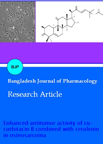
Cucurbitacin B (Figure 1), an oxygenated triterpene is isolated from Trichosanthes kirilowii Maximowicz (Cucurbitaceae family). The plant has a long traditional medicinal importance and is used for its anti-inflammatory, antidiabetic and abortifacient effects. Cucurbitacins are reported to exhibit in vivo anti-inflammatory activities, and preventive and curative effects against CCl4-induced hepatotoxicity (Chen et al., 2005). They also exhibit cytotoxicity and anti-cancer activity (Jayaprakasam et al., 2003). Cucurbitacin B exhibits antiproliferative effects and acts a dual inhibitor of the activation of both JAK2 and STAT3 in some malignancies (Sun et al., 2995). Recent, reports demonstrate that cucurbitacin B has antiproliferative activity against human breast cancer, glioblastoma multiforme, and myeloid leukemia cells (Wakimoto et al., 2008; Yin et al., 2008). It inhibits growth by cell cycle arrest in G2/M phase and increases apoptosis by inhibition of the JAK/STAT pathway (Toyonaga et al., 2003).
Figure 1: Structure of cucurbitacin B
Cerulenin has been isolated from Cephalosporium caerulens. It acts an inhibitor of fatty acid synthase (FASN) by reacting with the keto-acyl synthase domain of FASN. Various studies have been performed to investigate the anti-tumor activity of cerulenin against the malignant neoplasms (Elbaz et al., 2010; Wang et al., 2008). The present study demonstrates the effects of the combination of cucurbitacin B and cerulenin on the rate of osteosarcoma cell proliferation, suppression of SENP5 expression and induction of apoptosis.
Materials and Methods
Reagents and drugs
Cerulenin, cucurbitacin B and dimethyl sulphoxide were purchased from the Sigma (USA).
Cell growth assay
Human osteosarcoma cell line, Saos-2 was obtained from the American Type Culture Collection (Manassas, USA). The cells were grown in 10% fetal bovine serum-Dulbecco's modified Eagle's medium (FBS-DMEM) (HyClone Laboratories, USA) in an incubator with 5% CO2 atmosphere.
MTT assay
Saos-2 cells were distributed at a density of 2.5 x 10⁵ cells per well on to the 96-well tissue culture plates containing FBS-DMEM supplemented with 2 mM L-glutamine and cultured for 12 hours. To each of the well different concentrations of cerulenin, cucurbitacin B or combination of cerulenin and cucurbitacin B were added and incubated. Following incubation for 36 hours 3-(4,5-dimeth-ylthiazol-2-yl)-2,5-diphenyltetrazolium bromide (MTT) was put into each the of well and incubated again for 2 hours. Dimethyl sulfoxide was added to each well for dissolution of farmazan crystals formed. The microplate reader (SpectraMax Plus; Molecular Devices) was employed to measure the absorbance at 455 nm for each of the well three times. The IC50 values were determined by using Originpro 7.5 program from the plot of concentration verses viability curves. For all the combinations of cerulenin and cucurbitacin B, combination index (CI) was calculated which indicated the nature of interaction. The values of less than 0.95 indicated synergism, between 0.95 and 1.05 indicated antagonism and above 1.05 indicated additive interactions.
Analysis of apoptosis and cell cycle using fluorescence-activated cell sorting (FACS)
Saos-2 cells after incubation for 24 hours with various concentrations of cerulenin, cucurbitacin B and their combinations were analyzed using EPICS XL flow cytometer. The proportion of apoptotic cells and population of cells in different phases of cell cycle were determined using System II software. The equation 1 was used for the determination of Jin's Q value.
Q = E(A+B)/[EA + (1 - EA) x EB] -------------------- 1
Where, EA represents percentage of apoptotic cells at different doses of cerulenin, EB represents percentage of apoptotic cells at different doses of cucurbitacin B and E (A+B) stands for apoptotic cell percentage using different combinations of cerulenin and cucurbitacin B.
Western blot analysis
Saos-2 cells (2 x 10⁵) after incubation with cerulenin, cucurbitacin B and combination of cerulenin and cucurbitacin B were rinsed twice with PBS. The cells were then treated with 2 mL lysis buffer (50 mM Tris-HCl pH 7.4, 137 mM NaCl, 10% glycerol, 100 mM sodium vanadate, 1 mM PMSF, 10 mg/mL aprotinin, 10 mg/mL leupeptin, 1% NP-40, and 5 mM cocktail). The concentration of proteins in the cell lysates was determined by BCA method was. For the isolation of proteins electrophoresis on 10% polyacrylamide gel was performed. The proteins were then transferred on to the PVDF membrane Gelman Science (Ann Arbor, MI). The non-specific binding sites on the membranes were blocked by incubation with 5% non-fat dry milk overnight. The membrane was washed with TBST followed by incubation primary antibodies for 12 hours. After incubation the membrane was washed again and then incubated with secondary antibodies for 2 hours. Then X-ray autoradiography was performed and the gray scale images were analysed.
Terminal deoxynucleotidyl transferase dUTP nick end labeling (TUNEL) assays
The tumour tissues stored at -78 degree celcius under liquid nitrogen were embedded in paraffin and sliced into thin 2 µM sections. TUNEL assays was performed using Situ Cell Death Detection kit (Roche Diagnostics Corp., USA). The tissue sections after deparaffinization in xylene were rehydrated using gradient ethanol and then boiled with sodium chloridesodium citrate buffer (pH 7.0) for 30 min at 80 degree celcius. The sections were washed followed by 1 hour treatment with proteinase K. The sections were incubated with fluorescein-labeled deoxyuridine triphosphate (dUTP) and TUNEL reagents according to the manufacturer's instructions. The sections after washing were incubated with horseradish peroxidase (HRP)-conjugated fluorescein antibody followed by counterstaining with 0.5% methyl green. The apoptotic index (AI) was calculated by dividing the number of brown-stained cells with total number of tumor cells.
Statistical analysis
All the data expressed are the mean of ± SD. Student's t-test was used to analyse the significance. p<0.05 was considered to indicate a statistically significant result.
Results
Synergistic effect of cerulenin and cucurbitacin B on antitumor activity in Saos-2 cells
The results from MTT assay indicated that Saos-2 cells were sensitive to both cerulenin and cucurbitacin B. Both the agents caused an inhibition in growth of Saos-2 cells in a concentration-dependent manner (Figure 2A). The IC50 values of cerulenin and cucurbitacin B for antitumor activity against Saos-2 cells were 5.1 and 21.3 ug/mL, respectively. However, when Saos-2 cells were treated with a combination of cerulenin and cucurbitacin B in the proportion of 1:1, 1:2 and 2:1, the values for IC50 were 3.5, 7.2 and 8.6 ug/mL, respectively (Figure 2B). The calculated CI values for all the three combinations were <0.95, indicating that cerulenin and cucur-bitacin B exhibited synergistic effect on inhibition of growth in Saos-2 cells.
Figure 2: Inhibitory effect of cucurbitacin B, cerulenin or combination treatment on the growth of Saos-2 cells. (A) Cucurbitacin B and cerulenin both inhibited Saos-2 cell growth in a dose-dependent manner. (B) Cucurbitacin B was applied to Saos-2 cells adjunctively with cerulenin in a proportion of 1:1, 1:2 and 2:1. The adjunctive treatment also inhibited Saos-2 cell growth in a dose-dependent manner
Synergistic effect of cerulenin and cucurbitacin B on inhibition of SENP5 expression in osteosarcoma cell lines
The results from quantitative PCR and western blotting analysis are shown Figure 3A and B. It is clear from the figure that SENP5 is significantly overexpressed in Saos-2 osteosarcoma cell line compared with HOB cells (human osteoblasts isolated from normal human bone).
Treatment of Saos-2 osteosarcoma cell line with cucurbitacin B or cerulenin resulted in inhibition of SENP5 expression in a dose and time-dependent manner (Figure 3C, D). We treated osteosarcoma cells with 1:1, 1:2 and 2:1 proportion of cucurbitacin B and cerulenin. The results from RT-PCR analysis clearly demonstrated a significant inhibition of SENP5 expression on treatment 1:1, 1:2 and 2:1 proportion of cucurbitacin B and cerulenin. The values for IC50 were 4.7, 8.5 and 9.4 ug/mL, respectively. The calculated CI values for all the three combinations were <0.93, indicating that cerulenin and cucurbitacin B exhibited synergistic effect on inhibition of SENP5 expression in Saos-2 cells.
Figure 3: SENP5 is overexpressed in osteosarcoma cell lines. (A) mRNA expression and (B) protein levels of SENP5 in Saos-2 osteosarcoma cell lines. Inhibition of SENP5 expression by cucurbitacin B and cerulenin treatment. (C) Concentration-dependent inhibition of SENP5 expression in Saos-2 cells. (D). Time-dependent inhibition of SENP5 expression in Saos-2 cells
Synergistic effect of cerulenin and cucurbitacin B on induction of apoptosis in Saos-2 cells
The Saos-2 cell cultures were treated with a range of cucurbitacin B, cerulenin and their combination concentrations for 24 hours. The results from FACS analysis indicated a concentration dependent induction of apoptosis in Saos-2 cell (Figure 4). There was apoptosis in 17.3, 26.1 and 31.5% cells treated with 1, 5 and 10 μg/mL of cucurbitacin B, respectively. The Q-value for combination of 1 μg/mL cerulenin and 1 ug/mL cucurbitacin B was 1.2 which indicated synergistic effect between the two.
Figure 4: FACS analysis of U2-OS cells treated with epirubicin, cerulenin and the combination treatment for 24 hours. (A) Few apoptotic cells were noted in the control group. Yet, the percentage of apoptotic cells was (B) 31.5% when treated with 10 ug/mL epirubicin and (C) 38.8% when treated with 20 ug/mL cerulenin. (D) The percentage of apoptotic cells rose to 66.2% when treated with 10 ug/mL epirubicin in conjunction with 20 μg/mL cerulenin. FACS, fluorescence-activated cell sorting; PI, propidium iodide
Synergistic effect of cerulenin and cucurbitacin B on induction of apoptosis in tumor cells
Treatment of tumor tissues with cucurbitacin B, cerulenin or their combination induced apoptosis in tumor cells (Figure 5). However the AI calculated for tumor tissues treated with a combination of cucurbitacin B and cerulenin (26.2 ± 8.4%) was higher than that of the tissues treated separately with cucurbitacin B (14.5 ± 6.8%) or cerulenin (12.6 ± 7.5 %). The AI for untreated tumor tissues was 4.2 ± 1.5%.
Figure 5: TUNEL assays of tumor tissue slides. The apoptotic index was significantly higher than that in the control, cerulenin and epirubicin alone groups
Discussion
The results revealed that the use of the two agents in combination exhibited synergistic effect on antitumor activity, inhibition of SENP5 expression and induction of apoptosis in osteosarcoma cell lines. The IC50 values for cerulenin and cucurbitacin B combination in the proportion of 1:1, 1:2 and 2:1 were 3.5, 7.2 and 8.6 μg/mL, respectively. The calculated CI values for all the three combinations were <0.95, indicating that cerulenin and cucurbitacin B exhibited synergistic effect on inhibition of growth in Saos-2 cells.
The combination of cucurbitacin B and cerulenin also inhibited SENP5 expression in Saos-2 cells through synergistic effect. Among three proportions tested the calculated CI values for all the three were <0.93, indicating that cerulenin and cucurbitacin B exhibited synergistic effect on inhibition of SENP5 expression in Saos-2 cells. There was apoptosis in 17.3, 26.1 and 31.5% cells treated with 1, 5 and 10 ug/mL of cucurbitacin B, respectively. The Q-value for combination of 1 ug/mL cerulenin and 1 ug/mL cucurbitacin B was 1.2 which indicated synergistic effect between the two. AI calculated for tumor tissues treated with a combination of cucurbitacin B and cerulenin (26.2 ± 8.4%) was higher than that of the tissues treated separately with cucurbitacin B (14.5 ± 6.8%) or cerulenin (12.6 ± 7.5%). The AI for untreated tumor tissues was 4.2 ± 1.5%.
Conclusion
A synergistic effect exits between cerulenin and cucurbitacin B for anti-OS in vitro and a synergistic effect in vivo. It has been suggested that cerulenin combined with cucurbitacin B may be a potential treatment regimen for OS.
References
Cesari M, Alberghini M, Vanel D, et al., Periosteal osteosarcoma: A single-institution experience. Cancer 2011; 117: 1731-35.
Chen JC, Chiu MH, Nie RL, Cordell GA, Qiu SX. Cucurbitacins and cucurbitane glycosides: Structures and biological activities. Nat Prod Rep. 2005; 22: 386-99.
Elbaz A, Wu X, Rivas D, et al., Inhibition of fatty acid biosyn¬thesis prevents adipocyte lipotoxicity on human osteoblasts in vitro. J Cell Mol Med. 2010; 14: 982-91.
Jaffe N. Osteosarcoma: Review of the past, impact on the future. The American experience. Cancer Treat Res. 2009; 152: 239-62.
Janeway K, Gorlick R, Bernstein M. Osteosarcoma. In: Oncology of infancy and childhood. Orkin S, Fisher D, Look A, Lux S, Ginsburg D, Nathan D (eds). Philadelphia, Saunders Elsevier, 2009, pp 871-910.
Jayaprakasam B, Seeram NP, Nair MG. Anticancer anti-inflammatory activities of cucurbitacins from Cucurbita andreana. Cancer Lett. 2003; 189: 11-16.
Marina N, Gebhardt M, Teot L, Gorlick R. Biology and therapeutic advances for pediatric osteosarcoma. Oncologist 2004; 9: 422-41.
Menendez JA, Mehmi I, Atlas E, et al., Novel signaling molecules implicated in tumor-associated fatty acid synthase-dependent breast cancer cell proliferation and survival: Role of exogenous dietary fatty acids, p53-p21 WAF1/CIP1, ERK1/2 MAPK, p27KIP1, BRCA1, and NF-κB. Int J Oncol 2004; 24: 591-608.
Sun J, Blaskovich MA, Jove R, Livingston SK, Coppola D, Sebti SM. Cucurbitacin Q: A selective STAT3 activation inhibitor with potent antitumor activity. Oncogene 2005; 24: 3236-45.
Toyonaga T, Nakano K, Nagano M, Zhao G, Yamaguchi K, Kuroki S et al., Blockade of constitutively activated Janus kinase/signal transducer and activator of transcription-3 pathway inhibits growth of human pancreatic cancer. Cancer Lett. 2003; 201: 107-16.
Wang WQ, Zhao XY, Wang HY, et al., Increased fatty acid synthase as a potential therapeutic target in multiple myeloma. J Zhejiang Univ Sci B. 2008; 9: 441-47.
Wakimoto N, Yin D, O’Kelly J, et al. Cucurbitacin B has a potent antiproliferative effect on breast cancer cells in vitro and in vivo . Cancer Sci. 2008; 99: 1793-97.
Yin D, Wakimoto N, Xing H, et al. Cucurbitacin B markedly inhibits growth and rapidly affects the cytoskeleton in glioblastoma multiforme. Int J Cancer. 2008; 123: 1364-75.