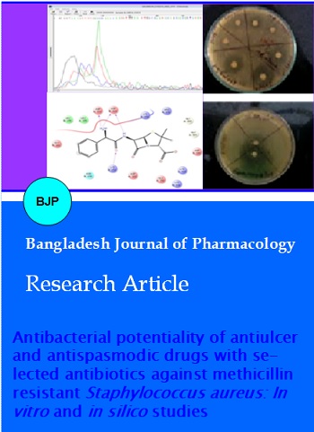
Antibacterial potentiality of antiulcer and antispasmodic drugs with selected antibiotics against methicillin resistant Staphylococcus aureus: In vitro and in silico studies
Abstract
In the present study, the antispasmodic drug mebeverine hydrochloride and the antiulcer drug troxipide were tested for their possible antibacterial properties in vitro. The antimicrobial assays of the above drugs were determined with ampicillin, penicillin and ciprofloxacin against sensitive and resistant strains and their resistance were confirmed through Polymerase Chain Reaction by identifying the presence of the mecA gene. A computer-aided method was used for screening the effectiveness of the drug interactions. Mebeverine and troxipide inhibited most of the sensitive and resistant strains tested in vitro from 32.5 to 125 µg/mL. The loss of structural alterations of the cell wall was analyzed by atomic force microscopy. In docking studies, troxipide and mebeverine were found to have substantial inhibition against penicillin binding protein 2a (IVQQ) and UDP-N-acetylglucosamine 1-carboxyvinyltransferase (2YVW) receptor proteins that seem to have interacted with most of the residues.
Introduction
Microbes such as bacteria, viruses, fungi and parasites can cause virulent and contagious diseases in individuals mainly through direct exposure to aerosols and contaminated materials (Barker and Jones, 2005). The resistance to infection depends upon the strength of the attack by the person's immune system (Carter, 2005). Antibiotics are able to control many bacterial infections like methicillin resistant Staphylococcus aureus (MRSA) yet they develop resistance to multiple drugs. Treatments have failed not only because of the development of resistance but also because of untoward reactions of the administered drugs in the infected individuals (Martins et al., 2008). Research reveals that there are some drugs that do not traditionally come under the category of antibacterial classification yet have moderate to powerful antibacterial action. They might have different pharmacological actions such as antihistaminic (El-Nakeeb et al., 2012), antipsychotic (Basu et al., 2005), antihypertensive (Dutta et al., 2005), antispasmodic (Karak et al., 2003), cardiovascular (Dasgupta et al., 2007; Kumar et al., 2004) and anti-inflammatory, such as the drug diclofenac sodium (Annaduri et al., 2008).
In the present study, we evaluated the antibacterial activity of troxipide and mebeverine in vitro and discovered a synergism between the selected drugs with antibiotics ampicillin, penicillin, and ciprofloxacin against a clinical isolate of S. aureus. In addition, we used a computer program for screening the existing drugs and the effectiveness of the drug interactions was tested with an in silico docking model with various receptor protein found on S. aureus strains. Nearly all these assumed targets were involved in more than one metabolic pathways of MRSA.
Materials and Methods
Bacterial strains
aureus NCIM 2079, K. pneumoniae NCIM 2719, and Enterobacter cloacae NCIM 2164 were obtained from NCIM, Pune. Clinical strain S. aureus, Escherichia coli and Enterococcus faecalis were obtained from KAP Viswanathan Medical College, Tiruchirappalli, Tamil Nadu, India. The strains were confirmed and stored at 4°C until use.
Drugs
Troxipide, mebeverine hydrochloride, metaclopropamide and aceclofenac were obtained as pure drugs from Sigma Aldrich, India and kept under refrigeration until use.
Media
Nutrient broth, Muller-Hinton broth, nutrient agar and Muller-Hinton agar were prepared and steam sterilized at 15 psi for 15 min by autoclaving.
Standardization of inocula
The selected strains were grown in Muller-Hinton broth overnight at 37°C in an incubator under standard conditions. The harvested cells of the exponential phase culture were suspended in sterile distilled water and adjusted to a turbidity of 0.5 McFarland standard using a spectrophotometer (Cary-60 UV-Visible, Agilent Technologies) at 625 nm.
Molecular identification of mecA gene using gene-specific primers
Total DNA isolation and PCR analysis
Total DNA was extracted from overnight cultures of the selected bacterial isolates using a DNA isolation kit (Qiagen) and suspended in 100 uL of elution buffer (10 mM Tris-HCl, pH 8.5) and quantified by measuring optical density at 260 nm UV-Thermo Scientific BIOMATE 35. PCR amplification was performed using a 50 uL reaction mixture containing 100 ng of template DNA, 20 μmol of gyrA and gyrB primers, 200 uM of dNTPs, 1.5 mM of MgCl2, 1U of Taq DNA polymerase (MBI Fermentas) and 10 μL of 10X Taq polymerase buffer. The sequences of the methicillin resistance gene (mecA) primers used were as follows:
mecA-F 5'-AAAATCGATGGTAAAGGTTGGC-3'
mecA-R 5'-AGTTCTGCAGTACCGGATTTGC-3'
Amplification was carried out with an initial denaturation at 94°C for 5 min followed by 35 cycles of denaturation at 94°C for 30 sec, annealing at 55°C for 30 sec, extension at 72°C for 1 min and final extension at 72°C for 5 min using a thermocycler (Eppendorf Personal Cycler, Germany). The amplified gene was analyzed on a 1% agarose gel for mecA amplicons in 1X TAE buffer at 50 V and was further confirmed by DNA sequencing with ABI PRISM 3730 Genetic Analyzer (Applied Biosystems) (Towner KJ et al., 1998, Trindade PA et al., 2003).
Checker-board method
To determine the interactions between the identified methicillin resistant bacteria and a combination of two drugs, the most frequently used procedure is the checker-board method. In this technique, aliquots of log-phase bacterial cultures (0.5 McFarland standard) were transferred to microtiter plates containing earlier-tested concentrations of drugs' ampicillin 25 to 800 µg/mL, troxipide and mebeverine 6.25 to 200 µg/mL. In the first row, ampicillin alone was distributed, whereas, in first column troxipide was added, and similarly, the other drugs were dispensed in micro well plates. The inoculated micro titer plates were incubated at 37°C for 36 hours (Kumar et al., 2004). The growth inhibition was measured by determining the absorbance at 530 nm in a multi-mode plate reader (Enspire, Perkin Elmer). From these readings, the synergistic and additive interactions were calculated (Mazumdar et al., 2003).
In vitro disc diffusion tests between the non-antibiotic drugs and the antibiotics
The drug-antibiotic combinations were tested by the disc diffusion technique with sterile filter paper discs (7.25 mm, Whatman No. 1) (Mazumdar et al., 2003). The filter paper was soaked in different concentrations (10, 20, 30, 40 and 50 µg) of drug. Sensitive and resistant strains were grown in sterile liquid media for 18 hours. Plates were prepared with Muller-Hinton agar and inoculated on the surface of which drug-soaked sterile disks were placed. The plates were incubated at 37°C for 36 hours and the zone of inhibition for each bacterial strain was measured. The experiments were performed in triplicates.
Atomic Force Microscopy (AFM) analysis
Atomic force microscopy was used to analyze the surface morphology and topology of the prepared samples mebeverine loaded MRSA cultures (Tyagi and Malika, 2010; Li et al., 2007). The images were taken using a Park Systems XE 100 (Germany). The samples were measured in the non-contact mode.
Methodology of docking
The chemical structures of troxipide, mebeverine and the reference drugs ampicillin and ceftrioxone were drawn using ChemSketch software version 12.01. Ceftriozone was selected for the docking studies as it is best drug of choice that effectively acts against MRSA. The target Protein Data Bank (PDB) structure with the IDs: Penicillin binding protein 2a from MRSA strain (1VQQ), UDP-N-acetylglucosamine 1-carboxyvinyl-transferase (2YVW), fructose 1,6-bisphosphate aldolase (4LV4) and potassium transporter gating component ktrA as a c-di-AMP receptor (4J7C) (Corrigan et al., 2013; Yadav et al., 2012) were downloaded from PDB and the ligand structures were imported to ChemSketch. The target was given as an input, and the binding site was specified and prepared. The ligand was docked with the target. Population size was set to 50, generations to 10 and the number of solutions to 1. The docking analysis was performed and the docked poses were further examined. The software includes GLIDE module version 5.9, Mastero 9.4, Quik prop -3.6 -Schrodinger, LLC, New York, NY, 2013- docking, Swiss PDB viewer-4.04- protein viewer, Pymol viewer 1.3 – image viewer, Marvin sketch 5.5- ligand structure (Tomasz and Alexander, 2005; Parasuraman et al., 2014; Perumal et al., 2014; Sabitha and Rajkumar, 2012).
Results
Table I shows the inhibitory activity of the antibiotics and drugs used. The MIC of ciprofloxacin is 3.95 to 15.625 µg/mL, and mebeverine and troxipide, 15.625 to 62.5 µg/mL against sensitive and resistant bacterial strains. The MIC of penicillin and ampicillin were 125-500 µg/mL against sensitive strains whereas it showed resistance against clinical strain S. aureus at 1000 µg/mL and its resistance was further identified with PCR analysis.
Table I:Minimum inhibitory concentrations (MICs) of the selected drugs and antibiotics by broth dilution method
| Drugs | Staphylococcus aureus NCIM 2079 (µg/mL) |
Clinical strain Enterococcus faecalis (µg/mL) | Klebsiella pneumoniae NCIM 2719 (µg/mL) | Enterobactor clocea NCIM 2164 (µg/mL) |
Clinical strain Staph aureus(µg/mL) |
|---|---|---|---|---|---|
| Ciprofloxacin | 15.6 | 7.8 | 3.95 | 3.95 | 15.6 |
| Penicillin | No activity | 500 | 125 | 125 | 1000 |
| Ampicillin | No activity | 125 | 125 | 125 | 1000 |
| Metaclopropamide | No activity | No activity | No activity | No activity | No activity |
| Mebeverine | 62.5 | 31.25 | 62.5 | 15.6 | 62.5 |
| Troxipide | 62.5 | 62.5 | 62.5 | 62.5 | 62.5 |
| Aceclofenac | No activity | No activity | No activity | No activity | No activity |
To identify the methicillin resistance of S. aureus, gene-specific primers were designed for amplification of the mecA gene by Polymerase Chain Reaction. The amplified region has been sequenced and confirmed that the 436 bases were found to be similar to the mecA gene of S. aureus strain S10215. This was done to confirm that the amplified sequence of the given sample was indeed, that of the mecA gene. The obtained sequences were searched by BLAST on the NCBI website, and they clearly belong to the taxa S. aureus penicillin binding protein (mecA) (details shown in Figure 1: A,B,C) which plays an important role in beta-lactamase resistance. Therefore, the resistive nature of the antibiotic was studied by using both disc diffusion and minimum inhibitory concentration methods with specific beta-lactamase antibiotics penicillin and ampicillin.
Table II. In checker board method the MRSA strain was used. When used alone, penicillin and ampicillin have an MIC of 1000 µg/mL against MRSA but when combined with troxipide 50 µg/mL and mebeverine 50 µg/mL the MIC reduced to 50 µg/mL showing the synergistic effect.
Table II: Synergistic interaction of combinations of antibiotics and non-antibiotics against methicillin-resistant S. aureus by checker board method
| Drugs | MIC (µg/mL) | Type of interaction |
|---|---|---|
| Penicillin | - | No activity |
| Penicillin + Troxipide | 50 | Synergy* |
| Penicillin + Mebeverine | 50 | Synergy* |
| Ampicillin | - | No activity |
| Ampicillin + Troxipide | 50 | Synergy* |
| Ampicillin + Mebeverine | 50 | Synergy* |
| Penicillin and ampicillin from 25-800 ug/mL combined with troxipide and mebeverine from 6.25 to 200 ug/mL; *Penicillin 200 ug/mL and troxipide 50 ug/mL showed synergistic effect; *Penicillin 100 ug/mL and mebeverine 50 ug/mL showed synergistic effect; *Ampicillin 200 ug/mL and troxipide 50 ug/mL showed synergistic effect; *Ampicillin 100 ug/mL and mebeverine 50 ug/mL showed synergistic effect | ||
In Table III and Figure 1D, the disc diffusion tests demonstrated the inhibitory effects of troxipide and mebeverine against MRSA and E. faecalis. Ciprofloxacin (10 µg/mL) alone shows an inhibitory zone of 36 mm, but when combined with troxipide (50 µg/mL) the inhibitory zone was increased to 48 mm, whereas with mebeverine (50 µg/mL) the diameter of zone of inhibition was 52 mm. Thus, the synergistic effects of combination of drugs were observed to some extent.
Table III: Zone of inhibition of bacterial growth with combinations of antibiotics and drugs
| Name of the organism | Antibiotics and Drugs | Zone of inhibition (mm) | ||||
|---|---|---|---|---|---|---|
| Concentration (µg/10 µL) | ||||||
| 10 | 20 | 30 | 40 | 50 | ||
| MRSA | Ampicillin | No activity | No activity | No activity | No activity | No activity |
| Mebeverine | 14 ± 0.1 | 14 ± 0.2 | 16 ± 0.2 | 12 ± 0.1 | 12 ± 0.1 | |
| Ampicillin + Mebeverine | 14 ± 0.1 | 14 ± 0.5 | 16 ± 0.1 | 16 ± 0.2 | 16 ± 0.2 | |
| Troxipide | 12 ± 0.3 | 14 ± 0.5 | 12 ± 0.2 | 15 ± 0.5 | 15 ± 0.5 | |
| Ampicillin + Troxipide | 12 ± 0.2 | 15 ± 0.2 | 12 ± 0.4 | 15 ± 0.5 | 15 ± 0.2 | |
| Enterococcus faecalis | Ciprofloxacin | 28 ± 0.3 | 30 ± 0.5 | 32 ± 0.2 | 33 ± 0.5 | 32 ± 0.5 |
| Mebeverine | 18 ± 0.3 | 20 ± 0.6 | 23 ± 0.3 | 19 ± 0.5 | 19 ± 0.5 | |
| Ciprofloxacin (10 µg/10 µL) + Mebeverine | 43 ± 0.2 | ND | ND | 43 ± 0.3 | 52* ± 0.7 | |
| Troxipide | 18 ± 0.3 | 20 ± 0.5 | 22 ± 0.2 | 24 ±0.5 | 26 ± 0.5 | |
| Ciprofloxacin (10 µg/10 µL) + Troxipide | 44 ± 0.3 | ND | ND | ND | 48 ± 0.5 | |
| Mebevereine + Troxipide | 23 ± 0.5 | 17 ± 0.4 | 22 ± 0.5 | 16 ± 0.3 | 15 ± 0.7 | |
| Data are mean ± standard error | ||||||
Figure 1: Bacterial genomic DNA (A); PCR amplification of mecA gene from Staphylococcus aureus (B), Lane M: 1 kBP DNA ladder; Lane 1: mecA gene from S. aureus; Sequence of mecA gene (C); Zone of inhibition of bacterial growth for various drugs and combinations of antibiotics and drugs (D), a. Antispasmodic drug mebevereine against E. faecalis, b. Antiulcer drug troxipide alone against E. faecalis, c. Antibiotics with antispasmodic drug against E. faecalis, d. Antibiotics with antiulcer drug against E. faecalis
In Figure 2, the atomic force microscopy observation of MRSA cells demonstrates structural alterations characterized by the loss of regular shape that might be possibly attributed to the inhibition of cell wall biosynthesis by mebeverine. The cell wall degradation is more pronounced in drug-treated MRSA cells.
Figure 2: The influence of mebeverine hydrochloride on methicillin-resistant S. aureus (MRSA) as studied by atomic force microscopy
Analyzing the docking results
The docking analysis was done for the selected drugs with target receptors penicillin binding protein 2a from MRSA strain (1VQQ), the UDP-N-acetylglucosamine 1-carboxyvinyltransferase (2YVW), the fructose 1,6-bisphosphate aldolase (4LV4), and the potassium transporter gating component ktrA as a c-di-AMP receptor (4J7C) (Fuda et al 2004; Corrigan et al., 2013; Tomasz et al., 2005) using docking software. The structures docked by GLIDE are generally ranked according to the glide scoring function. From the output folder, the binding energy for the best hydrogen pose of the ligand was noted and the docked images are shown (Figure 3). The hydrogen bond interactions between the ligand and binding site residues were analyzed.
Figure 3: Ampicillin (A), ceftrioxone (B), mebeverine (C), troxipide (D) with IVQQ (PE binding protein 2a from MRSA strain)
The docking score was exhibited between troxipide, mebeverine, ceftriaxone and ampicillin with penicillin binding protein 2a from MRSA strain (1VQQ), the UDP-N-acetylglucosamine 1-carboxyvinyltransferase (2YVW), the fructose 1,6-bisphosphate aldolase (4LV4), and the potassium transporter gating component ktrA as a c-di-AMP receptor (4J7C). These results can be correlated to the optimized binding of drugs to the active site of the receptor protein. Troxipide and mebeverine seem to have interacted with most of the residues with an optimum energy. The docking outcome of these ligands is given in Table IV. The interaction energy including van der Waals and electrostatic forces as well as intermolecular hydrogen bonding was calculated. The docking score widely ranges from -6.1727 to -5.42268 against IVQQ protein, -8.20542 to -4.6481 against 2YVW, -5.864 to -3.43802 against 4J7C and -3.36156 to -3.28715 against 4LV4. Table IV shows that the penicillin binding protein receptor (IVQQ) interactions with selected ligands and docking score values of -6.1727 for mebeverine and -5.42268 for troxipide were compared to the commercial antibiotics -5.73339 for ceftriazone and -5.76686 for ampicillin. These scores indicate that mebevereine has more interactions than ampicillin and ceftriazone. Ceftriaxone and ampicillin score value -8.2, -4.79 respectively against 2YVW were compared with mebeverine -6.297 indicates more interaction than ampicillin, troxipide exhibits similar interaction compared with ampicillin.
Table IV: Docking studies of Troxipide and Mebeverine hydrochloride with various receptor protein*
| Ligand | Receptor protein | Docking score | Glide energy | Interaction with H-bond side chain and backbone |
|---|---|---|---|---|
| Ampicillin | IVQQ(PBP) | -5.8 | -45.5 | Asp A323, Asp B323, Lys B153 |
| Ceftriaxone | IVQQ(PBP) | -5.7 | -62.8 | Lys A153, Arg A151,Thr A165, Asp B323 |
| Troxipide | IVQQ(PBP) | -5.4 | -36.0 | Glu A150, Asp B320, Asp A323 |
| Mebeverine | IVQQ(PBP) | -6.2 | -49.8 | Lys B153, Asp B323, Asp A323 |
| Ceftriaxone | 2YVW | -8.2 | -64.1 | Arg 329, Arg 129, Gln 303, Gln 133,Thr 172 |
| Ampicillin | 2YVW | -4.8 | -33.5 | Lys 31, Asn 32, Arg 129 |
| Mebeverine | 2YVW | -6.3 | -40.7 | Thr 170, Asp 311, Lys 31, Arg 100 |
| Troxipide | 2YVW | -4.6 | -31.0 | Asp 311 |
| Ceftriaxone | 4J7C | -5.9 | -56.9 | Asn A143, Lys B211, Lys B131, Asp B146,Tyr B144, Tsp B146, Tyr B144 |
| Troxipide | 4J7C | -4.0 | -33.2 | Asn B143, Asp A127 |
| Ampicillin | 4J7C | -3.9 | -37.2 | Tyr B144, Leu A142 |
| Mebeverine | 4J7C | -3.4 | -40.1 | Arg B120, Asn A143, Asp B127 |
| Ceftriaxone | 4LV4 | -3.4 | -26.6 | Asp 276, Ser 53, Asn 274, Gly 253 |
| Troxipide | 4LV4 | -4.7 | -33.2 | Hip 96 Glu 178 |
| Ampicillin | 4LV4 | -2.0 | -31.8 | Gly 253 |
| Mebeverine | 4LV4 | -3.3 | -34.6 | Glu 178, Gly 253 |
| *Penicillin binding protein 2a from MRSA strain (1VQQ), UDP-N-acetylglucosamine 1 carboxyvinyltransferase (2YVW), potassium transporter gating component ktrA as a c-di-AMP receptor (4J7C) and fructose 1, 6-bisphosphate aldolase (4LV4) Use one digit after dot | ||||
Discussion
Troxipide, which has a 3-(3,4,5-trimethoxybenzamido) piperidine chemical structure, is used in the treatment of gastroesophageal reflux disease. ME, 4-[ethyl-[1-(4-methoxyphenyl) propan-2-yl]amino]butyl 3,4-dimethoxybenzoatehydrochloride, which is used in the treatment of irritable bowel syndrome (IBS) and the associated abdominal cramping. Both were found to possess antibacterial activity against sensitive and resistant strains (Karak et al., 2003). The MIC was observed from 9 to 125 µg/mL. Treatment of S. aureus infection is a serious task due to wide spread resistance to beta lactam antibiotics.
In the present study, mebeverine and troxipide exhibited inhibitory action against MRSA especially mebeverine shows higher synergistic interaction with ampicillin than troxipide. Earlier studies revealed synergistic interaction between no antibiotic with non-antibiotic. When these drugs were used in combination there was an enhancement of the antibacterial capability against Gram (+)ve and Gram (-)ve micro organism. On the basis of MIC and disc diffusion method the antibacterial activity of mebeverine and troxipide and their following synergistic effect with an antibiotic ampicillin indicated that they act like non-antibiotics diclofenac sodium (Dutta et al., 2008) oxyfedrine (Mazumdar et al., 2003) omeprazole, ranitidine (Alkuraishy, 2011) dicyclomine (Karak et al., 2003), azelastin (El-Nakeeb et al., 2012; Akilandeswari et al., 2015).
However, bacterial growth inhibition for both troxipide and mebeverine was achieved after 36 hours compared with the conventional antibiotics. Though the mechanism of bacterial inhibition of non-antibiotics is not well-known, it could be due to multiple factors interfering with bacterial cell wall synthesis. Specifically multiple receptors found on the MRSA strain could be responsible for the reduction of MIC of two drugs in combination which were confirmed by docking analysis (Tomasz and Alexander, 2005; Parasuraman et al., 2014; Perumal et al., 2014; Sabitha and Rajkumar, 2012). We confirmed the efficacy of drug by performing an in silico docking study to obtain results with several receptor proteins responsible for the resistance of S. aureus. From these investigations, we have found that the MRSA strain has multiple receptors for resistance and if any one of the receptors interacts with the non-antibiotic compound, then it is seen to have antibacterial potency. When the compounds were docked against microbial receptors, the drugs exhibited stronger interactions with the potential target of MRSA receptors than the FDA-approved antibiotics ampicillin. Ligand interaction with the active site of the MRSA receptor protein with lower energy reveals higher binding affinity towards the active site of the receptor. These ligands might prove to be moderate inhibitors for MRSA infections, thus proving to be potentially beneficial for creating a novel antibacterial therapeutic molecule and the compounds mebeverine and troxipide were found to have substantial inhibition against IVQQ and 2YVW receptors.
Conclusion
The increasingly widespread emergence of bacterial resistance to multiple antibiotics might be overcomed partially by utilizing these non-antibiotics troxipide and mebeverine hydrochloride as alternative approaches, thereby reducing the additional usage of antibiotics.
References
Alkuraishy HM. In vitro antibacterial effects of selective histaminic receptor type 2 blockers: A novel study. Webmed Central Pharmacology. 2011; 2: WMC002636.
Akilandeswari K, Ruckmani K. Studies on antimicrobial potential of non-antibiotics on resistant bacteria: A review. J Young Pharmacists. 2015; 7: 63-68
Annaduri S, Basu S, Ray S, Dastidar SG, Chakrabarty AN. Antimicrobial activity of the anti-inflammatory agent diclofenac sodium. Indian J Exp Biol. 2008; 36: 86-90.
Barker J, Jones MV. The potential spread of infection caused by aerosol contamination of surfaces after flushing a domestic toilet. J Appl Microbiol. 2005; 99: 339-47.
Basu LR, Mazumdar K, Dutta NK, Karak P, Dastidar SG. Antibacterial property of the antipsychotic agent prochlorperazine, and its synergism with methdilazine. Microbiol Res. 2005; 160: 95-100.
Cappuccino J, Sherman N. Antimicrobial susceptibility procedure. In: Practical microbiology manual. 5th edn. New York, Benjamin/Cummings Science Publishing, 2002, pp 254-56.
Carter SJ. Cooper and Gunn's Tutorial pharmacy. 5th ed. New Delhi, CBS Publishers and Distributor, 2005, pp 121-25.
Corrigan RM, Campeotto I, Jeganathan T, Roelofs KG, Lee VT, Grundling A. Systematic identification of conserved bacterial c-di-AMP receptor proteins. Proc Natl Acad Sci. 2013; 110: 9084-89.
Dasgupta A, Jeyaseeli, Dutta NK, Mazumdar K, Karak P, Dastidar SG, Motohashi N Shirataki Y. Studies on the antimicrobial potential of the cardiovascular drug lacidipine. In vivo. 2007; 21: 847-50.
Dutta NK, Mazumdar K. Dastidar SG, Chakrabarty AN, Shirataki Y. Motohashi N. In vitro and in vivo antimycobacterial activity of an antihypertensive agent methyl-L-DOPA. In vivo. 2005; 19: 539-46.
El-Nakeeb MA, Abou-Shleib HM, Khalil AM, Omar HG, El-Halfawy OM. Reversal of antibiotic resistance in Gram-positive bacteria by the antihistaminic azelastine. APMIS. 2012; 120: 215-12.
Fuda C, Suvorov M, Vakulenko SB, Mobashery S. The basis for resistance to β-lactam antibiotics by penicillin binding protein 2a of methicillin-resistant Staphylococcus aureus. J Biol Chem. 2004; 279: 40802-06.
Karak P, Kumar KA, Mazumdar K, Mookerjee M, Dastidar SG. Antibacterial potential of an antispasmodic drug dicyclomine hydrochloride. Indian J Med Res. 2003; 118: 192-96.
Kumar KA, Mazumdar K, Dutta NK, Karak P, Dastidar SG, Ray R. Evaluation of synergism between the aminoglycoside antibiotic streptomycin and the cardiovascular agent amlodipine. Biol Pharm Bull. 2004; 27: 1116-20.
Li A, LeePY, HoB, Ding JL, Lim CT. Atomic force microscopy study of the antimicrobial action of sushi peptides on Gram negative bacteria. Biochimica Biophysica Acta Biomembranes. 2007; 1768: 411–18.
Martins M, Dastidar SG, Fanning S, Kristiansen JE, Molnar J, Pages JM. Potential role of non-antibiotics (helper compounds) in the treatment of multidrug-resistant Gram-negative infections: Mechanisms for their direct and indirect activities. Int J Antimicrob Agents. 2008; 31: 198–208.
Mazumdar K, Ganguly K, Kumar KA, Dutta NK, Chakrabarty AN, Dastidar SG. Antimicrobial potentiality of a new non-antibiotic: The cardiovascular drug oxyfedrine hydrochloride. Microbiol Res. 2003; 158: 259–64.
Parasuraman P, SureshR, PremnathD. Balancing anti-amyloid and anti-cholinesterase capacity in a single chemical entity: In silico drug design. Int J Pharm Pharm Sci. 2014; 6: 571-74.
Perumal P, Pandey VP, Parasuraman P. Docking studies on some novel piperidine analogues against DNA gyrase enzyme. Inventi Rapid Molecular Modeling. 2014; 1: 1-4.
Sabitha K, Rajkumar T. Identification of small molecule inhibitors against UBE2C by using docking studies. Bioinformation 2012; 8: 1047-58.
Tomasz AL, Alexander A. Role of penicillin-binding protein 2 (PBP2) in the antibiotic susceptibility and cell wall cross-linking of Staphylococcus aureus: Evidence for the cooperative functioning of PBP2, PBP4, and PBP2A. J Bacteriol. 2005; 5: 1815–24.
Towner KJ, Talbot DC, Curran R. Development and evaluation of a PCR-based immunoassay for the rapid detection of methicillin-resistant Staphylococcus aureus. J Med Microbiol. 1998; 47: 607–13.
Trindade PA, McCulloch JA, Oliveira GA, Mamizuka EM. Molecular techniques for MRSA typing: Current issues and perspectives. Braz J Infect Dis. 2003; 7: 32-43.
Tyagi AK, Malik A. In situ SEM, TEM and AFM studies of the antimicrobial activity of lemon grass oil in liquid and vapour phase against Candida albicans. Micron 2010; 41: 797-805.
Yadav PK, Singh G, Singh S, Gautam B, Saad EIF. Potential therapeutic drug target identification in community acquired-methicillin resistant Staphylococcus aureus (CA-MRSA) using computational analysis. Bioinformation 2012; 8: 664-72.