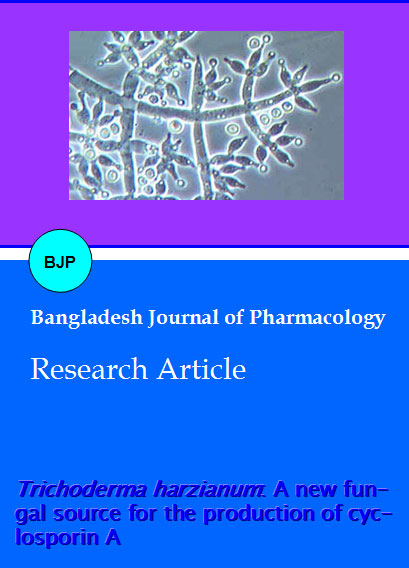
Trichoderma harzianum: A new fungal source for the production of cyclosporin
Abstract
Pure cultures of Trichoderma harzianum, T. aureoviride, T. reesei, T. koningii and T. hemantum were checked for their potential to produce cyclosporin A. Production medium used for drug production was consisted of: glucose, 5%; peptone, 1%; KH2PO4, 0.5%; KCL, 0.25% (w/v). Whereas, butyl acetate was used to extract the fermentation medium for cyclosporine A. This was then analyzed through high performance liquid chromatography and the chromatograms obtained were compared with that of cyclosporine and with the external standard cyclosporin A 98.5% pure. Only chromatogram of T. harzianum showed a peak at 2.78, which was comparable with both the standards. Mass spectroscopy of this peak showed [CsA + H] + ion of m/z 1203. The amount of drug calculated was 44.06 µg/mL.
Introduction
Cyclosporin A is a member of the group of cyclic undecapeptides with anti-inflammatory, mmunosuppressive, antifungal and antiparasitic properties (Sallam et al., 2005). It is used extensively in the prevention and treatment of graft-versus-host reactions in bone marrow transplantation and for the prevention of rejection of kidney, heart and liver transplants. Cyclosporin A is a major secondary metabolite usually produced by an aerobic filamentous fungus, Tolypocladium niveum (Park et al., 2006). Although cyclosporin A was initially developed as an antifungal antibiotic (Deo et al., 1984), it is currently prescribed as one of the most important immunosuppressive drugs for the treatment of organ transplants, as well as patients with autoimmune diseases, including AIDS, owing to its superior T-cell specificity and low levels of myelotoxicity (Borel, 1986). It consists of 11 amino acids with a molecular weight of 1202.6 and occurs as a white solid with a melting point of 148 to 151°C (natural) and 149 to 150°C (synthetic). It is stable in solution at temperatures below 30°C but is sensitive to light, cold, and oxidization (IARC, 1990).
The drug was first isolated from T. inflatum (Gams, 1971) and after that very few studies were planned to explore other sources for its production. Until now most of the research work related to cyclosporin A deals with T. inflatum and Aspergillus terreus. The present study was designed to explore Trichoderma species for this drug production.
Materials and Methods
Acquisition and maintenance of T. harzianum
Pure cultures T. harzianum (FCBP 140), T. aureoviride (FCBP 234), T. reesei (FCBP 271), T. koningii (FCBP 585) and T. hemantum (FCBP 907) were obtained from the First Fungal Culture Bank of Pakistan (FCBP) and main-tained on Malt extract agar (MEA) medium and preserved at 4ºC.
Seed inoculum preparation
Malt Yeast extract (MY) medium was selected as seed medium for inoculum preparation of selected fungi. MY medium is composed of malt extract 2%, yeast extract 0.4% and initial pH was adjusted to 5.7 using 1.0 M HCl. 50 mL of MY medium was prepared and poured in Erlenmeyer flasks (250 mL capacity) plugged with cotton plugs. These prepared medium flasks were sterilized by autoclaving at 121ºC and 15 lb/inch2 pressure for 15-20 min. By using cork borer, 0.8 cm disk of five days old cultures on MEA of Trichoderma species were inoculated in sterilized medium in flasks. The inoculated flasks were incubated on orbital shaker at 200 rpm for 72 hours at 30ºC (Borel et al., 1977). The inoculum of all the strains was prepared by the same procedure.
Cultivation
For cultivation 50 mL production medium specially design for cyclosporin A production, was prepared with following composition: Glucose, 5%; peptone, 1%; KH2PO4, 0.5%; KCL, 0.25% (w/v), at pH 5.3. According to the methodology of Agathos et al. (1986) 5 mL of seed inoculum from each isolate was introduced into 250 mL Erlenmeyer flasks containing 50 mL of production medium. The fermentation was continued at 28 ± 1ºC for 10 days, at 200 rpm.
Cyclosporin A extraction
Harvested fermentation medium was mixed with 30 mL of butyl acetate and stirred at 200 rpm for 24 hours at 30ºC. Organic layer was formed after 24 hours, which was separated by separating funnel and evaporated under vacuum till dryness. Dried extract was weighed and dissolved in 30 mL methanol.
Measurement of fungal biomass
The aqueous layer of cultivation medium containing fungal pellets was filtered for harvest of biomass. The filtration was performed by using Whatman filter paper No. 1. The fresh biomass was collected on the filter paper by using a conical funnel. Filters were slightly rinsed with water 1-2 times for the washing of media and weighed on digital weighing machine. The filters were dried in oven at 40ºC and reweighed after cooling. Following formula was used to calculate the dry biomass:
Fungal dry biomass = weight of filter paper - weight of dry filtrate.
Cyclosporin A analysis
Level of cyclosporin A in the crude extract was analyzed by high performance liquid chromatography (HPLC). HPLC was carried out using Hitachi system consisting of L-2100/2130 pump, L-2420 UV-VIS detector with a detection span from 190 to 900 nm.
Analysis was done using a C18 column with a 5 µm particle size, and acetonitrile:methanol:water (42.5:20:37.5, v/v) as mobile phase at a flow rate of 0.8 mL/min with UV detection at 215 nm.
Cyclosporin A confirmation
For initial confirmation of cyclosporin A, Sandimmun Neoral® capsule (Novartis) containing 100 mg of cyclosporin was used. For this purpose 100 µL from capsule was taken with the help of micropippet and dissolved in 10 mL of methanol. Then, HPLC of this solution was carried out according to the conditions mentioned above.
The retention time and peak area of the samples were compared for final confirmation and quantification, with the external standard cyclosporin A 98.5% pure (Sigma-Aldrich, Fluka). The standard was analyzed under same conditions as described.
Estimation of cyclosporin A
The cyclosporin A level in crude extracts was determined by the following formula: % cyclosporin A by weight =
As Wr Vs x purity of reference ArWsVr
Where, As is area of sample peak; Ar is area of reference peak; Wr is weight of reference material in grams; Ws is weight of sample in grams; Vs is volume of sample; Vr is volume of reference material.
The area of sample peaks and of reference peak was calculated from the chromatograms obtained by HPLC program LaChrom Elite.
Mass spectroscopy
The ESI/MS spectrum was obtained from single-quadrupole mass spectrometer. Sample was introduced using a silica capillary at a flow rate of 4.0 µL/min. The nebulizer gas was optimized and set at a rate of 1.6 L/min, and an electrospray potential of 4200 V was applied in the interface sprayer. A curtain gas of ultrapure nitrogen was pumped into the interface at the rate of 1.2 L/min.
Result and Discussion
Trichoderma as antagonists controlling wide range of pathogens are well documented (Ajith and Lakshmidevi, 2010). As cyclosporin A is also known for its antifungal properties, five species of Trichoderma were checked for their potential to produce this drug. The drug extracted in butyl acetate was confirmed as cyclosporin A by comparing chromatograms with Sandimmun Neoral capsules containing cyclosporin A as active ingredient and pure cyclosporin A. Sandimmun Neoral capsules showed a clear peak at 2.77 whereas peak at 2.8 was recorded by authentic compound (Figure 1, Figure 2). In a previous study, Sallam et al. (2003) recorded cyclosporin A peak at 3.03 which was produced by Aspergillus terreus following almost similar experimental plan as is described in this study.
Figure 1: HPLC chromatogram of Sandimmun Neoral capsule extracted at 215 nm showing cyclosporin A peak at 2.77
Figure 2: HPLC chromatogram of Sandimmun Neoralâ capsule extracted at 215 nm. Showing cyclosporin A peak at 2.768
Among five tested Trichoderma species only T. harzianum (FCBP 140) isolated from air showed a comparable peak at 2.78 (Figure 3). The ESI-MS analysis showed the ion [CsA + H]+ at m/z 1203. The estimated yield of cyclosporine A was 44.1 µg/mL with a 0.26 g/50 mL of fungal biomass. No such peak was recorded in other tested species. However, this quantity was found lower to that recorded by earlier workers in the most studied fungal strain i.e., T. inflatum. As Dreyfuss et al. (1976) and Agathos et al. (1986) found a very good yield of 105.5 mg/L and Balakrishnan and Pandey (1996) showed that T. inflatum can produce higher levels of cyclosporin A up to 183 mg/L.
Figure 3: HPLC chromatograms of T. harzianum extracted at 215 nm. Peaks appeared at 2.779 in chromatogram identified as cyclosporin A
Beside T. inflatum so far this drug has been reported to be produced from Aspergillus terreus, Fusarium solani and Fusarium oxysporum (Sallam et al., 2003). This study confirms T. harzianum as a new fungal source of cyclosporine A production.
References
Agathos SN, Marshell JW, Maraiti C, Parekh R, Modhosingh C. Physiological and genetic factors for process development of cyclosporin fermentation. J Industrial Microbiol. 1986; 1: 39-48.
Ajith PS, Lakshmidevi N. Effect of volatile and non-volatile compounds from Trichoderma spp. against Colletotrichum capsici incitant of Anthracnose on Bell peppers. Nat Sci. 2010; 8: 265-69.
Borel JF, Feurer C, Magnee AC, Stahelin H. Effects of the new anti-lymphocytic peptide cyclosporin A in animals. Immunology 1977; 32: 1017-25.
Borel JF. Cyclosporin and its future In: Cyclosporin. Borel JF (ed). Progress in Allergy. Vol. 38. Switzerland, Karger, Basel, 1986, pp 9-18.
Balakrishnan K, Pandey A. Growth and Cyclosporin A production by an indigenously isolated strain of Tolypocladium inflatum. Folia Microbiol. 1996; 41: 401-06.
Deo YM, Gaucher GM. Semi-continuous and continuous production of penicillin G by Penicillium chrysogenum cells immobilized in κ-carrageenan beads. Biotechnol Bioeng. 1984; 2: 285-95
Dreyfuss M, Harri E, Hofmann H, Kobel H, Pache W. Tscherter H. Cyclosporin A and C: New metabolites from Trichoderma polysporum. European J Appl Microbiol. 1976; 3: 125–33.
Gams W. Tolypocladium. Eine Hyphomycetengattung mit geschwollenen Phialiden. Persoonia. 1971; 6: 185–91.
IARC. Pharmaceutical Drugs. IARC Monographs on the evaluation of carcinogenic risk of chemicals to humans. International Agency for Research on Cancer, 1990, pp 415.
Park NS, Park HJ, Han K, Kim ES. Heterologous expression of novel cytochrome P450 hydroxylasegenes from Sebekia benihana. J Microbiol Biotechnol. 2006; 16: 295-98.
Sallam LA, El-Refai AM, Hamdi AH, El-Minofi A, Abd-Elsalam. Studies on the applications of immobilization technique for the production of cyclosporin A by a local strain of Aspergillus terreus. J Gen Appl Microbial. 2005; 51: 143-49.
Sallam LRA, Abdel-Monem H, El-Refai, Abdel-Hamid A, Hassaan A, El-Minofi, Ibrahim S, Abdel-Salam. Role of some fermentation parameters on cyclosporin A production by a new isolate of A. terreus. J Gen Appl Microbial. 2003; 49: 321- 28.

