Antimicrobial activity of Leucas clarkei
Abstract
The antimicrobial potency of the whole plant of Leucas clarkei have been studied using the soxhlet extracts of petroleum ether, benzene, chloroform and ethanol extract against Gram-positive bacteria (Two strains), Gram- negative bacteria (Two strains) and two fungi strains by disc diffusion method. Microdilution methods, for the determination of minimal inhibition concentration (MIC) and the minimal bactericidal and fungicidal concentration (MBC, MFC). The ethanol extract at a concentration of 30 to 60 µg/disc and chloroform extract at a concentration 60 µg/disc showed significant activity against all the bacteria and fungus. All the extracts of L. clarkei have got moderate action but chloroform and ethanol extracts have got significant activity against Candida krusei, Candida albicans, Escherichia coli, Salmonella typhi, Staphylococcus aureus, and Bacillus subtilis. This may be due to phytochemicals such as phytosterols, alkaloid, tannins, phenolic compounds and flavonoids present in the extracts.
Introduction
Nature has been a source of medicinal agents for thousands of years and an impressive number of modern drugs have been isolated from natural sources; many of these isolations were based on the uses of the agents in traditional medicine (Owoabi, 2007; Cragg and Newman, 2001). This plant based traditional medicine system continues to play an essential role in health care, with about 80% of the worlds inhabitants relying mainly on traditional medicines for their primary health care (Farnsworth et al., 1985). According to World Health Organization, medicinal plants would be the best source to obtain a variety of drugs. Therefore, such plants should be investigated to better understand their properties, safety and efficacy (Doughari et al., 2008; Nascimento et al., 2000). Recently, scientific interest in medicinal plants has burgeoned due to the increased efficiency of plant derived drugs and raising concern about the side effects of modern medicine. The efficacy of current antimicrobial agents has been reduced due to the continuing emergence of drug resistant organisms and the adaptations by microbial pathogens to commonly used antimicrobials. Therefore, the search for new drugs from plants continues to be a major source of commercial drugs. Plant based antimicrobials represent a vast untapped source of medicines even after their enormous therapeutic potential and effectiveness in the treatment of infectious disease; hence, further exploration of plant antimicrobials needs to occur. The screening of plant extracts and their products for antimicrobial activity has shown that, higher plants represent potential sources of novel antibiotic prototypes (Aliero and Afolayan, 2006; Afolayan, 2003).
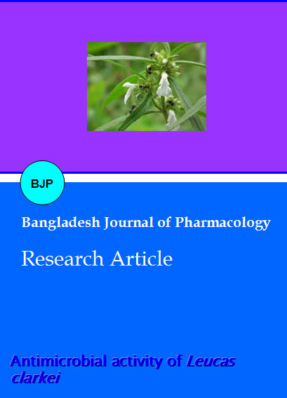
Plants of genus Leucas (Lamiacae) have been widely employed by the traditional healers to cure many diseased conditions which insinuated that this genus have immense potential for the discovery of new drugs or lead molecules. The genus Leucas comprises of about 80 species (Hedge, 1990). The highest species diversity has been found in East Africa (Ryding, 1998). In India, 43 species are available (Mukerjee, 1940). Plants of genus Leucas are generally shrubs, subshrubs, annual herbs or perennial herbs with woody root and/or stem-base. Leaves are opposite, entire or with spiky lobes, oval shaped with tapered end, petiolated or sometimes without intervening stalk. The axillary or terminal inflorescence is usually with indeterminate augmentation. Bracteoles are roughly erect. The calyx shape varies within the genus (often tuberlar shape) sometimes calyx enlarges into fruits. Calyx comprises of five connate sepals (one upper, two lateral and two lower) and 5-20 secondary lobes. Whitish hairs are generally present on the outer surface of the upper lip of the corolla, though yellowish cream color or red hair can also be present in some species (Bentham, 1835).
Leucas clarkei (Saxena, 1950 and Hooker, 1885) (Lamiaceae) is a rare species of Odisha which grow as annual herbs in most tropical countries and is widely distributed throughout India. The Tribal of Orissa uses this plant for the treatment of pain caused by obstruction in menstrual flow, epilepsy, sore throat and infectious diseases. It is widely used for healing of wounds (Haines, 1924). The aim of present study is under taken to evaluate its phytochemical and antimicrobial potential of whole plant extracts using disc diffusion assay for bacterial and fungi.
Materials and Methods
Materials
Solvents such as petroleum ether, benzene, chloroform and ethanol used were of analytical grade and obtained commercially from Merck-Limited, India, Mumbai.
Collection and identification of plant material
The plant L. clarkei were collected in the month of October from Bolangir, Odisha, India. The plant material was taxonomically identified and authenticated by Dr. Uma Devi, Head, Department of Botany, Govt. Women's College, Sambalpur, Odisha. A voucher specimen (GWC/B-315/09) has been deposited in the Herbarium of the Department of School of Pharmaceutical Education & Research, Berhampur University, India for future reference.
Preparation of plant material
The whole plant was first sun dried for several weeks, crushed by hands and dried again. Then the crushed parts of the plant were ground into coarse powder with the help of a mechanical grinder. By using the concept of the nature of solubility and distribution of the active ingredients, powdered material (1,500 g) was packed in soxhlet apparatus (Reuben et al., 2008; Harborne, 1998) and extracted successively with petroleum ether (60-80) (43 g) to deffat the drug, petroleum ether was removed from the powdered defatted drug which was then extracted with benzene (39 g), chloroform (41 g) and 95% of ethanol (43 g) as increasing polarity. The whole each mixture then underwent filtration through what man filter paper. The filtrates (petroleum ether, benzene, chloroform and ethanol filtrate) obtained were evaporated by rotary evaporator at 5 to 6 rpm and at 40ºC temperature. It rendered a gummy concentrates. The gummy concentrate was designated as crude extract which was then freeze dried and preserved at 4ºC.
Phytochemical screening
Qualitative phytochemical tests for the identification of alkaloids, flavonoids, steroids, glycosides, saponins, tannins and terpenoids were carried out for all the extracts by appropriate method (Sazada et al., 2009; Sofowora, 1993; Harborne, 1993; Kulkarni, 2006; Ndukwe et al., 2006).
The freshly prepared extracts of L. clarkei were qualitatively tested for the presence of chemical constituents. Phytochemical screening of the extracts was performed using the following reagents and chemicals: Alkaloids with Wagner reagent, flavonoids with the use of concentrated hydrochloric acid, tannins with 5% ferric chloride, saponins with ability to produce suds, gum with Molish reagents and concentrated sulfuric acid, steroids with sulfuric acid, reducing sugar with the use alpha-napthol and sulfuric acid and terpenoids with chloroform and concentrated hydrochloric acid.
Microbial cultures
The following microorganisms were used to test the activity of the extracts. Pure isolates of two Gram positive bacteria such as Escherichia coli (MTCC-443), Salmonella typhi and two Gram negative bacteria such as Staphylococcus aureus (MTCC-1430), Bacillus subtilis (MTCC-441) and fungi Candida albicans (MTCC-183), Candida krusei were collected from the Department of Life Science, Sambalpur University, Odisha, India.
Culture media
Nutrient broth, nutrient agar and Sabouraud dextrose agar (SDA) were purchased from the Himedia.
Reference antibiotics
Amoxycillin and Miconazole were used as reference antibiotics (RA) against bacteria and yeast, respectively. This was collected as standard sample from Medico Remedies Pvt. Ltd, Maharashtra, India.
Inoculum preparation
Nutrient broth and SDA were used for growing and diluting the microorganism suspensions. Inoculums from bacterial cultures were prepared by picking colonies from 24 hours old cultures. Colonies were suspended in 5 mL of a solution containing 0.145 mol of saline per liter. The density was adjusted by spectrophotometer to that of a 0.5 McFarland standard at a wavelength of 530 nm to yield a stock suspension of 1 x 10⁶ to 5 x 10⁶ cells per mL. Two different inoculums sizes were evaluated: 1 x 10⁶ to 5 x 10⁶ cells per ml and a 1:100 dilution containing 1 x 10⁴ to 5 x 10⁴ cells per mL. Plates were swabbed in three directions.
Fungal cultures were aseptically inoculated on petri dishes containing autoclaved, cooled, and settled SDA medium. The petri dishes were incubated at 31ºC for 48 hours to give white round colonies against a yellowish background. These were aseptically subcultured on SDA slants. The yeast colonies from SDA slants were suspended in sterilized 0.9% sodium chloride solution (normal saline), which was compared with McFarland solution. According to the manufacturer's directions, 1 mL of yeast suspension in normal saline was added to 74 mL of sterile medium and kept at 45°C to give a concentration of 2 x 10⁷ cells/mL.
Determination of MIC
Double strength nutrient agar was prepared in 500 mL of distilled water which was swirled and mixed thoroughly by heating to allow uniform dissolution, after which 5 mL of it was dispensed into universal bottles and sterilized in an autoclave at 121°C for 15 min. The agar was allowed to cool to 45ºC and each graded solution was then mixed gently with molten double strength nutrient agar in a petri dish and allowed to solidify for one hour. Extracts' concentrations of 100, 90, 80, 70, 60, 50, 40, 30, 20, 10 and 5 ug/mL (Ndukwe et al., 2006) respectively were prepared by serial dilution. Each plate was divided into six equal sections and labeled accordingly to correspond to six test organisms. Two 6 mm diameter paper discs (Whatman No. 1) were placed aseptically into each labeled section of the plate using sterilized forceps. With an automatic micropipette, 20 uL of each bacterial suspension was taken and transferred aseptically and carefully into each appropriate pre-labeled paper disc on the agar plates. The plates were incubated for 24 hours at 37°C after which they were observed for growths or death of the test organisms. In contrast, fungal cultures were incubated at 31°C for 48 hours in SDA medium. The lowest concentration inhibiting growth was taken as the minimum inhibitory concentration (MIC). The average of 3 values was calculated and that was the MIC for the test material.
Determination of MBC and MFC
This was carried out to know if the organisms could be killed completely or their growths could only be inhibited. Another set of plates of nutrient agar were prepared and sterilized in an autoclave as earlier described. The paper discs in all the plates from the MIC tests were reactivated. Emphasis was mostly paid to the MIC plates and the preceding plates. The reactivation was done in a mixture of 0.5% egg lecithin and 3% Tween 80 solution in a test tube. The reactivated organisms were sub-cultured into appropriately labeled quadrants of the sterilized nutrient agar plates using wire loop into each test tube and streaking uniformly on the labeled quadrants. This was then incubated for 24 hours at 37°C after which they were observed for growths (Ndukwe et al., 2006; Navarro et al., 1996). In contrast, fungal cultures were incubated at 31°C for 48 hours in SDA medium. The MBC was the quadrant with the lowest concentration of the extract without growth.
Disc diffusion assay (Navarro et al., 1998; Swain et al., 2008)
Stock solutions of different extracts of L. clarkei were prepared separately by initially dissolving 0.05 g of the extract in 0.5 mL of DMSO to obtain a stock solution of concentration 100 mg/mL. From this stock solution, concentrations of 1 mg/mL were prepared by serial dilution. From the above stock solution further dilutions were made to get 10, 20, 30, 40, 60 ug/mL working solutions. The cork and bore diffusion method of Bauer et al. (1966) and Barry and Thornsberry (1985) were used in the antimicrobial screening. Nutrient agar was inoculated with a microbial cell suspension (200 uL in 20 mL of medium) and poured into sterile petri dishes followed by cross-streaking with the same wire loop to achieve uniform spread on the plate. Sterile filter paper discs 6 mm in diameter were impregnated with 20 uL of each extract concentration (30 and 60 ug/ mL), which were prepared using the same solvents employed to dissolve the plant extracts, and placed on the inoculated agar surface. Standard 6 mm discs containing rifampicin 30 ug/disc was used as positive control. Negative controls were made using paper discs loaded with 20 uL of the solvents. After pre-incubation for 2 hours in a refrigerator the plates were incubated overnight at 37°C for 18-24 hours. In contrast, fungal cultures were incubated at 31°C for 48 hours in SDA medium using miconazole 10 ug/disc (Sur-Altiner et al., 1999). Negative controls were made using paper discs loaded with 20 uL of the solvents. At the end of the incubation period antimicrobial activity was evaluated by measuring the zones of inhibition.
Result and Discussion
Antimicrobial studies are conducted to investigate the antimicrobial potency of L. clarkei extracts against a few selected strains of bacteria and fungi. Before performing the antimicrobial screening it was important to check the MIC for bacteria. The determination of MBC and MFC were carried out to check whether the organisms could be killed completely or their growths only be inhibited. The ethanol and chloroform extract of L. clarkei exhibited significant activity against all the tested bacteria and fungi.
The zones of inhibition ranged from 4-20 mm for different extracts against bacteria and fungi. The alcoholic and chloroform extract had promising MIC values against all tested bacteria and fungi (Table I). The resistance of bacteria and fungi towards different drugs can be due to modification of the target site, by pass of pathways, decreased uptake (reduced intrace llular concentration of the antimicrobial agent, either reducing membrane permeability or by active efflux pump), enzymatic inactivation or modification of the drug, or over production of the target. The antimicrobial effects of L. clarkei may be attributed to various photochemical contained in its extracts, such as phytosterols, alkaloid, tannins, phenolic compounds and flavonoids (Table II).
Table I:Antibacterial and antifungal activity of Leucas clarkei (Whole plant)
| Microorganisms | Different extracts of Leucas clarkei | Amoxycillin (µg/disc) | Miconazole (µg/disc) | |||||||
|---|---|---|---|---|---|---|---|---|---|---|
| Petroleum ether (µg/disc) | Benzene (µg/disc) | Chloroform (µg/disc) | Ethanol (µg/disc) | |||||||
| 30 | 60 | 30 | 60 | 30 | 60 | 30 | 60 | 30 | 10 | |
| Bacteria | ||||||||||
| Escherichia coli | + | ++ | - | - | - | + | ++ | +++ | +++ | NT |
| Salmonella typhi | - | + | - | - | - | + | ++ | +++ | +++ | NT |
| Staphylococcus aureus | - | + | - | + | + | ++ | ++ | +++ | +++ | NT |
| Bacillus subtilis | - | ++ | + | + | + | + | + | ++ | +++ | NT |
| Fungi | ||||||||||
| Candida krusei | + | ++ | - | - | + | + | ++ | +++ | NT | +++ |
| Candida albicans | + | ++ | - | - | + | ++ | ++ | +++ | NT | +++ |
| Experiments were done in triplicate. Disc diameter = 4 mm; Diameter of zone of inhibition: - <4; + = 5-10; ++ = 11-15; +++ >16; NT = not tested. DMF had not shown any antimicrobial activity against the tested organisms | ||||||||||
Table II: Preliminary phytochemical analysis of Leucas clarkei
| Constituents | Petroleum ether extract | Benzene extract | Chloroform extract | Ethanol extract |
|---|---|---|---|---|
| Alkaloid | ||||
| Dragendorff's test | + | + | + | - |
| Meyer's test | + | + | + | - |
| Carbohydrates | ||||
| Mohch's test | - | - | - | - |
| Burford's test | - | - | - | - |
| Fehling (reducing sugar) | - | - | - | - |
| Fehling (combined reducing sugar) | - | - | - | - |
| Cardiac glycosides | ||||
| Killer-killanis test | - | - | + | + |
| Flavonoid | ||||
| Shinoda's test | - | - | + | + |
| FeCl3 test | - | - | + | + |
| Saponins | ||||
| Frothing test | - | + | + | + |
| Terpene and Steroids | ||||
| Salkowski test | + | + | + | - |
| Libarman-Burchard's test | + | + | + | |
| Tanins | ||||
| FeCl3 test | - | - | + | + |
| Lead acetate test | - | - | + | + |
| (+) = Present & (-) = Absent | ||||
Conclusion
Acknowledgements
The authors are thankful to Prof. A. K. Satpathy, Director and Prof. S. K. Mohapatra, Principal, Gayatri College of Pharmacy, Sambalpur, Odisha, India for providing necessary facilities to carry out this work.
References
Afolayan AJ, Extracts from the shoots of Arctotis arctotoides inhibit the growth of bacteria and fungi. Pharm Biol. 2003; 41: 22-25.
Aliero AA, Afolayan AJ. Antimicrobial activity of Solanum tomentosum. Afr J Biotechnol. 2006; 5: 369-72.
Bentham G. The genera and species of the plants of the order Labiatae with their general history, characters, affinities and geographical distribution. London, James Ridgway and Sons, 1835, p 602.
Cragg GM, Newman DJ. Medicinals for the Millennia. NY Acad Sci. 2001; 953: 3-25.
Doughari JH, El-Mahmood AM, Tyoylna I. Antimicrobial activity of leaf extracts of Senna obtusifolia (L). Afr J Pharm Pharmacol. 2008; 2: 7-13.
Farnsworth NR, Akerele O, Bingel AS. The value of plants used in traditional medicine for drug discovery. WHO. 1985; 63: 965-81.
Haines. Botany, Bihar & Orissa, 1924, p 750.
Harborne JB. Phytochemical methods. A guide to modern techniques of plant analysis. 3rd ed. London, Chapman and Hall, 1998, p 302.
Harborne JB. Phytochemistry. London, Academic Press, 1993, pp 89-131.
Hedge IC. Labiatae. In: Flora of Pakistan. Ali SI, Nasir YJ (ed). University of Karachi, Karachi, 1990, p 192.
Hooker JD. Flora of Brit.ish India, 1885, p 688.
Kulkarni SK. Handbook of experimental pharmacology. 3rd ed. Delhi, Vallabh Prakashan, 2006, pp 128-31.
Mukerjee SK. A revision of the Labiatae of the Indian Empire. Records of Botanical Survey of India, 14, 1940, pp 1-205.
Nascimento GGF, Lacatelli J, Freitas PC, Silva GL. Antibacterial activity of plant extracts and phytochemicals on antibiotic resistant bacteria. Braz J Microbiol. 2000; 31: 886-91.
Navarro V, Villarial ML, Rojas G, Lozoya X. Antimicrobial evaluation of some plants used in Mexican traditional medicine for the treatment of infectious diseases. J Ethnopharmacol. 1996; 53: 143-47.
Ndukwe IG, Habila JD, Bello IA, Okoh JI. Phytochemical and antimicrobial studies on the leaves and stem of Desmodium scorpiurus Der (sw). Afr J Biotechnol. 2006; 5: 1789-91.
Owoabi J, Omogbai EKI, Obasuyi O. Antifungal and antibacterial activities of the ethanolic and aqueous extract of Kigella africana (Bignoniaceae) stem bark. Afr J Biotechnol. 2007; 6: 882-85.
Reuben KD, Abdulrahman FI, Akan JC, Usman H, Sodipo OA, Egwu GO. Phytochemical screening and in vitro antimicrobial investigation of the methanolic extract of Croton Zambesicus Muell ARG, stem bark. Eur J Sci Res. 2008; 23: 134-40.
Ryding O. Phylogeny of the Leucas Group (Lamiaceae). Syst Bot. 23, 1998, pp 235-47.
Saxena HO, Brahman M. The flora of Orissa. 1950, p 1457.
Sazada S, Verma A, Rather AA, Jabeen F, Meghvansi MK. Preliminary phytochemicals analysis of some important medicinal and aromatic plants. Adv Biol Res. 2009; 3: 188-95.
Sofowora H. Screening plants for bioactive agents. In: Medicinal plants and traditional medicine in Africa. 2nd ed. Ibadan, Nigeria, Spectrum Books Ltd., 1993, pp 134-56.
Sur-Altiner D, Gürkan E, Mutlu G, Tuzlaci E, Ang O. The antifungal activity of Pancratium maritimum. 1999; 70: 187-89.
Swain SR, Sinha BN, Murthy PN. Antiinflammatory, diuretic and antimicrobial activity of Rungia pectinata Linn. and Rungia repens Nees. Indian J Pharma Sci. 2008; 70: 679-83.