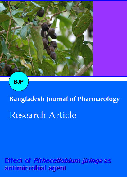
Effect of Pithecellobium jiringa as antimicrobial agent
Abstract
Pithecellobium jiringa is Malay traditional local delicacy. There is no local data on antimicrobial nature of P. jiringa is available. The objective of our study was to evaluate the antimicrobial activity of P. jiringa. Leaves, pods and seeds of P. jiringa were extracted using methanol. Ten test microorganisms were used in the study. The disc diffusion assay was used to determine the sensitivity of the samples, while the liquid dilution method was used for the determination of the minimal inhibition concentration (MIC). Chloramphenicol was used as a reference standard. The results revealed that all extracts of P. jiringa showed the antimicrobial and antifungal activities against the test organisms. Amongst the active extracts, the minimal inhibition concentration (MIC) determination showed that the extract of P. jiringa leaf was the most active against S. aureus, S. epidermidis and M. gypsum (100 mg/mL). The results provided evidence that the studied plants extract might be potential sources of new antimicrobial drug.
Introduction
Plants of the genus Pithecellobium mostly grow in southeast Asia (Lee et al., 1992). Pithecellobium belongs to the subfamily Mimosoideae. Mimosoideae is from family of Leguminosae (Allen & Allen, 1981). The Leguminosae is one of the largest families of flowering plants with 18,000 species classified into around 650 genera in the world (Harborne, 1994). There are 70 genera and 270 species of Leguminosae can be found in Malaysia (Corner, 1988).
In Malaysia, there are 12 species of Pithecellobium (Corner, 1988). Pithecellobium jiringa, one of the species of Pithecellobium is Malay traditional local delicacy. (Mohamed et al., 1987). Pithecellobium jiringa is known as jering in Malaysia. In Indonesia, it is known as djenkol (Areekul et al., 1976). P. jiringa seed is eaten raw or half boiled with rice and many believe that it has medicinal values. However, there is no local data on antimicrobial nature of P. jiringa is available. In the present study, antimicrobial activity of leaves, pods and seeds of P. jiringa were investigated.
Materials and Methods
Plant material
The leaves, pods and seeds of P. jiringa were collected from Air Itam Dam, Penang, Malaysia.
Preparation of the extract
One kilogram of coarsely powdered dry leaves, pods and seeds of P. jiringa were successively extracted using a Soxhlet apparatus with 95% methanol as solvent, at 55°C for 3 hours. The resultant extracts were then concentrated to dryness in a rotary evaporator under reduced pressure at a temperature of 40°C. The stock solution containing 1,000 mg/mL (w/v) were prepared in the 95% methanol and then further diluted in distilled water.
Microorganisms studied
Ten test microorganisms were used namely, Klebsiella sp., Pseudomonas aeruginosa, Salmonella typhi, Staphylococcus aureus, Staphylococcus epidermidis, Bacillus subtilis, Candida albicans, Microsporum canis, Microsporum gypsum and Trichophyton rubrum. The microorganisms were obtained from Microbiology Lab, School of Biology, Universiti Sains Malaysia, Penang, Malaysia.
Antimicrobial activity test
The agar disc diffusion method was used to determine the sensitivity of the samples. The bacterial strains were grown on nutrient agar while fungal strains were grown on sabouraud dextrose agar. The plates were prepared by pouring 30 mL of media into sterile 90 mm Petri dish. The sterile Whatman no. 1 filter papers (6 mm diameter) were placed on the spread-surface of the Petri dishes and 3 uL of dried extract was spotted on each of the filter papers. Blank discs impregnated with sterile distilled water were used as negative controls, and discs of chloramphenicol (30 mg) as positive control. The plates were incubated overnight at 37°C for bacterial strain and 30°C for 3 days for fungal stain. Each experiment was repeated at least three times and the mean of the diameter of the inhibition zones was calculated. Antimiorobial activities were indicated by clear zones of growth inhibition.
Determination of the minimum inhibitory concentrations (MIC)
Minimum inhibitory concentrations (MIC) were determined by the liquid dilution method. The estimate of the MIC was carried by using P. jiringa leaf extract against the organisms. Dilution series were set up with 0.5, 1.5, 8.5, 15.0, 30.0, 55.0, 100.0, 130.0, 165.0, 250.0, 350.0 and 500.0 mg/mL of Sabouraud glucose broth medium. Chloramphenicol was used as a reference standard. The lowest concentration which did not show any growth of the tested microorganism after macroscopic evaluation was determined as the MIC. All data represent at least three replicated experiments per microorganism.
Effect of P. jiringa leaf extract on B. subtilis
Two milliliter of B.subtilis suspension was added into nutrient broth containing P. jiringa leaf crude extract to give a final dilution of 250 mg/mL. The mixture was then incubated at 30°C for 24 hours. Sterile distilled water was used as control. A few drops of the test mixture were also fixed for light and scanning electron microscopy studies.
Result and Discussion
Herbal medicine has been practiced especially in developing countries for many years. It is used for the traditional treatment of health problems (Cragg and Newman, 2002). Numerous studies have been conducted with the extracts of various plants, screening antimicrobial activity as well as for the discovery of new antimicrobial compounds (Cowan, 1999; Gordon and David, 2001; Parekh et al., 2006). Scientific experiments on the antimicrobial properties of plant components were first documented in the late 19th century (Zaika, 1975).
In recent years, antimicrobial resistance among pathogens is becoming a serious concern especially due to the indiscriminate use of antimicrobial drugs (El-Mahmood et al., 2008). Furthermore, antibiotics are sometimes associated with adverse effects to the host. Thus, there is a need to develop alternative antimicrobial drugs from medicinal plants for the treatment of infections (Cordell, 2000).
The results of the antimicrobial activity tests of P. jiringa extracts (Table I) revealed that all extracts of P. jiringa showed the antimicrobial and antifungal activities against the test organisms. The diameters of growth inhibition area of extracts studied were in the range 10.5-25.5 mm. No activity was seen against Trichophyton rubrum and Microsporum canis. Our results showed a remarkable antibacterial activity of P. jiringa leaf extract compared to P. jiringa pod and seed extracts.
Table I: Antimicrobial activities of the leaf, pod and seed of P. jiringa extracts
| Test micro-organisms | Zone of inhibition (mm) | ||
|---|---|---|---|
| Leaf | Pod | Seed | |
| Bacteria | |||
| Klebsiella pneumonia | 24.0 | 20.5 | 15.3 |
| Pseudomonas aeruginosa | 18.0 | 20.5 | 10.5 |
| Salmonella typhi | 25.0 | 17.7 | 17.2 |
| Staphylococcus aureus | 25.5 | 20.9 | 12.5 |
| Staphylococcus epidermidis | 24.1 | 16.5 | 13.1 |
| Bacillus subtilis | 21.3 | 21.7 | 15.2 |
| Fungi | |||
| Microsporum canis | - | - | - |
| Microsporum gypsum | 15.3 | 13.2 | 5.2 |
| Trichophyton rubrum | - | - | - |
| Yeast | |||
| Candida albicans | 19.7 | 20.7 | 9.4 |
Table II showed the result of the minimal inhibition concentration (MIC) of P. jiringa leaf extract. The results of the MIC on Gram-positive and negative bacteria as well as on fungi confirmed the antimicrobial potency of P. jiringa leaf extract as previously observed by the disc diffusion assay. The MIC values ranging between 100 and 220 mg/mL and maximum zone of inhibition ranging between 10.3 and 17.0 (mm). Amongst the active extracts, the MIC determination showed that P. jiringa leaf extract was the most active against S. aureus, S. epidermidis and M. gypsum (100 mg/mL). Figure 1 showed comparison of MIC values and maximum zone of inhibition of S. aureus when using P. jiringa leaf extract and chloramphenicol. It could pointed out that the inhibition zones obtained in the liquid dilution method of P. jiringa leaf extract against S. aureus are comparable to those shown by chloramphenicol. However, it was observed that the inhibition zones of the control antibiotic 30 times higher compared to those shown by P. jiringa leaf extracts. The MIC determination is important in giving a guideline to the choice of an appropriate and effective dose of a therapeutic substance.
Figure 1: Comparison of zone of inhibition of chloramphenicol and P. jiringa leaf extract against S. aureus
Table II: Minimal inhibition concentration (mg/mL) of P. jiringa leaf extract on test microorganisms
| Microorganisms | Minimal inhibition concentration (MIC) (mg/mL) | Maximum zone of inhibition (mm) |
|---|---|---|
| Bacteria | ||
| Klebsiella pneumonia | 100 | 10.3 |
| Pseudomonas aeruginosa | 100 | 12.5 |
| Salmonella typhi | 130 | 12.8 |
| Staphylococcus aure- | 165 | 15.0 |
| Staphylococcus epi- dermidis | 220 | 12.2 |
| Bacillus subtilis | 130 | 13.3 |
| Fungi | ||
| Microsporum gypsum | 220 | 17.0 |
| Yeast | ||
| Candida albicans | 100 | 12.2 |
Microscopic observation on the effect of the extract on P. jiringa was done by scanning electron microscopy studies (Figure 2). The most prominent change seen was the morphology of the bacteria. The untreated culture revealed abundant cylindrical shaped bacteria, whereas the treatment culture showed the shrunken and collapsed bacteria. This phenomenon could be due to the leakage of the cell wall or perhaps some alteration in the membrane permeability and resulting in the loss of the cytoplasm. This could lead to the loss in rigidity of the bacteria and finally cause the death of the cells.
Figure 2: Scanning electron microscopy study of B. subtilis after exposure to 250 mg/mL of P.jiringa leaf extracts for 24 hours. (A) Untreated culture (control), showing many cylindrical shaped of bacteria (2000x). (B) Treated culture, showing the collapse and shrunken bacteria (5000x)
To the best of our knowledge, the antimicrobial activity of P. jiringa is being reported for the first time. The present study provides an important basis for the use of extracts from P. jiringa for the treatment of infections associated with the studied microorganisms. The results provided evidence that the studied plant extracts might be potential sources of new antimicrobial drug. However, it is important to point out that further work is needed to find out the active and pure compounds from the crude extracts which can then be tested for antibacterial and antifungal activities.
Conclusion
This study shows that methanol extracts of leaves, pods and seeds of P. jiringa have antibacterial and antifungal activities against the test pathogens.
References
Allen ON, Allen EK. The leguminosae: A source book of characteristics, uses and modulation. Wisconsin, Wisconsin Press, 1981.
Areekul S, Kirdudom P, Chaovanapricha K. Studies on djenkol bean poisoning (djenkolism) in experimental animals. Southeast Asian J Trop Med Public Health. 1976; 7: 551–58.
Cragg GM, Newman DJ. Drugs from nature: Past achievements, future prospects. In: Iwu MM, Wootton JC (eds). Ethnomedicine and drug discovery. Amsterdam, Elsevier Science, 2002, pp 23-37.
Cordell GA. Biodiversity and drug discovery a symbiotic relationship. Phytochemistry 2000; 55: 463-80.
Corner EJH. Wayside trees of Malaya. Vol 1. Singapore, Government Printing Office, 1988, pp 396–464.
Cowan MM. Plant products as antimicrobial agents. Clin Microbiol Rev. 1999; 12: 564–82.
El-Mahmood AM, Doughari JH, Ladan N. Antimicrobial screening of stem bark extracts of Vitellaria paradoxa against some enteric pathogenic microorganisms. Afr J Pharm Pharmacol. 2008; 2: 89-94.
Gordon MC, David JN. Natural product drug discovery in the next millennium. Pharmaceut Biol. 2001; 139: 8–17.
Harborne JB. Phytochemistry of the Leguminosae. In: Phytochemical dictionary of the Leguminosae. Bisby F.A. (ed) London, Chapman & Hall, 1994.
Lee MW, Morimoto S, Nonaka GJ, Nishioka J. Flavon 3-ol gallates and proanthocyanidines from Pithecellobium lobatum. Phytochemistry. 1992; 31: 2117-20.
Mohamed S, Rahman MSA, Sulaiman S. Some nutritional and anti-nutritional components in jering Pithecellobium-jeringa, keredas Pithecellobium-microcarpum and petai Parkia-speciosa. Pertanika 1987; 10: 61–68.
Parekh J, Karathia N, Chanda S. Screening of some traditionally used medicinal plants for potential antibacterial activity. Indian J Pharm Sci. 2006; 68: 832-34.
Zaika LL. Spices and herbs: Their antimicrobial activity and its determination. J Food Safety. 1975; 9: 97–118.

