N-acetyl glucosamine improves intestinal mucosal barrier function in rat
Abstract
Our study investigated the effect of N-acetylglucosamine (GlcNAc) on the intestinal mucosal barrier function in rats. Rats were randomly assigned into normal control group, diarrhea-predominant irritable bowel syndrome (IBS-D) group and GlcNAc group. IBS-D was introduced into the IBS-D group without any treatment. The GlcNAc group were treated with GlcNAc. Microvilli and tight junctions of intestinal epithelial cells were detected. The D-lactic acid level and diamine oxidase (DAO) activity in the serum were determined. Compared with normal rats, microvilli were sparsely distributed on the intestinal epithelial cells, the tight junction gap also widened, and D-lactic acid level and DAO activity were significantly higher in the IBS-D group. After GlcNAc treatment, the microscopic structure of the intestinal mucosa became largely normal, and the level of D-lactic acid and the DAO activity were lowered. In conclusion, GlcNAc can effectively improve the intestinal mucosal barrier dysfunction, perhaps through enhancing the cellular metabolism.
Introduction
The intestinal mucosa mainly functions to form a barrier that separates luminal contents from the interstitium. Intestinal epithelial cells communicate with each other via the intercellular junctions, and the glycocalyx on the epithelial cells is also a part of the intestinal mucosal barrier. In particular, the gastrointestinal epithelial lining consists of a monolayer of columnar cells that are held together by circumferential intercellular junctions to form a selectively permeable barrier to the luminal contents (Clayburgh et al., 2004). Among the intercellular junctions, tight junctions typically locate at the apical end of the lateral intercellular space. The tight junctions is a highly dynamical structure that may regulate the paracellular movement of fluid and solutes and other physiological processes such as glucose absorption (Turner et al., 1997). It undergoes rapid changes in response to inflammation (Turner et al., 1997; Berkes et al., 2003), and a number of pathogens and bacterial toxins may influence the epithelial permeability by modulating the tight junctions (Berkes et al., 2003; Fasano and Nataro, 2004; Al-Sadi et al., 2010). The intestinal epithelial barrier dys-function, including the abnormalities in tight junctions and epithelial permeability, has been observed in a number of intestinal disorders such as diarrhea-pre-dominant irritable bowel syndrome (IBS-D) (Dunlop et al., 2006) and inflammatory bowel disease (IBD).
To date, few effective drugs or agents have been developed to repair the defective intestinal barrier. Previous clinical trials suggest that N-acetyl glucosamine (GlcNAc), a monosaccharide derivative of glucose, may have therapeutic potential for IBS. Since GlcNAc has been shown to play important roles in regulating the cell distribution, stabilizing mast cells, repairing the gastric mucosal barrier (Liu et al., 2007) and enhancing the intestinal respiratory enzyme activity (Liu et al., 2007), the present study aimed to investigate the role of GlcNAc in repairing the intestinal mucosal barrier in a rat IBS-D model.
Materials and Methods
Animals: Wistar rats weighing 200-220 g were purchased from the Animal Research Institute of Third Military Medical University. Animals were grown in an environment at 24 ± 2ºC with the humidity of 45-60%, and were given ad libitum access to food and water.
Randomly selected rats were monitored for defecation frequency and fecal water content for 3 days. These rats were intraperitoneally injected with 1 mL of 10 mg/mL sodium cefuroxime once daily for 3 consecutive days. Seven days after the last injection, Sichuan pepper immersion fluid was intragastrically administered twice daily for 3 consecutive days. The ratio of fluid volume (mL) to the body weight (g) was 1:8. Fecal water content was subsequently recorded. Animals in which the fecal water content significantly increased following sodium cefuroxime treatment, returned to normal level after termination of drug treatment, and reincreased in response to sodium cefuroxime treatment were selected as IBS-D rats.
The experimental design has been previously described (Liu et al., 2007). Briefly, 24 IBS-D rats were divided into 2 equal groups. In the IBS-D group, rats had no treatment. In the GlcNAc group, rats were orally given 2 mL of 32.5 mg/mL GlcNAc thrice daily for 7 consecutive days.
Reagents: GlcNAc was purchased from Jiangsu Jiangshan Pharmaceutical Co., Ltd., Cefuroxime sodium was from William Glaxo. D-lactic acid detection kit was purchased from Biopharm (Germany). Other reagents included diamine oxidase (DAO) detection kit, adlitteram (diagnostic laboratories), and 2.5% glutaraldehyde.
Instruments: A video microsystem was composed of multifunctional microscope (Olympus, Japan), JVC TK-C1381camera, JVC TM-1700PN-S monitor, VS2000 acquisition card equipped with the 1394 acquisition card program, Sharp and VCRs. Others included: 3111REL #5 carbon dioxide incubator (Forma Scientific); a TGL-16G high-speed centrifuge (Shanghai Medical Analytical Instrument Factory); Bio-RAD 550 Microplate Reader; TECNAI-10 transmission electronic microscope (PHILIPS, Netherlands); DU-800UV spectrophotometer (Beckman USA).
Transmission electron microscopy (TEM): Colon specimens were collected from rats in different groups at the indicated time points, fixed in 2.5% glutaraldehyde and processed for TEM. The microvilli and tight junctions were observed under a transmission electron microscope.
Measurement of serum D-lactic acid level and DAO activity: In the presence of D-lactate dehydrogenase, D-lactic acid is oxidized by nicotinamide adenine dinucleotide (NAD) into pyruvate, generating NADH. To measure the serum level of D-lactic acid, the absorbance was detected at 334, 340 and 365 nm using the D-lactic acid detection kit. Serum DAO activity was measured according to the manufacturer's intructions.
Results
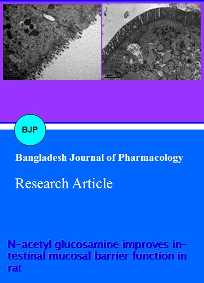
From the 2nd day of the experiment, IBS-D rats were either untreated or treated with GlcNAc for 7 days. On the 14th day, ileocecal mucosa was collected from animals in different groups, and the changes in microvilli and tight junction were observed under a transmission electron microscope. The microvilli were sparsely distributed on the intestinal epithelial cells of IBS-D rats and the tight junction gap was widened when compared with the control group (Figure 1). In the GlcNAc treated rats, the morphology of the microvilli and tight junction was similar to that in the control group. These results indicate that GlcNAc significantly improves the microvilli and tight junction, two components crucial for intestinal mucosal barrier function.
Figure 1: Electron microscopy of cell membrane and microvilli (Mv) in the intestinal mucosa of IBS-D rats with or without GlcNAc treatment. Arrow: Tight junction
Serum was collected on the 7th and 14th day. Serum D-lactic acid level and DAO activity were subsequently determined via biochemical assays.
Serum D-lactic acid level is commonly used as an indicator of intestinal permeability (Nielsen et al., 2011). D-lactic acid is a metabolic product of intestinal flora, and does not enter the circulation under normal conditions. The increase in D-lactic acid in the blood serves as a marker for enhanced intestinal mucosal permeability. The IBS-D rats displayed enhanced serum D-lactic acid level as compared to normal rats (p<0.05; Table I). After treatment with medium-dose GlcNAc, serum D-lactic acid level returned to nearly normal level on the 7th day. At 14 days, there was no significant difference (p>0.05) in the D-lactic acid level between the IBS-D group and the GlcNAc group. These results indicated that the intestinal permeability was improved following GlcNAc treatment, suggesting the improvement in intestinal mucosal barrier.
Table I:Serum D-lactic acid level after GlcNAc treatment
| Group | 7 days | 14 days |
|---|---|---|
| GlcNAc | 9.4 ± 0.7 | 9.0 ± 0.3 |
| IBS-D | 13.1 ± 1.9 | 10.5 ± 0.9 |
| control | 8.5 ± 0.4 | 8.3 ± 0.4 |
| n = 6; data are mean ± SD (µg/mL) | ||
Serum DAO is a marker for mammalian intestinal mucosa. Following the damage to the mucosal barrier, DAO activity which is related to the impaired synthesis of nucleic acids and proteins is increased. As shown in Table II, the IBS-D rats exhibited significantly higher DAO activity than those in the control group. In contrast, GlcNAc treatment markedly reduced the DAO activity in a time-dependent manner, and the serum DAO activity returned to nearly normal level on the 14th day. There was no significant difference in the DAO activity between the GlcNAc group and the control group (p>0.05).
Table II: Serum DAO activity after GlcNAc treatment
| Group | 7 days | 14 days |
|---|---|---|
| GlcNAc | 1.1 ± 0.2 | 0.7 ± 0.1 |
| IBS-D | 3.7 ± 0.2 | 3.0 ± 0.2 |
| Control | 0.6 ± 0.1 | 0.6 ± 0.1 |
| n = 6; data are mean ± SD (µg/mL) | ||
Discussion
In this study, TEM showed marked abnormality in the microvilli and tight junction in rats with IBS-D. Furthermore, the serum DAO activity and D-lactate level were also significantly enhanced in the IBS-D rats. Previous studies have shown that the increase in D-lactate level and DAO activity reflects the increase in the intestinal permeability and decline in the anabolic metabolism respectively (Nielsen et al., 2011; Tsujikawa et al., 1999). Under normal conditions, the intestinal epithelial tissues maintain the structural integrity by continuous cell proliferation, supported by robust synthetic activities in epithelial cells. However, following inflammation or stress, animals display reduction in both respiratory metabolism and ATP content (Liu et al., 2007). In the present study, results also demonstrated the increase in serum DAO activity in IBS-D rats. The compromise in the synthetic ability of intestinal epithelial cells may impair the cell proliferation, leading to the widening of intercellular gaps, the mucosal barrier dysfunction, and eventually the increase in intestinal mucosal permeability.
The IBS is a chronic, relapsing, and variably disabling bowel disorder characterized by the presence of abdominal pain or discomfort in association with altered bowel habits. IBS is further subtyped into one of IBS with constipation (IBS-C), IBS with diarrhea (IBS-D), or mixed IBS (IBS-M) based on the predominant stool pattern. (Longstreth et al., 2006; Sadik et al., 2010) Abdominal discomfort is the most common symptom in IBS-D patients, likely as a result of mucosal barrier dysfunction (Fennessy and Warrillow, 2012). In the pilot clinical trials, results showed the effectiveness of GlcNAc in IBS-D patients. Structurally, GlcNAc is an acetylated derivative of glucosamine (Ciszewicz et al., 2007). There is evidence showing that GlcNAc can promote the proliferation of bacillus bifidus and regulate the intestinal microecology (Garrido et al., 2012). The regulation of intestinal microecology has been found to be crucial for the maintenance of integrity of gastrointestinal barrier (Khailova et al., 2008). GlcNAc is also a precursor in the synthesis of glycol-proteins. GlcNAc serves as a monosaccharide in the initiation of synthesis of 50% glycoproteins (Zawalich et al., 1979), which is helpful for the maintenance and supplement of glucose based structure on the intestinal epithelial cells. In addition, the glucose based structure is also an important component of intestinal barrier. Once entering the cells, GlcNAc can enter the hexosamine biosynthetic pathway (HBP) via direct phosphorylation and then modify the proteins in the cytoplasm and nucleus by O-GlcNAc, which enhance the capability of proteins and cells to adapt to the environmental stress (Hart et al., 2007; Chatham et al., 2008). In addition, it also efficiently maintains the normal structure (including microstructure) and normal metabolism. These may explain why the intestine of IBS rats treated with GlcNAc had normal ultrastructure under an electron microscope and these rats had normal metabolism (DAO) and structure in intestinal epithelial cells. The O-GlcNA modification of proteins in cells may efficiently regulate the phosphorylation of serine and threonine, which is important for the normal function of tight junction proteins. Studies also demonstrate that GlcNAc can inhibit the production of IL-1 and IL-6 (Shikhman et al., 2001), two cytokines detrimental to intestinal barrier function (Al-Sadi et al., 2010). This might be a major reason for the stabilization of intestinal barrier. Taken together, GlcNAc can repair the intestinal mucosal barrier function. The improvement of intestinal barrier function reduces the penetration of intestinal endotoxin and metabolites from foods and bacteria, which attenuates the stimulation to submuco sal nerve endings and mast cells leading to the improvement of symptoms in IBS-D patients.
Our results showed that GlcNAc was able to improve the damage to the microvilli and tight junction of intestinal epithelial cells. Moreover, the serum D-lactic acid level and DAO activity also concomitantly enhanced following the treatment, suggesting that the improvement of mucosal barrier dysfunction may be due to the enhanced intestinal mucosa cellular activities.
Conclusion
Our results provide experimental evidence for a possible role of GlcNAc in the treatment of abdominal discomfort, and suggest putative mechanisms underlying the improvement of mucosal barrier dysfunction by GlcNAc in IBS-D.
Contribution
Yanxia Liu and Weiwei Xu contributed equally in this study.
Acknowledgment
We would also like to appreciate Mr. Qianglin Duan for critical reading of the manuscript.
References
Al-Sadi R, Ye D, Said HM, Ma TY. IL-1β-Induced Increase in Intestinal Epithelial Tight Junction Permeability Is Mediated by MEKK-1 Activation of Canonical NF-κB Pathway Original Research Article. Am J Pathol. 2010; 177: 2310-22.
Berkes J, Viswanathan VK, Savkovic SD, Hecht G. Intestinal epithelial responses to enteric pathogens: Effects on the tight junction barrier, ion transport, and inflammation. Gut. 2003; 52: 439-51.
Chatham JC, Nöt LG, Fülöp N, Marchase RB. Marchase, Hexosamine biosynthesis and protein O-glycosylation: The first line of defense against stress, ischemia, and trauma. Shock 2008; 29: 431-40.
Ciszewicz M, Wu G, Tam P, Polubinska A, Breborowicz A. Changes in peritoneal mesothelial cells phenotype after chronic exposure to glucose or N-acetylglucosamine. Translational Res. 2007; 150: 337-42.
Dunlop SP, Hebden J, Campbell E, Naesdal J, Olbe L, Perkins AC, Spiller RC. Abnormal intestinal permeability in subgroups of diarrhea-predominant irritable bowel syndromes. Am J Gastroenterol. 2006; 101: 1288-94.
Fasano A, Nataro JP. Intestinal epithelial tight junctions as targets for enteric bacteria-derived toxins. Adv Drug Deliv Rev. 2004; 56: 795-807.
Fennessy GJ, Warrillow SJ. Gastrointestinal problems in intensive care. Anaest Int Care Med. 2012; 13: 152-57.
Garrido D, Ruiz-Moyano S, Mills DA. Release and utilization of N-acetyl-d-glucosamine from human milk oligosaccharides by Bifidobacterium longum subsp. infantis. Anaerobe 2012; 18: 430-35.
Hart GW, Housley MP, Slawson C. Cycling of O-linked beta-N-acetylglucosamine on nucleocytoplasmic proteins. Nature. 2007; 446: 1017-22.
Liu JK, Hu JC, Huang M. Study on the Influence of AWA on ATP Content of Intestinal Epithelia of IBS Rats. Biowave Sci Appl Oncol. 2007; 1: 29-31.
Liu JK, Hou WN, Wang J. Experimental study on choosing stimulating methods of establishing animal model. Biowave Sci Appl Oncol. 2007; 1: 15-17.
Longstreth GF, Thompson WG, Chey WD, Houghton LA, Mearin F, Spiller RC. Functional bowel disorders. Gastroenterology. 2006; 130: 1480-91.
Ludmila Khailova, Andrew Maynard, Katerina Dvorak, Kelly M. Arganbright, Melissa Halpern, Toshi Kinouchi, Masako Yajima, Bohuslav Dvorak. W1774 Bifidobacterium bifidum improves intestinal barrier function in experimental necrotizing enterocolitis. Gastroenterology 2008; 134: A712-13.
Liu Jk, Wu XL, Chen J, Xu QW. Effect of GlcNAc against activation of bacterial cryptic growth cells to mast cells. Acta Acad Med Mil Tert. 2007; 29: 579-81.
Nielsen C, Lindholt JS, Erlandsen EJ, Mortensen FV. D-lactate as a marker of venous-induced intestinal ischemia: An experimental study in pigs. Int J Surg 2011; 9: 428-32
Sadik R, Bjornsson E, Simren M. The relationship between symptoms, body mass index, gastrointestinal transit and stool frequency in patients with irritable bowel syndrome. Eur J Gastroenterol Hepatol. 2010; 22: 102–08.
Shikhman AR, Kuhn K, Alaaeddine N, Lotz M. N-acetylglucosamine prevents IL-1-mediated activation of human chondrocytes. J Immunol. 2001; 166: 5155– 60.
Schenk M, Mueller C. The mucosal immune system at the gastrointestinal barrier. Best Pract Res Clin Gastroenterol. 2008; 22: 391-409.
Turner JR, Rill BK, Carlson SL, Carnes D, Kerner R, Mrsny RJ, Madara JL. Physiological regulation of epithelial tight junctions is associated with myosin light-chain phosphorylation. Am J Physiol. 1997; 273: C1378-85.
Tsujikawa T, Uda K, Ihara T, Inoue T, Andoh A, Fujiyama Y, Bamba T. Changes in serum diamine oxidase activity during chemotherapy in patients with hematological malignancies. Cancer Lett. 1999; 147: 195-98.
Zawalich WS, Dye ES, Matschinsky FM. Metabolism and insulin releasing capabilities of glucosamine and N-acetylglucosamine in isolated rat islets. Biochem J. 1979; 180: 145–52.