Anti-tumor activity of cabozantinib by FAK down-regulation in human oral squamous cell carcinoma
Abstract
Cabozantinib is a tyrosine kinase inhibitor involved in inhibition of cell proliferation and colony formation. We studied anti-cancer properties of cabozantinib in oral squamous cell carcinoma cells. The viability of BHY and HSC-3 cells decreased with increase in cabozantinib concentration and time. The proliferation of cell lines was affected by increasing concentration of cabozantinib from 0.3 to 1.2 uM after 48 hours of treatment. The expression of MET and phosphorylated MET was not affected by cabozantinib treatment. Cabozantinib-treated cells when compared to control, showed concentration-dependent increase in BHY and HSC-3 cells during G2/M phase and decrease in S phase with an increase in cabozantinib concentration. Annexin-V/propidium iodide double staining showed that cells with annexin-V increased with the increase in cabozantinib concentration. The expression of apoptosis- related proteins cleaved caspase-3 and cleaved-PARP were increased with increase in cabozantinib concentration. It was also found that suppression of FAK activation and expression was dose-dependent. The results from this study revealed that cabozantinib can be useful in developing a drug for effective treatment of oral squamous cell carcinoma cells.
Introduction
Oral squamous cell carcinoma is one of the most known cancers in the universe, with a degree of 300,000 new cases identified in 2012 (Ferlay et al., 2012). The analysis underlying the cause of oral squamous cell carcinoma is not completely understood but it is believed that alcohol, tobacco usage, chewing the betel are the major influences which are involved in the formation of carcinogenesis (Chen et al., 2008). The collection of genetic alteration leads to uncontrolled proliferation in the mutated oral squamous cell lines whereas the damage in the genetic components leads to an uncontrolled multiplication of mutant oral keratinocyte cells (Chianeh and Prabhu, 2014). The cellular vital response functions of both chemotherapy and irradiation are the cause of apoptosis (Eriksson and Stigbrand, 2010; Jonathan et al., 1999; La Vecchia et al., 2004; Ocker and Hopfner, 2012; Ricci and Zong, 2006).
The treatment possesses multi-modal activities comprises of radiotherapy, surgery, and chemotherapy (Seiwert and Cohen, 2005) but all these methods are still not satisfactory due to their adverse effects.
Cabozantinib is considered as a multi-targeted tyrosine kinase receptor with chiefly possessing MET, and vascular endothelial growth factor (VEGFR2) inhibition characteristics. MET is a hepatocyte growth factor which is widely expressed in adult tissues and incriminated in postnatal physiological and pathophysiological events. Cabozantinib is a TKI which targets three related pathways in MET, MTC, VEGFR2, and RET (Yakes et al., 2011). Herein, we showed the anti-tumor effects of cabozantinib on two Oral squamous cell carcinoma cell lines (BHY and HSC-3), and probe into the potential underlying mechanisms.
Materials and Methods
Materials
Recombinant Human Hepatocyte Growth Factor (HGF), Dubecco's modified Eagle's medium (DMEM), fetal bovine serum and MTT were purchased from Life Techologies (Carlsbad, USA). Cabozantinib (Tradename: COMETRIQ) was purchased from Exelixis (San Francisco, USA). Crystal violet was purchased from Solarbio (Beijing, China). Propidium iodide, RNase, annexin VFICT/propidium iodide apoptosis detection kit and antibodies to MET, phosphorylated MET, cyclin B1, phosphorylated histone H3, cyclin A, cleaved caspase-3, cleaved PARP-1, FAK and phosphorylated FAK, beta-actin, FAK specific siRNA were purchased from Cell Signaling Technology (Danvers, USA). PVDF membrane and chemiluminescent reagents were from EMD Millipore (Massachusets, USA). Antibody to pan-caspase inhibitor z-VAD-FMK was purchased from Santa-Cruz Biotechnology (USA)
Cell lines and cell culture
The human oral squamous cell carcinoma cell lines BHY and HSC-3 were purchased from American Type Culture Collection (ATCC, USA). BHY and HSC-3 cells were routinely cultured with DMEM supplemented with 10% heat-inactivated fetal bovine serum. Cell cultures were maintained at 37°C incubator with an atmosphere of 5% CO2. To treat the cells with cabozantinib, cells were grown up to 60% confluence. The cells were then treated with various concentration of cabozantinib ranging from 0 to 1.0 µM for 0-48 hours. Cells treated with DMSO were used as the control for comparison. After the cells were treated with cabozantinib, trypsin was used to detach the cells and collected for posterior analysis.
MTT assay
The inhibition of cell growth by cabozantinib was studied in BHY and HSC-3 cells. Various concentrations of the cabozantinib for 24, 48 and 72 hours and the viability of the cells was assayed by MTT. Cell viability assay was carried out in a 24-well plate. BHY and HSC-3 cells at a density of 5,000 cells/well were grown in a 24-well plate (Corning, USA). After treatment with drug, the culture medium was replaced with fresh medium containing 10% MTT (5 mg/mL in PBS) and incubated at 37ºC for 4 hours. Then, the medium with MTT was discarded and DMSO was added to dissolve the newly formed formazan crystals. Absorbance was measured in a microplate reader (Sunrise, Tecan) at a 540 nm wavelength. The cell viability was calculated by normalizing the absorbance to control and the results were represented as a percentage of the control.
Colony formation assay
The colony formation of BHY and HSC-3 cells after treatment with cabozantinib was studied. Various concentrations of the cabozantinib for 24, 48 and 72 hours and the viability of the cells was assayed by MTT. 6-well plates (1,000 cells/well) at single cell density were used for colony forming assay. BHY and HSC-3 cells were pre-cultured for 24 hours. The cells were treated after 24 hours with the various concentration of cabozantinib or DMSO from 0 to 1.0 µM for additional 48 hours. The medium was replaced with fresh medium to allow the cell growth for 10 days. Methyl alcohol was used to fix the cells for 15 min and stained with crystal violet for 30 min. Colonies consisting of more than 50 cells were considered for counting. The colony forming ability of the cells treated with cabozantinib was normalized with the control cells without drug treatment.
Flow cytometry analysis
To know further about the inhibitory effect of cabozantinib on cell growth, flow cytometry was used to measure the cell cycle progression by DNA quantification. BHY and HSC-3 cells were pre-cultured for 24 hours in DMEM medium. Cells were treated with cabozantinib or DMSO and incubated for 24 hours. The cells were harvested by using trypsin and washed with PBS twice. The cells were fixed with ice-cold 75% ethanol at 20°C overnight and stained with 500 mL of propidum iodide at a concentration of 50 mg/mL containing 0.1% RNase (1 mg/mL) for 15 min in dark at room temperature for cell cycle assay. Annexin-V/propidum iodide double staining was used to study the role of cabozantinib in inducing apoptosis in oral squamous carcinoma cancer cells by quantifying the cells that undergo apoptosis. For apoptosis assay, the cabozantinib-treated cells were suspended in 100 mL of annexin-V-FITC binding buffer and 5 mL of annexin V-FITC and 5 mL of propidum iodide were used for staining in dark at room temperature. The stained cells were analyzed by flow cytometry (BD FACSCanto II, BD Biosciences, USA). The results were shown as mean values from three independent value.
Western blot analysis
To elucidate the role of cabozantinib in the cell cycle, the expression of G2/M phase regulatory proteins (Vermeulen et al., 2003) cyclin B1, cyclin A and phosphor-histone H3 by Western blot. The cells after the drug treatment were harvested and lysed using RIPA buffer with protease inhibitor cocktail (Sigma-Aldrich, USA). The protein concentration was quantified using BCA protein assay kit (Thermo Scientific, USA). The cell extracts were denatured and analyzed by SDS-PAGE. 12% SDS-PAGE was used to separate the proteins and transferred to PVDF membrane. The proteins in the membrane were blocked with 5% non-fat milk, and incubated with the corresponding primary. The membrane with proteins was then washed thrice with TBS and 0.05% Tween-20. After washing, it was incubated with HRP-conjugated secondary antibody raised in goat at 1:5000 dilution. Membranes were incubated with chemiluminscent substrate for 5 min and scanned with Image Quant LAS4000 (GE Life Sciences, USA). The density of the bands was estimated by standard methods and normalized by division with the density of control protein bands. All the experiments were done in triplicate.
SiRNA mediated FAK knockdown
Focal Adhesion Kinase, FAK is a non-receptor tyrosine kinase protein usually overexpressed and highly activated in most of the solid-tumors like oral squamous cell carcinoma (Kornberg, 1998; de Vicente et al., 2013). FAK as a protein with multiple functions helps in regulating the signals from extracellular environment such as integrins and growth factor receptors. It is also involved in cell mitosis and organizing the cytoskeleton and acts as a protein scaffold (Schaller, 2010). So, the anti-tumor function of cabozantinib through inhibition of cabozantinib was tested. SiRNA for FAK or control were transfected into cells using lipofectamine 2000. The FAK knockdown was carried out according to manufacturer's protocol. After 48 hours of transfection, total cell lysates were prepared to assess the knockdown efficiency by Western blot. Meanwhile, after 24 hours of transfection, cells were treated with or without cabozantinib for additional 24 hours. The viability of the cells were assessed by MTT assay. The cell viability was calculated by normalizing the absorbance to control and the results were represented as the percentage of the control.
Statistical analyses
All experiments were done in triplicate. Graphpad prism software was used to analyze the data. Student's t-test or one-way ANOVA was used to differentiate between mean values. P-values of <0.05 were considered to be significant.
Results
Cell growth and proliferation in BHY and HSC-3 cells
Figure 1A shows that BHY and HSC-3 cells treated with cabozantinib had decreased cell viability with response to increase in concentration and time. The IC50 values of cabozantinib in BHY cells at 24, 48 72 hours were 0.72, 0.53 and 0.24 uM respectively. For HSC-3 cells, the IC50 values were 0.62, 0.39 and 0.25 uM at 24, 48, 72 hours respectively.
Figure 1B shows that cells treated with cabozantinib at increasing concentration of 0.3, 0.6 and 1.2 uM could inhibit cell colony formation dependent on its concentration at 48 hours
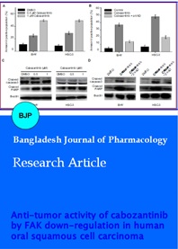
Cabozantinib has been reported to selectively inhibit MET, so whether the cytotoxic effect of cabozantinib is dependent on HGF/MET pathway inhibition was examined. BHY and HSC-3 cells with the various concentration of cabozantinib treated for 24 hours and MET and phosphorylated expression levels were identified by western blot. Figure 1C shows that MET and phosphorylated MET expression were not affected by the drug treatment in both BHY and HSC-3 cells. Meanwhile, BHY and HSC-3 cells pre-treated with HGF (15 ng/mL) for 6 hours and treated with 0.5 mM cabozantinib for another 24 hours. Figure 1D shows that HGF pretreatment did not affect the antiproliferative effect of cabozantinib in both the cell lines.
Figure 1: Cabozantinib effect on cells proliferation and colony formation. (A) Cells proliferation after cabozantinib treatment by MTT. (B) Colony formation analysis after treatment with cabozantinib for 48 hours. (C) Western blotting of MET expression in cabozantinib treated cells for 24 hours with beta-actin as control. (D) Effect of HGF treatment with cabozantinib on BHY and HSC-3 cells. Data was obtained from mean of triplicate experiments
Effect of cabozantinib in cell cycle arrest
Figure 2A shows that when compared to control cells, cabozantinib-treated cells in G2/M phase showed concentration-dependent increase in BHY and HSC-3 cells while a decrease in S phase accordingly with the concentration of cabozantinib. Figure 2B shows cyclin B1 and phosphor-histone H3 expression increased with increase in the concentration of cabozantinib expression, whereas the cyclin A expression was decreased.
Figure 2: Cell cycle analysis in cabozantinib-treated cells. (A) Histogram analysis of cell cycle distribution after 0.5 or 1 µM cabozantinib treatment. (B) Immunoblotting of proteins involved in cell cycle regulation. beta-Actin was used as total control. Data was obtained from three independent experiments
Apoptotic signaling pathway
Figure 3A shows that total amount of cells with annexin-V increased with corresponding increase in cabozantinib concentration in both BHY and HSC-3 cells. Immunoblotting analysis revealed the expression levels of proteins associated with apoptosis such as cleaved-caspase-3 and cleaved-PARP were increased after treating with cabozantinib in a concentration-dependent manner as shown in Figure 3C. BHY and HSC-3 cells pre-treated with pan-caspases inhibitor z-VAD (30 mM) for 4 hours and then treated with 2mM of cabozantinib for 24 hours. Figure 3B shows that in both cell lines cabozantinib induced apoptosis were reduced by pan-caspase inhibitor, z-VAD. Meanwhile, z-VAD also blocked the cleavage of caspase-3 and PARP- mediated by cabozantinib (Figure 3D).
Figure 3: Cabozantinib induced apoptosis. (A) Quantitative analysis of apoptotic cells. (C). Immunoblotting of proteins associated with apoptosis with beta-actin as control. (B) z-VAD, a caspase inhibitor reduces cabozantinib induced apoptosis. (D) Western blotting of cleaved caspase-3 and PARP induced by cabozantinib and z-VAD. Data was obtained from three independent experiments
Down-regulation of FAK
Figure 4A shows that decrease in total expression of phosphorylation at Tyr397 of FAK in both the cell lines with an increase in cabozantinib concentration. Through specific siRNA, FAK was knocked down in both BHY and HSC-3 cells resulted in decreased expression of total FAK and phosphorylated levels Tyr397 as shown in Figure 4B. Figure 4C shows the cell proliferation was reduced in FAK knockdown cells in both the cell lines. Moreover, knockdown of FAK can make the cancer cells more vulnerable to cabozantinib treatment as seen in Figure 4D.
Figure 4: Suppression of FAK signaling pathway by cabozantinib treatment. (A) Western blotting of FAK and phosphorylated FAK expression by 0.5 or 1 uM of cabozantinib for 24 hours with DMSO as negative control. (B) Western blotting of expression of FAK and phosphorylated FAK in FAK siRNA transfected cells or control siRNA. (C) Cell proliferation after FAK knockdown by MTT assay. (D) Cell proliferation detection affected by FAK knockdown and cabozantinib treatment by MTT assay
Discussion
We studied the anticancer activity of cabozantinib and the mechanism in which cabozantinib acts on OSCC cell lines BHY and HSC-3 cells. It was found that there was strong cytotoxic activity by cabozantinib on proliferation and colony formation in both oral squamous cell carcinoma cell lines. Meanwhile, the total expression and MET phosphorylation were not affected by cabozantinib at the concentrations used for cytotoxicity. HGF, the ligand of MET, when treated with the cells, did not attenuate the inhibitory effect cabozantinib. This proves that cabozantinib can be used as an effective chemotherapeutic compound against oral squamous cell lines through MET independent signaling pathway. Some of the other MET inhibitors such as PHA-665752 and crizotinib caused cell cycle arrest in G0/G1 phase (data not shown), whereas cabozantinib treatment had caused a decrease in G2/M in both BHY and HSC-3 cells. The decrease in cyclin A and increase in cyclin B1 prove that cabozantinib-treated cells exit in G2 phase and enter into the mitotic phase. The cell cycle analysis reveals that both BHY and HSC-3 cells exit in G2 phase and undergoes mitotic inhibition after cabozantinib treatment This was further evaluated by the up-regulation histone H3 phosphorylation which usually gets phosphorylated at serine during cell mitosis by Aurora B (Hirota et al., 2005).
The mode of action of chemotherapeutic drugs in anti-cancer treatment is targeting the cell apoptosis. Caspase enzymes plays an important role in apoptosis regulation extrinsically (Ola et al., 2011). Caspase cascade was cleaved one by one after it gets the apoptotic signal from death receptor which then leads to activation of effector caspases especially caspase-3. Then the cellular substrates were cleaved by activated effector caspases such as poly-ADP-ribose polymerase and apoptotic signals were amplified to induce cell apoptosis (Porter and Janicke, 1999). This study showed that cabozantinib induced cleavage of caspase-3 in a concentration dependent manner. When pan-caspases inhibitor z-VAD was used these effects were attenuated which indicates the important role of death receptor-mediated caspases activation in cabozantinib-induced apoptosis. The results suggest that caspases dependent apoptosis can be induced by cabozantinib in oral squamous carcinoma cells.
Many studies are providing satisfactory evidence that FAK overexpression and activation resulted in increases cell survival rate and proliferation, angiogenesis and metastasis and tumor invasion (Miyazaki et al., 2003; McLean et al., 2005). A small molecule found as FAK scaffolding inhibitor Y15 can decrease cancer growth in thyroid cancer and leads to synergistic action with other chemotherapeutic agents (O'Brien et al., 2014). Some of the other FAK inhibitors such as VS-4718, PF-00562271 and VS-6063 have been tested clinically and have shown promising results in cancer patients.
So, it is clear that FAK signaling suppression can be targeted as an efficient way of cancer treatment (Lee et al., 2015). The current study showed that the overall expression and phosphorylation of FAK was affected by cabozantinib treatment with response to concentration. siRNA-mediated silencing of FAK enhanced the anti-tumor activity of cabozantinib and reduced the cell proliferation showing that cabozantinib treatment is associated with FAK signaling pathway and not MET signaling pathway in OSCC cells. So the results proved that FAK signaling pathway can be blocked by cabozantinib treatment which can be a part of anti-tumor function of cabozantinib. To the best of our knowledge, this has to be the first report on FAK signaling inhibition by cabozantinib. This can give new insights to understand the molecular mechanism of cabozantinib. It is still unclear whether cabozantinib interacts directly or indirectly with FAK and blocks its activation and has to be explored further.
Conclusion
Cabozantinib treatment can be used to suppress cell growth, affects cellular mitosis and also induces apoptosis in human oral squamous carcinoma cells with suppression of FAK signaling pathway.
References
Chen YJ, Chang JT, Liao CT, Wang HM, Yen TC, Chiu CC, Lu YC, Li HF, Cheng AJ. Head and neck cancer in the betel quid chewing area: Recent advances in molecular carcinogenesis. Cancer Sci. 2008; 99: 1507-14.
Chianeh YR, Prabhu K. Biochemical markers in saliva of patients with oral squamous cell carcinoma. Asian Pac J Trop Dis. 2014;4 Suppl 1: S33-40.
de Vicente JC, Rosado P, Lequerica-Fernández P, Allonca E, VillallaÃn L, Hernández-Vallejo G. Focal adhesion kinase overexpression: Correlation with lymph node metastasis and shorter survival in oral squamous cell carcinoma. Head Neck. 2013; 35: 826-30.
Eriksson D, Stigbrand T. Radiation-induced cell death mechanisms. Tumour Biol. 2010; 31: 363-72.
Ferlay J, Soerjomataram I, Ervik M, Forman D, Bray F, Dikshit R, Elser S, Franken NA, Rodermond HM, Stap J, Haveman J, van Bree C. Clonogenic assay of cells in vitro. Nat Protoc. 2006; 1: 2315-19.
Hirota T, Lipp JJ, Toh BH, Peters JM. Histone H3 serine 10 phosphorylation by aurora B causes HP1 dissociation from heterochromatin. Nature 2005; 438 (7071): 1176-80
Jonathan EC, Bernhard EJ, McKenna WG. How does radiation kill cells? Curr Opin Chem Biol. 1999; 3: 77-83.
Kornberg LJ. Focal adhesion kinase expression in oral cancers, Head Neck. 1998; 20: 634-39.
La Vecchia C, Lucchini F, Negri E, Levi F. Trends in oral cancer mortality in Europe. Oral Oncol. 2004; 40: 433–39.
Lee BY, Timpson P, Horvath LG, Daly RJ. FAK signaling in human cancer as a target for therapeutics. Pharmacol Ther. 2015; 146: 132-49.
Mathers C, Rebelo M, Parkin DM. GLOBOCAN. In: Estimated cancer incidence, mortality, and prevalence worldwide in 2012. 2012.
McLean GW, Carragher NO, Avizienyte E, Evans J, Brunton VG, Frame MC. The role of focal-adhesion kinase in cancerda new therapeutic opportunity. Nat Rev Cancer. 2005; 5: 505-15.
Miyazaki T, Kato H, Nakajima M, Sohda M, Fukai Y, Masuda N, Manda R, Fukuchi M, Tsukada K, Kuwano H. FAK overexpression is correlated with tumour invasiveness and lymph node metastasis in oesophageal squamous cell carcinoma. Br J Cancer. 2003; 89: 140-45.
O'Brien S, Golubovskaya VM, Conroy J, Liu S, Wang D, Liu B, Cance WG. FAK inhibition with small molecule inhibitor Y15 decreases viability, clonogenicity, and cell attachment in thyroid cancer cell lines and synergizes with targeted therapeutics. Oncotarget. 2014; 5: 7945-59.
Ocker M, Hopfner M. Apoptosis-modulating drugs for improved cancer therapy. Eur Surg Res. 2012; 48: 111-20.
Ola MS, Nawaz M, Ahsan H. Role of Bcl-2 family proteins and caspases in the regulation of apoptosis. Mol Cell Biochem. 2011; 351: 41-58.
Porter AG, Jänicke RU. Emerging roles of caspase-3 in apoptosis. Cell Death Differ. 1999; 6: 99-04.
Ricci MS, Zong WX. Chemotherapeutic approaches for targeting cell death pathways. Oncologist 2006; 11: 342-57.
Schaller md, Cellular functions of FAK kinases: Insight into molecular mechanisms and novel functions. J Cell Sci. 2010; 123:1007-13
Seiwert TY, Cohen EE. State-of-the-art management of locally advanced head and neck cancer. Br J Cancer. 2005; 92(8): 1341-48.
Vermeulen K, Van Bockstaele DR, Berneman ZN. The cell cycle: A review of regulation, deregulation and therapeutic targets in cancer, Cell Prolif. 2003; 36: 131-49.
Yakes FM, Chen J, Tan J, Yamaguchi K, Shi Y, Yu P, Qian F, Chu F, Bentzien F, Cancilla B, Orf J, You A, Laird AD, Engst S, Lee L, Lesch J, Chou YC, Joly AH. Cabozantinib (XL184), a novel MET and VEGFR2 inhibitor, simultaneously suppre-sses metastasis, angiogenesis, and tumor growth. Mol Cancer Ther. 2011; 10: 2298-08.