Development of a novel antimicrobial peptide AWRK6
Abstract
We have previously identified an antimicrobial peptide called dybowskin2-CDYa (Dy2) in the cutaneous secretion from the Chinese frog Rana dybowskii. In this study, we used Dy2 as a template to prepare some novel peptides with improved stability and hydrophobicity. The antimicrobial activities exerted by these peptides against Gram positive and Gram negative bacteria were evaluated by MIC and CFU assays under physiological conditions. One peptide, AWRK6, was 2-4 fold more potent than Dy2, and was toxic to most of the bacterial strains tested, exhibiting a faster killing rate. AWRK6 was most potent in alkaline environments, and this was related to the more highly organized secondary structure of the peptide, as revealed by circular dichroism spectroscopy. Furthermore, AWRK6 was more resistant than Dy2 to degradation by trypsin. The improved properties of AWRK6 suggested that the peptide could potentially be further developed into an antibiotic.
Introduction
Increases in antibiotic resistance among pathogenic bacteria and the declining rate of conventional antibiotic development are two major concerns in modern medicine. These two factors, therefore, become the driving force in the search for novel drugs against pathogenic bacteria (Shlaes and Spellberg, 2012). There is an urgent need to find new types of antibiotics to maintain our existing capability to treat infectious diseases, especially those caused by multidrug-resistant (MDR) organisms (Afacan et al., 2012). Antimicrobial peptides (AMPs) are important and highly conserved components of innate immune responses in most multicellular organisms. They provide fast-acting, non-selective protection against microbial infections (Boman, 1995; Zasloff, 2002). Amphibian skin has proven to be one of the richest sources of AMPs. Hundreds of amphibian AMPs have been isolated to date and many of these can be isolated in large quantities. They are effective against a broad range of microorganisms at micromolar concentrations. However, none of the peptide has been developed for clinical use (Conlon and Sonnevend, 2010).
Dybowski's frog (Rana dybowskii) lives in stagnant ponds in the valley groves of northeastern China as well as in Korea and Japan. Its skin has been used extensively in Traditional Chinese Medicine for many centuries to promote wound healing. The antimicrobial components in the skin secretions of Dybowski's frog may contribute to its efficacy. To understand the biodiversity and nature of antimicrobial peptides from this frog, we have previously used cDNA-sequencing and mass-spectrometry approaches to analyze the skin and its secretions, and identified dybowskin2-CDYa (Dy2), a novel antibacterial peptide (SAVGRHSRRFGLRKHRKH, GenBank number: ACF08009.1) having low sequence conservation with antibacterial peptides from other amphibians (Jin et al., 2009a). Dy2 has several characteristics that favor its development as a peptide antibiotic, and these two characteristics are the net positive charge and high pI (12.60) of the peptide, which enable it to inhibit the growth of many Gram negative and Gram positive pathogens (Jin et al., 2009a; Jin et al., 2009b) and yet exert only low toxicity to mammalian cells. Opportunities for further improvement of the peptide are suggested by the presence of arginine residues. It has been previously shown that substituting Lys for Arg in cationic peptides can enhance their antimicrobial activity by about 2-fold while reducing their hemolytic activity (Yang et al., 2003).
In this study, we developed a novel peptide called AWRK6 by enhancing its hydrophobicity and stability through alteration of the amino acid sequence of Dy2. The antimicrobial activity of AWRK6 was tested against several Gram positive and Gram negative bacteria. The structure and various physical properties of the peptide were also characterized, and the mechanism of action against Staphylococcus aureus was investigated.
Materials and Methods
Bacterial strains Escherichia coli (ATCC 25922), Pseudomonas aeruginosa 01 (ATCC 15692), and S. aureus (ATCC 29213) were purchased from ATCC. Pseudomonas aeruginosa 508 was a kind gift from Dr. D. Radzioch (Department of Medicine, McGill University and MUHC, Montreal, Canada). S. aureus 8325-4, Δhla, Δhlb and Newman (WT strain) were kindly provided by Dr. D. Nguyen (Department of Medicine, McGill University and MUHC, Montreal, Canada).
Biochemical synthesis of peptides
Peptides were synthesized by GL Biochem Ltd. (Shanghai) using a solid-phase synthesis protocol. Synthesis was performed at the 0.5 mM scale with a coupling time of 1 hour and O-benzotriazole-N,N,N',N'- tetramethyluronium-hexafluoro phosphate (HBTU) /N-hydroxybenzotriazole (HOBt) as coupling reagents followed by a 30 min deprotection reaction. The Kaiser test was performed after the coupling step and the coupling reaction was repeated. The peptides were cleaved from the resin by treatment with 95% trifluoroacetic acid for 1 hour at 0°C. The crude peptides were subjected to further purification via sephadexTM-15 column chromatography using 10% (v/v) phosphoric acid/double distilled water as the eluent. The sequences of the synthesized peptides were confirmed by reversed-phase HPLC and electro-spray ionization mass spectrometry (HPLC-ESI-MS).
Minimal inhibitory concentration assay
Stock cultures of bacterial strains were grown in Mueller-Hinton Broth (MHB) for overnight at 37°C. The bacterial cells were harvested from the cultures by centrifugation, washed with PBS (pH 7.2, 10 mM) and then resuspended in MHB to give a working concentration of 1 x 10⁵ cfu/mL. MIC was evaluated using a modified method designed for cationic antimicrobial peptides (Giacometti et al., 2000). Briefly, the antimicrobial peptide was dissolved in 0.01% acetic acid containing 0.2% BSA and then subjected to a series of double dilutions to obtain concentrations ranging from 512 ug/mL down to 0.98 ug/mL, and 10 µL of each diluted peptide was dispensed into 96-well plates. Aliquots of bacterial suspension (90 μL) were then dispensed into each well containing the peptide and incubated at 37°C for overnight, and the OD of the plate was then measured at 650 nm using a Synergy Mx microplate reader (BioTek Instruments). The MICs for each of the peptides was determined for each bacterial strain. Each experiment was performed at least three times.
Hemolysis activity assay
The hemolytic activity of the peptide was measured using human red blood cells (hRBCs) as described previously (Jin et al., 2009a). Briefly, hemolysis was measured by incubating hRBCs in PBS containing the peptide at 37°C for 10 min. The hRBCs accounted for 10% of the volume in the final reaction mixture and the concentration of peptide was 100 mg/mL. After centrifugation at 1,000 xg for 10 min, the OD of the supernatant at 350 nm was measured. Hemolytic activity was expressed as the absorbance relative to that obtained by treatment of hRBCs with 0.1% Triton X-100 (100%) and PBS, which was used as negative control (0%). This experiment was performed in triplicate.
Antimicrobial activity assay
Bacterial cultures were prepared from cells taken from a fresh TSB culture plate. Aliquot of the overnight culture was then diluted into fresh TSB medium and grown to mid-logarithmic phase. The cells were harvested and resuspended in sterile 10 mM PBS (pH 7.2) to a concentration of 3 x 10⁶ cfu/mL. The bacterial suspension was then diluted to 3 x 10⁴ cfu/mL with 100 mM sodium phosphate buffer containing 1% TSB buffered at different pHs and the cell suspension was placed in a microcentrifuge tube. The peptide was added to the cells to a final concentration of 100 ug/mL. After incubating for a specific time (0-180 min, with shaking at 220 rpm) at 37°C, the cells were harvested and the CFU was determined, by placing 10 µL of a diluted sample on TSB plate and incubated at 37°C for overnight. The number of colonies appearing on the plate was counted and cell viability was expressed as percentage of the control (0 hour incubation) at each pH value tested.
Circular dichroism (CD) spectroscopy
CD spectra were recorded with a Chirascantm CD spectrometer (Applied photophysics, USA) in the range of 180-260 nm. For each experiment, three scans were averaged and corrected by subtracting the contributions by phosphate buffer and solvents without peptides. The peptide was dissolved in 10 mM sodium phosphate buffer having different pHs (pH 7, 8 and 9) and 30 mM SDS to a final concentration of 0.5 mg/mL. CD spectral data were plotted and analyzed for secondary structure using CDNN, a deconvolution program which is based on neural network theory (www.photophysics.com).
Susceptibility to proteolytic degradation
To examine the susceptibility of the peptides to proteolysis, the peptide was diluted in 10 mM phosphate buffer (pH 7.5) to a final concentration of 2 mg/mL and incubated with trypsin at 37°C for up to 4 hours using an enzyme-peptide molar ratio of 1:4 x 10⁶. After incubation, the sample was resolved by SDS-PAGE using 18% gel. The gel was visualized by staining with Coomassie R-250 (Bio-Rad) and the peptide bands were quantified by densitometry using Ezquantgel software (Ezquant, Israel).
Transmission electron microscopy (TEM)
The effect of the peptide on the morphology of S. aureus was assessed by SEM. S. aureus was cultured in LB medium to a log phase, and then harvested by centrifugation at 1,000 x g for 10 min. The cells were washed twice with PBS, and resuspended in sterile PBS to a concentration of 106 cfu/mL. One microliter of AWRK6 (10 mg/mL) was added to 1 mL of bacterial suspension and cultured for 1-4 hours at 37°C. For the negative, control, the same volume of PBS was added instead of the peptide. After incubation, the bacterial cells were harvested by centrifugation, washed twice with sterile PBS and fixed for 3 hours with 3% glutaraldehyde. They were then washed again with PBS (two washes) and were fixed again for 4 hours with 1% osmic acid, followed by further washes in PBS. After that, the cells were dehydrated sequentially with 30%, 50%, 70%, 90% and 100% ethanol, and further dehydrated in different ratios of ethanol: Acetone (3:1, 1:1 and 1:3) for 20 min, and then washed with pure acetone for 20 min. The bacterial cells were first embedded in a mixture of acetone and Epon812 embedding fluid (2:1) for 3-4 hours at room temperature, then in acetone and Epon812 embedding fluid (1:2) for overnight at room temperature, and finally in pure Epon812 embedding liquid for 2-3 hours at 37°C. The embedded cells were dried at 45°C for 12 hours and then at 60°C for 48 hours. Ultrathin slices were prepared from the embedded cells and double stained with 3% acetic acid uranium and lead citrate. These slices were subjected to transmission electron microscopy using a JEM-2100 electron microscope (Japanese Electronics Co., Ltd.).
Results
Modifications of the peptide
To improve the binding affinity of Dy2, all of its Arg residues were replaced with Lys, which selectively bind anionic liposomes with higher affinity compared to the binding with zwitterionic liposomes (Yang et al., 2003). In addition, an Ala residue situated in the cationic region of Dy2 was substituted with Trp to increase the hydrophobicity of the peptide (Rose and Wolfenden, 1993). These modifications yielded an analogue peptide (Designated as AWRK6: SAVGRHGRRFGLRKHRKH) with a much lower instability index than Dy2 (Table I). Other predicted properties of AWRK6 that might affect the function of the peptide are also shown in Table II. AWRK6 also became more hydrophobic after substituting Arg with Lys, as shown by the grand average of hydropathicity (GRAVY) index. The calculated pI value of AWRK6 was slightly lower than the value of Dy2, but there was no difference in the net positive charge (+10) between the two peptides. The alpha-helical structures of Dy2 and AWRK6 were predicted using DSC (Discrimination of protein secondary structure class) (King and Sternberg, 1996; Combet et al., 2000). The alpha-helical content of AWRK6 predicted by Network Protein Sequence Analysis (http://npsa-pbil.ibcp.fr) was longer than that of Dy2 (66.7% vs 61.1%). Taken together, these analytical and computational results suggested that modifying the sequence of Dy2 improved the stability and helical propensity of the peptide. Since the activity of an antimicrobial peptide often increases with increased helical content, we next examined whether AWRK6 has greater antimicrobial activity than Dy2.
Table I:Forecast feature of dybowskin-2CDYa and AWRK6
| Mm | PI | GRAVY | Instability index | Net charge | Lipid soluble index | |
|---|---|---|---|---|---|---|
| Dybowskin-2CDYa | 2155.5 | 12.6 | -1.7 | 52.2 | 10 | 43.3 |
| AWRK6 | 2130.5 | 10.8 | -1.6 | -6.7 | 10 | 37.8 |
Table II: MICs of dybowskin-2CDYa and AWRK6
| Organisms | MIC50 (ug/mL) | |||
|---|---|---|---|---|
| Dyboswkin-2CDYa | AWRK6 | |||
| Polystyrene | Polypropylene | Polystyrene | Polypropylene | |
| Gram negative | ||||
| E. coli ATCC | 128 | 64 | 64 | 32 |
| P. aeruginosa 01 | 128 | 64 | 32 | 16 |
| P. aeruginosa 508 | >512 | >512 | >512 | >512 |
| Gram positive | ||||
| S. aureus (ATCC 29213) | 64 | 32 | 16 | 4 |
| S. aureus (Newman strain) | 64 | 32 | 16 | 4 |
| S. aureus (WT8325-4) | 128 | 64 | 32 | 8 |
Antimicrobial and hemolytic activity
Antimicrobial activity was examined against a diverse set of Gram positive and Gram negative bacteria using the MIC assay (Table II). Firstly, the MIC was performed according to the procedures outlined by the National Committee for Clinical Laboratory Standards (NCCLS). Both Dy2 and AWRK6 were more potent in killing Gram positive than Gram negative strains. Importantly, AWRK6 exhibited a 2-4 fold increase in killing activity against most of the strains tested compared to Dy2. Since cationic peptides bind to polystyrene surface, MIC values were examined using polypropylene plates (Giacometti et al., 2000). Using polypropylene plates for culturing the bacteria improved the MIC50 of both peptides by 2-4 fold, although the killing of Gram negative strains remained moderate (Table II). These MIC50 data indicated that modifying Dy2 by increasing its helical propensity resulting in improved antimicrobial activity against both Gram positive and Gram negative bacteria.
To investigate the cytotoxicity of AWRK6, the hemolytic activity of the peptide was measured using fresh hRBC. AWRK6 exhibited similar hemolytic activity as Dy2, being negligible even at concentrations >1000 ug/mL (Figure 1). Thus, the modification did not increase the cytotoxicity of the peptide, and the threshold concentrations of both peptides for hemolysis were significantly higher (10-20 fold) than the MIC50 for bacteria. The data, therefore, suggested the existence of a therapeutic window for AWRK6, where there is antimicrobial activity without cytotoxicity.
Figure 1: Hemolytic activity of Dy2 and AWRK6 against human red blood cells
Fresh hRBC suspension was incubated with each peptide. LL-37 was used as control. The data are the means ± SDs from three replicates or three experiments
Peptide modification induced rapid killing
The kinetics of killing exhibited by AWRK6 and Dy2 were compared by counting the CFU after exposing the bacteria to each peptide. Dy2 reduced the viability of P. aeruginosa by up to 60% after 180 min of incubation, whereas AWRK6 achieved a similar level of killing within 60 min (Figure 2A). AWRK6 showed complete killing of P. aeruginosa within 180 min, and also reduced the exposure time needed to kill S. aureus compared to Dy2 (Figure 2B). Rapid killing displayed by these peptides was consistent with the basic concept that alpha-helical peptides kill bacteria quickly via poreforming mechanisms (Brogden, 2005). The strong bactericidal effect obtained during short incubation was partially reversed after a 6 hours incubation period (data not shown). Rapid killing kinetics followed by partial recovery may explain the modest MIC50 values, since the incubation time for MIC determination was much longer than for the CFU assay. Taken together, the result suggested that AWRK6 required lesser time to exert its bactericidal effect than Dy2, both in the killing of P aeruginosa and S. aureus.
Figure 2: Growth inhibition of Pseudomonas a. and Staphylococcus a. by Dy2 and AWRK6
Bacterial suspension was diluted in each micro tube with 10 mM sodium phosphate buffer containing 0.1% TSB (pH 7.4). After incubating with either Dy2 or AWRK6 at the indicated concentrations at 37°C with shaking at 220 rpm for 3 hours, the number of viable cells was estimated by counting colonies. A. Inhibition of P. aeryginosa; B. Inhibition of S. aureus. Data are the means ± SEMs from six experiments
Peptide modification increased antimicrobial activity
To test if the antimicrobial activity of AWRK6 also increases in a dose-dependent manner, P. aeruginosa and S. aureus were exposed to different concentrations of the peptide for 3 hours and then plated on TSB plates. The number of colonies appearing on the plate were counted after 3 hours. Compared to the antimicrobial activity of Dy2, AWRK6 was more potent in killing both Gram positive and Gram negative bacteria (Figure 3).
Figure 3: Time-dependent inhibition of bacterial growth by Dy2 or AWRK6
A.P. aeruginosa was incubated with 128 μg/mL of either Dy2 or AWRK6 for the indicated time intervals (0-3 hours). B. S. aureus was incubated with 64 ug/mL of either Dy2 or AWRK6 for the indicated time intervals. Samples were collected at the indicated time and their CFUs were determined. Data are the means ± SEMs from four experiments
Improved antimicrobial activity in alkaline solutions
One of the factors that determine the activity of an antimicrobial peptide is its net positive charge, which promotes its binding to negatively charged phosphorlipid head groups on bacterial membranes, a process that may depend on pH (Li et al., 2010; Mao et al., 2013). Dy2 and AWRK6 each contains three histidine residues (pKa ~6.1), and therefore their net charge may be pH-dependent. To evaluate this, the activity of each peptide was tested in solutions having different pH values (6.5, 7.5 and 8.5). Both peptides inhibited bacterial growth effectively at concentrations above their respective MIC values in 3 hours. Surprisingly, the antimicrobial activity of AWRK6 was more potent at alkaline than at acidic pH, whereas Dy2 also showed similar pH-dependent killing at concentrations above its MIC value. Although the calculated charge on each peptide would decrease with increasing pH, the antimicrobial activity of either peptide was increased at alkaline pH, contrary to previous studies (Li et al., 2010). We estimated the net peptide charge of AWRK6 and Dy2 using a calculator provided by Gale Rhodes (University of Southern Maine). There was a small difference in net charge between Dy2 and AWRK6 at pH 8; however, this effect was too small to significantly affect killing activity.
CD spectroscopy at different pHs
To examine the conformational differences between AWRK6 and Dy2, the secondary structures of the two peptides were assessed by CD spectroscopy. The spectra of the peptides were determined as a function of pH since they showed a higher potency in alkaline solution. The spectra showed that both peptides contained predominantly random coil structure (Red dotted lines in Figures 4). However, when the peptides were placed in 30 mM SDS micelles and 50% TFE solution, they exhibited more ordered secondary structure, in particular alpha helical and beta-strand contents. As the pH of the solution became more alkaline, the contents of alpha helical and beta-strand in the peptides would increase. At pH 8.5, Dy2 displayed more alpha helical and beta-strand structures, and this pH-dependent increase in alpha helical structure was also observed for AWRK6. A larger fraction of AWRK6 exhibited alpha helical structure in alkaline pH (See Figure 4). The percent of alpha helical content in AWRK6 was maximal at pH 8.5 (up to 40%). As observed for other antimicrobial peptides, alpha-helicity obtained by deconvolution of the CD spectra correlated with antimicrobial activity against both Gram positive and Gram negative bacteria strains. These results indicated that the structural conformations of AWRK6 and Dy2 tended to be pH-dependent, with AWRK6 adopting a more helical structure in the presence of SDS micelles.
Figure 4: CD spectroscopy of Dy2 and AWRK6
CD spectra of Dy2 (A) and AWRK6 (B) determined at a concentration of 500 μg/mL in 10 mM sodium phosphate buffer having different pH. As a control, potassium phosphate buffer was used as a function of the pH 6.5, 7.5 and 8.5 and in dWater (dashed line). All solutions were examined in the presence of 30 mM SDS to mimic the interaction of peptides with bacterial cell membrane. Percentage of estimated secondary structure of Dy2 (C) and AWRK6 (D). Data are the means ± SEMs from experiment repeated 5 times
Susceptibility to proteolytic degradation
A major challenge for the clinical application of AMPs is their susceptibility to proteolytic degradation since they can be inactivated by the many proteases present in human tissues. To examine their protease susceptibility, the peptides were incubated with trypsin at a concentration of 0.15 units/mL in 10 mM phosphate buffer for up to 4 hours and the extent of peptide degradation was evaluated by SDS-PAGE using Tricine-gradient gels (18% acrylamide). As shown in Figure 5, the bands corresponding to AWRK6 and Dy2 remained relatively unchanged for at least 4 hours in the absence of trypsin. In the presence of trypsin, degradation of the peptides became obvious after 4 hours of incubation, with AWRK6 being more resistant to degradation than Dy2. Degradation of the peptides was abolished by the addition of protease inhibitors.
Figure 5: Susceptibility of Dy2 and AWRK6 to proteolysis
Each peptide was incubated with trypsin without or with protease inhibitor(P.I.)in 10 mM Phosphate buffer (pH 7.5) for the indicated time. After incubation the samples were evaluated by SDS-PAGE using Tris Tricine gels (18% acrylamide)
Transmission electron microscopy
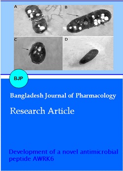
The effect of AWRK6 on the outer membrane of bacteria was examined by transmission electron microscopy (TEM). Typical TEM images of S. aureus in the absence and presence of AWRK6 are shown in Figure 6. Prior to incubation with 10 mg/mL AWRK6 the outer membrane of S. aureus was smooth (Figure 6A). Exposure to AWRK6 for 20 min induced obvious structural alterations in the outer membrane of S. aureus (Figure 6B). Following 2 hours of incubation with AWRK6, the outer membrane was ruptured or damaged and there was evidence of leakage of inner cytoplasmic contents (Figure 6C). The TEM images were therefore consistent with the rapid killing observed in the CFU assay and further substantiated the antimicrobial activity of AWRK6.
Figure 6: Transmission electron microscopy micrographs of S. aureus incubated with AWRK6
S. aurerus cells incubated in 10 mM PBS (pH 7.2) containing the peptide. Image was taken at the start of the incubation (A), 20 min into the incubation (B), 2 hours into the incubation (C), and 4 hours into the incubation (D). Arrow indicates structural damage on the cell membrane
Discussion
It is well established that the killing activity and specificity of antimicrobial peptides depend on their physiochemical properties such as size, net charge, conformational structure and hydrophobicity (Mao et al., 2013). AMP binds to negatively charged phosphorlipids in the outer membrane of bacterial cells through electrostatic and/or hydrophobic interaction (Matsuzaki, 1999). Although various physiochemical properties can affect the activity and specificity of AMP binding to membranes, it has been proposed that the net charge and hydrophobicity are the main factors conferring selectivity for the bacterial outer membrane (Zasloff et al., 2002). Among the 18 amino acid residues in Dy2, 10 are positively charged, suggesting a strong electrostatic interaction with the outer membrane of the bacterial cells.
One of our strategies to improve the killing activity of this AMP was to increase its hydrophobicity. This was done by replacing the Ala residue in Dy2 with Trp, which has a higher hydrophobicity index value at physiological pH (Trp:100, Ala:41) while maintaining the same net charge (Monera et al., 1995). In addition to enhancing the hydrophobicity of the peptide, replacement of Ala with Trp would be expected to facilitate killing since Trp-rich antimicrobial peptides such as indolicidin and tritrpticin have been shown to possess strong antimicrobial activity against both Gram positive and Gram negative bacteria while exhibiting low hemolytic activity (Yang et al., 2003; Yang et al., 2002; Ando et al., 2010). Fluorescence-quenching experiments have suggested that Trp residues help the peptides to penetrate the negatively charged lipid vesicles (Jing et al., 2003a; Jing et al., 2003b; Jing et al., 2006).
The stability of AMPs in a physiological environment is also important for therapeutic applications and many of the AMPs have low stability in vivo (Lohan and Bisht, 2013). Previous studies have demonstrated that buforin II, cecropin P1, indolicidin, magainin II exhibit lower killing activity in vivo than in vitro (Lohan and Bisht, 2013). Therefore, it became important that the modification of Dy2 would involve increasing its stability by substituting Arg with Lys residues. By applying this modification to Dy2, we obtained AWRK6, which displayed significantly higher stability (instability coefficient of -6.74. In addition, replacing Arg with Lys is expected to improve the stability of the peptide, rendering it more resistant to proteolysis by proteases, including trypsin, which has lower affinity for Lys (Craik et al., 1985). The result of the protease (trypsin) susceptibility assay showed that AWRK6 was more resistant to trypsin than Dy2, which was consistent with our expectations.
The discrepancy between the MIC test and CFU assay results could be due to the different incubation times; the long incubation time in the MIC test may have resulted in the underestimation of their antimicrobial potency. Rapid killing would be consistent with a membrane-active mode of antimicrobial action (Zasloff et al., 2002). The TEM result supported the fast (2-3 hours) antimicrobial action of AWRK6.
Based on the finding that Dy2 and AWRK6 were both more potent at alkaline pH, the conformation of the peptides was studied under different pH conditions using CD spectroscopy. As the pH became increasingly alkaline, the estimated secondary structure of Dy2 and AWRK6 became more alpha-helical in the membrane-mimicking micellar environment. These results were consistent with the observation that both peptides were more potent in alkaline pH. Indeed, our data suggested that the antimicrobial activity of Dy2 was lower than that of AWRK6 because at high pH there may be a greater propensity for AWRK6 to form a helical structure in the membrane. Alpha-helical peptides have been reported to act as membrane disruptive peptides because of they can cause the formation of pores within the bacterial cell membranes, a phenomenon consistent with the TEM results of AWRK6 (Brogden, 2005).
Conclusion
We have developed a new antimicrobial peptide AWRK6 via modification of the sequence of the existing peptide Dy2. The modification altered the physical properties of the peptide and improved its antimicrobial activity, turning it into a more stable peptide while maintaining a low toxicity.
Acknowledgement
We thank Dr. Alan K Chang from School of Life Science, Liaoning University for revising the language of the manuscript.
References
Afacan NJ, Yeung AT, Pena OM, Hancock RE. Therapeutic potential of host defense peptides in antibiotic-resistant infections. Curr Pharm Design. 2012; 18: 807-19.
Ando S, Mitsuyasu K, Soeda Y, Hidaka M, Ito Y, Matsubara K, Shindo M, Uchida Y, Aoyagi H. Structure-activity relationship of indolicidin, a Trp-rich antibacterial peptide. J Pept Sci. 2010; 16: 171-77.
Boman HG. Peptide antibiotics and their role in innate immunity. Annu Rev Immunol. 1995; 13: 61-92.
Brogden KA. Antimicrobial peptides: Pore formers or metabolic inhibitors in bacteria? Nat Rev Microbiol. 2005; 3: 238-50.
Combet C, Blanchet C, Geourjon C, Deleag, G. NPS@: Network protein sequence analysis. Trends Biochem Sci. 2000; 25: 147-50.
Conlon JM, Sonnevend A. Antimicrobial peptides in frog skin secretions. Methods Mol Biol. 2010; 618: 3-14.
Craik CS, Largman C, Fletcher T, Roczniak S, Barr PJ, Fletterick R, Rutter WJ. Redesigning trypsin: Alteration of substrate specificity. Science 1985; 228: 291-97.
Giacometti A, Cirioni O, Barchiesi F, Del Prete MS, Fortuna M, Caselli F, Scalise G. In vitro susceptibility tests for cationic peptides: Comparison of broth microdilution methods for bacteria that grow aerobically. Antimicrob Agents Chemother. 2000; 44: 1694-96.
Jin LL, Li Q, Song SS, Feng K, Zhang DB, Wang QY, Chen YH. Characterization of antimicrobial peptides isolated from the skin of the Chinese frog, Rana dybowskii. Comp Biochem Physiol B. 2009a; 154: 174-78.
Jin LL, Song SS, Li Q, Chen YH, Wang QY, Hou ST. Identification and characterisation of a novel antimicrobial polypeptide from the skin secretion of a Chinese frog (Rana chensinensis). Int J Antimicrob Agents. 2009b; 33: 538-42.
Jing W, Demcoe AR, Vogel HJ. Conformation of a bactericidal domain of puroindoline a: Structure and mechanism of action of a 13-residue antimicrobial peptide. J Bacteriol. 2003a; 185: 4938-47.
Jing W, Hunter HN, Hagel J, Vogel HJ. The structure of the antimicrobial peptide Ac-RRWWRF-NH2 bound to micelles and its interactions with phospholipid bilayers. J Pept Res. 2003b; 61: 219-29.
Jing W, Svendsen JS, Vogel HJ. Comparison of NMR structures and model-membrane interactions of 15-residue antimicrobial peptides derived from bovine lactoferricin. Biochem. Cell Biol. 2006; 84: 312-26.
King RD, Sternberg MJ. Identification and application of the concepts important for accurate and reliable protein secondary structure prediction. Protein Sci. 1996; 5: 2298-310.
Li L, He J, Eckert R, Yarbrough D, Lux R, Anderson M, Shi W. Design and characterization of an acid-activated antimicrobial peptide. Chem Biol Drug Des. 2010; 75: 127-32.
Lohan S, Bisht GS. Small cationic antimicrobial peptidomimetics: Emerging candidate for the development of potential anti-infective agents. Curr pharm Design. 2013; 19: 5809-23.
Mao Y, Niu S, Xu X, Wang J, Su Y, Wu Y, Zhong S. The effect of an adding histidine on biological activity and stability of Pc-pis from Pseudosciaena crocea. PloS one. 2013; 8: e83268.
Matsuzaki K. Why and how are peptidelipid interactions utilized for self-defense? Magainins and tachyplesins as archetypes. Biochim Biophys Acta. 1999; 1: 1462-70.
Monera OD, Sereda TJ, Zhou NE, Kay CM, Hodges RS. Relationship of sidechain hydrophobicity and alpha-helical propensity on the stability of the single-stranded amphipathic alpha-helix. J Pept Sci. 1995; 1: 319-29.
Rose GD, Wolfenden R. Hydrogen bonding, hydrophobicity, packing and protein folding. Annu Rev Biophys Biomol Struct. 1993; 22: 381-415.
Shlaes DM, Spellberg B. Overcoming the challenges to developing new antibiotics. Curr Opin Pharmacol. 2012; 12: 522-26.
Yang ST, Shin SY, Lee CW, Kim YC, Hahm KS, Kim JI. Selective cytotoxicity following Arg-to-Lys substitution in tritrpticin adopting a unique amphipathic turn structure. FEBS lett. 2003; 540: 229-33.
Yang ST, Yub Shin SY, Kim YC, Kim Y, Hahm KS, Kim JI. Conformation-dependent antibiotic activity of tritrpticin, a cathelicidin-derived antimicrobial peptide. Biochem Biophys Res Commun. 2002; 296: 1044-50.
Zasloff M. Antimicrobial peptides of multicellular organisms. Nature 2002; 415: 389-95.