Lactucin induces potent anti-cancer effects in HL-60 human leukemia cancer cells by inducing apoptosis and sub-G1 cell cycle arrest
Abstract
The main purpose of the present study was to examine the antitumor and apoptotic effects of lactucin in HL-60 human leukemia cancer cells. MTT assay was used to examine the cytotoxic effects of lactucin while as phase contrast, fluorescence and transmission electron microscopy techniques were used to evaluate the apoptotic effects of lactucin in these cells. Flow cytometry was used to assess the effects of lactucin on cell cycle phase distribution. The results indicate that lactucin induced potent, time- and dose-dependent antitumor effects. The microscopic techniques showed that lactucin induced characteristic features of apoptosis including cell shrinkage, membrane blebbing, appearance of vacuoles, swelling of mitochondria and endoplasmic reticulum. Viable cells are stained green, early apoptotic cells are stained yellow, while as late apoptotic cells are stained reddish orange. Flow cytometry revealed that lactucin induced sub-G1 cell cycle arrest.
Introduction
Leukemia, is a group of cancers that generally begin in the bone marrow and lead to high numbers of abnormal white blood cells. These white blood cells are not completely formed and are known as blasts or leukemia cells. Various risk factors have been found to initiate leukemia which involve both inherited and non-inherited factors. In 2012, leukemia developed in more than 300,000 people worldwide and resulted in 265,000 deaths. It is the most common category of tumors in youngsters, with three quarters of leukemia cases in children being the acute lymphoblastic type (Hutter, 2010; World Health Organization, 2014). Four main classes of leukemia have been identified namely lymphoblastic leukemia, chronic lymphocytic leukemia, chronic myeloid leukemia and acute myeloid leukemia (Vardiman et al., 2009). Treatment of leukemia involves individual therapeutic options like chemotherapy, radiotherapy or their combination. Bone marrow transplant, targeted therapy as well as palliative or supportive care are also needed in some cases. It must be emphasized here that most cases of leukemia are treated by using chemotherapeutic drugs mostly combined into a multidrug dose therapy (Hoffbrand et al., 2006).
Apoptosis, which is a highly specialized biochemical process, is very central for sustaining the physiological equilibrium of multi cellular organisms. The process is characterized by various morphological changes including DNA fragmentation, chromatin condensation, nuclear fragmentation, cellular shrinkage, apoptotic body formation, membrane blebbing etc. Apoptosis helps in eradicating damaged cells which may get cancerous. Various cell signalling pathways have been identified which can control the apoptosis process. The process of apoptosis may be initiated by either intrinsic or the extrinsic pathways (Kerr et al., 1972; Hengartner, 2000). The aim of the present investigation was to evaluate the anticancer and apoptotic effects of lactucin in HL-60 human leukemia cancer cells and study the mechanistic details involved.
Materials and Methods
Chemicals and other related reagents
Lactucin, 3-(4,5-dimethylthiazol-2-yl)-2, 5-diphenyltetrazolium-bromide (MTT) were procured from Sigma-Aldrich (USA). Lactucin was dissolved in dimethyl sulfoxide (DMSO, Sigma-Aldrich) to get a 100 mM stock solution, which was diluted in the medium to yield the anticipated concentration. An equivalent volume of DMSO in complete culture medium was used as the vehicle control. To exclude the cytotoxicity of DMSO, the final concentration of DMSO for all experiments was kept at less than 0.2%. Minimum Essential Medium (MEM) and RPMI, fetal bovine serum (FBS), penicillin, streptomycin, trypsin, phosphate-buffered saline (PBS) with calcium chloride and magnesium chloride were obtained from Hangzhou Sijiqing Biological Engineering Materials Co., Ltd. (China). Propidium iodide, acridine orange, were purchased from Boster Biological Technology Co., Ltd. (China).
Cell line and culture conditions
TheHL-60 human leukemia cancer cell line was purchased from Shanghai Institutes for Biological Sciences, Chinese Academy of Sciences (China). The cells were cultured in Minimum Essential Medium (MEM) and RPMI supplemented with 10% (v/v) fetal bovine serum (FBS) under humidified atmosphere of 5% CO2 at 37°C. The medium was replaced every 3 days. Cells were subcultured every 3 days.
MTT cell viability assay
The HL-60 human leukemia cancer cells were seeded on a 96-well plates at 2 x 10⁵ cells per well. After 24 hours the cells were treated with lactucin at several doses (0, 5, 25, 50, and 100 µM). After incubation times of 24 and 48 hours MTT solution (10 µL) was added. The formazan crystals thus formed were dissolved with DMSO and the absorbance was measured on a microplate reader (FLUO Star Optima, Germany).
Phase contrast microscopy for morphological evaluation
HL-60 human leukemia cancer cells were trypsinized followed by centrifugation at 12,000 rpm. After this the dilution of cells was done for counting in hemocytometer. The cells were then seeded in 6-well plates at a density of 2 x 10⁶ cells/mL and then incubated for 24 hours. Afterwards, the cells were subjected to treatment with numerous concentrations of lactucin (0, 5, 50 and 100 uM) for 48 hours. Subsequent to drug treatment, culture plates were observed using an inverted phase contrast microscope (Olympus, Japan) and images were captured. DMSO was used as a negative control.
Fluorescence microscopic assay for apoptosis
HL-60 human leukemia cancer cells were seeded on a chamber slide (Thermo Scientific Nunc Lab Tek II) at cell density of 2 x 10⁶ cells per chamber. The cells were treated with 0, 5, 50 and 100 µM lactucin for 48 hours. Subsequently, 20 ug/mL of acridine orange and 10 ug/mL of propidium iodide were added to each chamber. It was then observed under fluorescence microscope (Olympus IX-70, Japan).
Transmission electron microscopic study of cellular apoptosis
Following treatment by various doses of lactucin (0, 5, 50 and 100 µM), HL-60 human leukemia cancer cells were harvested, washed three times with PBS and then fixed with 3.0% glutaraldehyde in 0.2 mol/L PBS for 60 min at 37°C and post fixed with 2.5% osmium tetraoxide for 20 min. The cells were again washed, then gradually dehydrated in a series of 60-100% ethanol and then embedded in Epon 812 resin. The ultrathin sections were made with a microtome and these sections were stained with lead acetate and uranyl acetate. The cells were finally observed under a transmission electron microscope.
Cell cycle analysis assay
The effect of lactucin on the cell cycle was evaluated by flow cytometry using propidium iodide as a fluorescent probe. HL-60 human leukemia cancer cells at a density of 2 x 10⁶ cells/mL were plated in a 6-well plates. The cells were exposed to various doses of lactucin (0, 5, 50 and 100 µM) in a humidified atmosphere of 5% CO2 for 48 hours. The cells were harvested and washed with PBS twice and then fixed in cold ethanol at 4°C overnight. The cells were then stained with propidium iodide (PI) solution and 30 mg/mL RNase for 20 minutes in dark and then analyzed by flow cytometry (FACS Calibur, USA).
Statistical analysis
Each experiment was performed in triplicate. The results were expressed as means ± SD. Significant differences were determined using the one-way analysis of variance (ANOVA). Statistical significance was defined as p<0.05. Each experiment was performed at least in triplicate.
Results
Lactucin shows potent antitumor activity in HL-60 human leukemia carcinoma cells
The chemical structure of lactucin is shown in Figure 1. The antitumor effect of lactucin in HL-60 human leukemia carcinoma cells is shown in Figure 2. Lactucin induced significant cytotoxic effects in these cancer cells in a dose- and time-dependent manner. The cytotoxic effects were measured at 12, 24 and 48 hours time intervals. The cytotoxicity increased with the increase in the incubation time to which the cells were exposed.
Figure 1: Chemical structure of lactucin
Figure 2: Anti-proliferative effects of lactucin in HL-60 human leukemia cancer cells. Data are shown as the mean ± SD of three independent experiments. ap<0.05, bp<0.01 vs 0 µM (control)
Phase contrast microscopy evaluation of lactucin-induced apoptosis in HL-60 cells
As shown in Figure 3, as compared to the untreated control cells, which showed normal morphology, lactucin-treated HL-60 cells showed change in cellular morphology characterized by the appearance of a large number of cytoplasmic vacuoles. The size and the number of these vacuoles increased with increasing dose of lactucin. Further morphological changes that occurred after lactucin treatment included cell shrinkage, rounding of cells, detachment of cells from one another and from the substratum.
Figure 3: Phase contrast microscopic evaluation of the effect of lactucin on the morphology of HL-60 human leukemia cancer cells. The cells were treated without (A) and with 5 (B), 50 (C) and 100 (D) Îœm of lactucin for 48 hours. The untreated control cells adhere well, exhibiting normal morphology
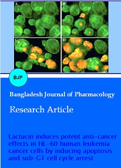
Fluorescence microscopy evaluation of lactucin induced apoptosis in HL-60 cells
Next, the effect of lactucin on apoptosis induction in HL-60 human leukemia carcinoma cells was verified using fluorescence microscopy and acridine orange/ethidium bromide staining. The results of this assay are presented in Figure 4A-D and designate that lactucin induced apoptotic morphological features including membrane blebbing, chromatin condensation, nuclear fragmentation, and appearance of apoptotic bodies. Viable cells are stained green, early apoptotic cells are stained yellow, while as late apoptotic cells are stained reddish orange. Control cells (untreated) only show green fluorescence indicating all cells are live (Figure 4A) while as the lactucin-treated cells show yellow and reddish orange fluorescence. The late apoptotic features appeared only at higher lactucin doses (Figure 4B-D).
Figure 4: Evaluation of apoptosis induced by different doses of lactucin in HL-60 human leukemia cancer cells using fluorescence microscopy. The cells were treated without (A) and with 5 (B), 50 (C) and 100 (D) µ of lactucin for 48 hours and then stained with acridine orange and ethidium bromide. Green fluorescence (A and B) indicate normal cells while as orange fluorescence (C and D) shows apoptotic cells
Transmission electron microscopy evaluation of lactucin induced apoptosisin HL-60 cells
Transmission electron microscopy results further established that lactucin induces apoptosis in HL-60 human leukemia carcinoma cells. The results which are shown in Figure 5A-D disclose that as compared to the untreated group (Figure 5A), 5, 50 and 100 µM lactucin-treated cells showed cells with injured mitochondria and nonexistence of nuclear membrane. The key feature of lactucin treated cells was the advent of vacuoles in the cytoplasm and their number which increased with increasing lactucin dose. The results also showed that swelling of the mitochondria and endoplasmic reticulum (ER) was detected in the cells treated with lactucin at increasing doses.
Figure 5: Ultrastructural examination of HL-60 human leukemia cancer cells using transmission electron microscopy (TEM). Untreated control cells (A) exhibited normal cell morphology including normal nuclei, cytoplasm and cell organelles. Lactucin-treated cells (B, C and C represent 5, 50 and 100 µM dose of lactucin) indicated the appearance of autophagic vacuoles characteristic of autophagy process. The number and size of these autophagic vacuoles increased with increasing lactucin-dose, arrows represent vacuoles. Magnification x4000
Lactucin induces sub-G1 cell cycle arrest
Further experiments using flow cytometry along with propidium iodide as fluorescent probe indicated that lactucin had a significant effect on cell cycle phase distribution in HL-60 human leukemia carcinoma cells. More specifically, lactucin led to the sub-G1 cell cycle arrest in a dose-dependent manner. As compared to the untreated control cells (Figure 6A) which showed no signs of apoptotic cells, the lactucin-treated cells with 5, 50 and 100 µM dose led to increase of cells from 23.5% to 34.2% and 53.8% respectively.
Figure 6: Effect of lactucin on the cell cycle phase distribution in HL-60 human leukemia cancer cells. The cells were treated without (A) and with 5 (B), 50 (C) and 100 (D) µM dose of lactucin for 48 hours and then analyzed by flow cytometry
Discussion
The main objective of the present study was to evaluate the antitumor and apoptotic effects of lactucin in HL-60 human leukemia carcinoma cells. Lactucin induced significant cytotoxic effects in these cancer cells in a dose- and time-dependent manner. The cytotoxicity increased with the increase in the incubation time to which the cells were exposed. Phase contrast microscopy revealed that lactucin-treated HL-60 cells showed change in cellular morphology characterized by the appearance of a large number of cytoplasmic vacuoles. The size and the number of these vacuoles increased with increasing dose of lactucin. Control cells (untreated) only showed green fluorescence indicating that cells are not dead while as the lactucin-treated cells show yellow and reddish orange fluorescence. The late apoptotic features appeared only at higher lactucin doses. Transmission electron microscopy revealed that lactucin-treated cells led to the formation of vacuoles in the cytoplasm and their number increased with increasing lactucin dose. TEM results also showed that lactucin treatment led to swelling of the mitochondria and endoplasmic reticulum (ER). Flow cytometry indicated that lactucin had a significant effect on cell cycle phase distribution in HL-60 human leukemia cells. Lactucin induced dose-dependent sub-G1 cell cycle arrest in these cells.
Lactucin belongs to the sesquiterpene lactone group of naturally occurring compounds with a wide variety of pharmacological effects including anticancer effects. Sesquiterpene lactones constitute a large and diverse group of biologically active plant compounds. Sesquiterpene lactones are found in a large number of plants including Chrysanthemum, Wormwood, Laurus nobilis, Chamomile, Pyrethrum etc (Kreuger et al., 2012; Chadwick et al., 2013; Dall' Acqua et al., 2006). Lactucin has been reported to show analgesic and sedative properties in mice. It has also been reported to exhibit antimalarial activity (Wesolowska et al., 2006; Bischoff et al., 2004). Lactucin has been previously reported to have been extracted from Mulgedium tatarica extract and the extract was found to be very active in killing cancer cells. The various chemical constituents were found to be lactucopicrin, 11,13-beta-dihydrolactucopicrin, lactucin and 11, 13-beta-dihydrolactucin (Ren et al., 2005).
Conclusion
To the best of our knowledge, there are no reports on the anti-cancer activity of lactucin against HL-60 human leukemia cancer cells. Further, the apoptotic effects as well as the cell cycle arrest inducing effects of lactucin were also not reported earlier.
References
Bischoff TA, Kelley CJ, Karchesy Y, Laurantos M, Nguyen-Dinh P, Arefi AG. Antimalarial activity of lactucin and lactucopicrin: Sesquiterpene i from Cichorium intybus L. J Ethnopharmacol. 2004; 95: 455-57.
Chadwick M1, Trewin H, Gawthrop F, Wagstaff C. Sesquiterpenoids lactones: Benefits to plants and people. Int J Mol Sci. 2013; 14: 12780-805.
Dall'Acqua S, Viola G, Giorgetti M, Loi MC, Innocenti G. Two new sesquiterpene lactones from the leaves of Laurus nobilis. Chem Pharm Bull. 2006; 54: 1187-89.
Hengartner MO. The biochemistry of apoptosis. Nature 2000; 407: 770-76.
Hoffbrand AV, Moss PAH, Pettit JE. Essential haematology. Blackwell. 5th edn. 2006.
Hutter JJ. Childhood leukemia. Pediatr Rev/American Academy of Pediatrics. 2010; 31: 234-41.
Kerr JF, Wyllie AH, Currie AR. Apoptosis: A basic biological phenomenon with wide-ranging implications in tissue kinetics. Br J Cancer. 1972; 26: 239-57.
Kreuger MR, Grootjans S, Biavatti MW, Vandenabeele P, D'Herde K. Sesquiterpene lactones as drugs with multiple targets in cancer treatment: Focus on parthenolide. Anticancer Drugs. 2012; 23: 883-96.
Ren YL, Zhou YW, Chen XZ, Ye YH. Discovery, structural determination and anti-cancer activities of lactucin-like guaianolides. Lett Drug Des Discov. 2005; 2: 444.
Vardiman JW, Thiele J, Arber DA, Brunning RD, Borowitz MJ, Porwit A, Harris NL, Le Beau MM, Hellstrom-Lindberg E, Tefferi A, Bloomfield CD. The 2008 revision of the World Health Organization (WHO) classification of myeloid neoplasms and acute leukemia: Rationale and important changes. Blood 2009; 114: 937-51.
Wesolowska A, Nikiforuk A, Michalska K, Kisiel W, Chojnacka-Wojcik E. Analgesic and sedative activities of lactucin and some lactucin-Like guaianolides in mice. J Ethnopharmacol. 2006; 107: 254-58.
World Health Organization. World Cancer Report. 2014.