Dracorhodin perchlorate induces apoptosis in bladder cancer cells through Bcl-2, Bcl-XL, survivin down-regulation and caspase-3 activation
Abstract
Urinary bladder cancer is one of the most commonly diagnosed urological malignancies worldwide. Dracorhodin perchlorate, anthocyanin red pigment, has been recently shown to induce apoptotic cell death in several types of cancer cells. However, there is no report elucidating its effect on bladder cancer T24 cells. In this study, for the first time, we investigated the effects of dracorhodin perchlorate on the cell viability and apoptosis in human bladder cancer T24 cells. DNA flow cytometric analysis demonstrated that dracorhodin perchlorate markedly rendered apoptosis of T24 cells in a time-dependent manner. Dracorhodin perchlorate significantly induced the dissipation of mitochondrial membrane potential in T24 cells. Furthermore, dracorhodin perchlorate-induced apoptosis was regulated by activation of caspase-3 and down-regulation of antiapoptotic proteins, Bcl-2, Bcl-XL, and survivin in T24 cells. These in vitro results suggested that dracorhodin perchlorate should be further examined for in vivo activity and molecular mechanism in human bladder cancer.
Introduction
Urinary bladder cancer is one of the most commonly diagnosed urological malignancies worldwide. More than 12 million new cases of cancer occur annually worldwide. Of those 5.4 million occur in developed countries and 6.7 million in developing countries (Ploeg et al., 2009). Bladder cancer is the fourth and eighth most common cancer in men and women in the United States, respectively. Bladder cancer afflicts about 60,000 Americans annually (Ploeg et al., 2009). Bladder cancer is quite common in the U.S., representing about 6 percent of all new cancer cases among men and 2 percent among women. In 2012, approximately 37,510 new urinary bladder cancer cases will be diagnosed and 14,880 will die in the United States (Siegel et al., 2012). In recent days, bladder cancer is usually cured with surgery, chemotherapy, and combination of chemotherapy and radiotherapy, but they have associated limitations (Ploeg et al., 2009). Prevailing treatment options have limited therapeutic success in human bladder cancer. Hence, the current therapy for bladder cancer is not satisfactory and better therapeutic options are immediately required to develop a more effective therapy for bladder cancer.
In the last few decades, herbal medicines have been proven to be an important source of novel agents with a pharmaceutical potential. Many herbal medicines, such as paclitaxel, camptothecin, vinca alkaloids, and etoposide hold great potential as promising agents for the treatment of cancer (Cragg and Newman, 2005). Natural products have traditionally provided a rich source of drugs for many diseases, including cancer, and plants are an important source of novel natural products (Amin et al., 2009). David Newman and Gordon Cragg (Newman and Cragg, 2007) found that of 155 FDA-approved small molecule anti-cancer drugs, 47% were either natural products or analogues inspired by them. Dracorhodin perchlorate is an analogue of the antimicrobial anthocyanin red pigment, dracorhodin, which is isolated in the exudates of the fruit of Daemonorops draco (Rao et al., 1982), known as "dragon's blood" in traditional Chinese medicines. An understanding of the molecular mechanism underlying dracorhodin perchlorate is just now emerging. In 2011, it was reported that dracorhodin perchlorate supperssed proliferation and induced apoptosis in human prostate cancer cells (He et al., 2011). Other studies have shown that dracorhodin perchlorate inhibited cell growth and triggered apoptosis in melanoma and leukemic cancer cells (Xia et al., 2005; Xia et al., 2006). Previously, we demonstrated that dracorhodin perchlorate altered cell cycle distribution and induced apoptosis in the human gastric adenocarcinoma T24 cell line (Rasul et al., 2012a). However, the effects of dracorhodin perchlorate on human bladder cancer SGC-7901 cells were still unknown. The objective of this study was to investigate if the antiproliferative effect of dracorhodin perchlorate was similar for human bladder cancer cell line T24 cells. Therefore, the present study was undertaken to explore the effects of dracorhodin perchlorate on the proliferation of T24 cells and its possible mechanism. Results showed that dracorhodin perchlorate effectively inhibited the proliferation of T24 cells through inducing the apoptosis, which was regulated by activation of caspase-3 and down-regulation of Bcl-2, Bcl-XL, and survivin.
Materials and Methods
Chemicals and reagents
Dracorhodin perchlorate was purchased from Tauto biotech Co., Ltd. (Shanghai, China) and purity (>98%) was determined by HPLC. Fetal bovine serum was purchased from Hangzhou Sijiqing Biological Engineering Materials Co., Ltd. Cell culture mediums (DMEM), propidium iodide (PI), and dimethylsulfoxide (DMSO) were purchased from Sigma. Cleaved caspase-3 antibody was purchased from Beyotime Institute of Biotechnology (Shanghai, China). Rabbit polyclonal anti-human Bcl-2, Bcl-XL, and survivin antibodies were purchased from Boster Biological Technology Co., Ltd. (Wuhan, China). Mouse anti-beta-actin, secondary anti-mouse, and anti rabbit antibodies were purchased from Santa Cruz Biotechnology (CA, USA). Rhodamine123 was purchased from Eugene Co. (Oregon, U.S.A.).
Cell culture
Human bladder cancer T24 cells were maintained in DMEM supplemented with 10%. All culture media were also supplemented with 100 units/mL penicillin and 100 µg/mL streptomycin at 37°C in a humidified atmosphere with 5% carbon dioxide and 95% air.
Cell proliferation assay
The effects of dracorhodin perchlorate on cell viability were evaluated by MTT assay as previously described (Rasul et al., 2011a). After 24 and 48 hours of exposure to different concentrations (0, 10, 30, 100 and 300 µM) of dracorhodin perchlorate, 10 µL of MTT was added to each well and incubated for further four hours. After the removal of medium, 150 µL DMSO was added to each well. The absorbance was recorded at the wavelength of 570 nm in the microplate reader and the inhibition ratio (I%) was calculated.
Flow cytometric analysis of apoptosis
T24 cells were treated with 100 µM dracorhodin perchlorate for 12, 24 and 48 hours, the cells were then collected, washed, and resuspended in phosphate buffered saline (PBS). The apoptotic cell death rate was examined with annexin V-FITC and PI double staining using the Annexin V-FITC Apoptosis Detection kit according to the manufacturer's instructions. After staining the cells with Annexin V-FITC/PI, flow cytometric analysis was performed and data were analyzed using Cell Quest software.
Determination of mitochondrial transmembrane potential (ΔΨm)
To determine the changes in ΔΨm, T24 cells were stained with Rho-123 as previously described (Rasul et al., 2011a). After treatment with 100 µM of dracorhodin perchlorate for 12, 24 and 48 hours, Rho-123 fluorescence was measured in flow cytometry with excitation and emission wavelengths of 488 and 530 nm.
Western blot analysis
T24 cells were exposed to dracorhodin perchlorate of the specified concentrations for the specified duration, and were then analyzed through Western blotting. Western blot was performed as we previously described (Rasul et al., 2011b). Briefly, T24 cells were collected and centrifuged. Cell pellets were resuspended in the lysis buffer and lysed on ice for 30 min. After centrifugation for 15 min, the supernatant fluids were collected and the protein content of the supernatant was measured by a NanoDrop 1000 spectrophotometer (Thermo Scientific, USA). The protein lysates were separated by electrophoresis on a 12% SDS-polyacrylamide gel and transferred to a PVDF membrane (Amersham Biosciences, Piscataway, NJ). The membranes were subsequently blocked with 5%skimmed milk for 2 hours. The membranes were then probed with specific primary antibodies overnight at 4°C, incubated with the relevant secondary antibodies, and signals were detected using ECL plus chemiluminescence kit on X-ray film.
Statistical analysis of data
For the statistical analysis of the data, the comparison between results from different groups was done with Student's t-test. The level of statistical significance was regarded as p<0.05. All experiments were repeated at least three times. Data were presented as mean ± SD.
Result and Discussion
The aim of the present study was to elucidate the cytotoxic effects of dracorhodin perchlorate in human bladder cancer T24 cells and to delineate the underlying mechanisms of dracorhodin perchlorate-induced apoptosis in bladder cancer T24 cells. The structure of dracorhodin perchlorate is shown in Figure 1. To evaluate the effects of dracorhodin Perchlorate on the proliferation of T24 cells, MTT assay was performed. T24 cells were treated with dracorhodin perchlorate at the concentrations of 0, 10, 30, 100 or 300 for 24 and 48 hours respectively. Dracorhodin perchlorate inhibited the growth of T24 cells in a dose- and time-dependant manner (Figure 2). Morphological changes were observed under phase contrast microscopy after treating cells with dracorhodin perchlorate resulting in the decreased number of cells as compared to control group and cells become rounded and shrinked, which were polygonal in untreated cells (data not shown). These results reveal that dracorhodin perchlorate can act as growth inhibitor of human bladder cancer T24 cells in a similar fashion as described in previous studies dealing with various other types of cancer cells including melanoma cells (Xia et al., 2005), leukemic cancer cells (Xia et al., 2006), prostate cancer cells (He et al., 2011) and gastric cancer cells (Rasul et al., 2012a).
Figure 1: The chemical structure of dracorhodin perchlorate
Figure 2: Dracorhodin perchlorate inhibited the cell growth and induced cell death. T24 Cells were treated with indicated doses of dracorhodin perchlorate for 24 and 48 hours and cell viability was measured by MTT assay. Data are expressed as Mean ± SD (n = 3). Columns not sharing the same superscript letter differ significantly (p<0.05)
Apoptosis, autophagy, and necrosis are the major types of cell death (Leist and Jaattela, 2001). Among the three major pathways of cell death, apoptosis is most well planned and orderly mode of cell death (Elmore, 2007;
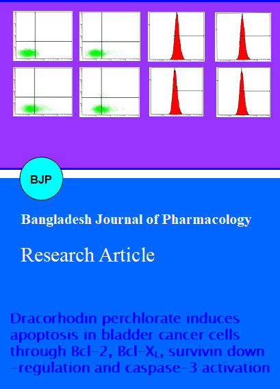
Hengartner, 2000). More than 50% of neoplasms undergo aberrations in the apoptotic machinery which leads to abnormal cells proliferation (Mashima and Tsuruo, 2005; Pommier et al., 2004). The regulation of apoptosis is, therefore, the most important in the treatment of cancer (Fulda, 2010; Lawen, 2003; Reed, 2002). Accumulated evidences indicated that most of chemotherapeutic agents halt tumor cells proliferation via induction of apoptosis (Rasul et al., 2013; Rasul et al., 2012a; Rasul et al., 2011a; Rasul et al., 2012b; Rasul et al., 2011d; Rasul et al., 2012b). We examined whether dracorhodin perchlorate inhibited cell growth T24 cells through the induction of apoptosis. Dracorhodin perchlorate-induced apoptosis was determined by flow cytometric analysis. Cells were seeded in the 12 well plates. After incubation of cells with or without dracorhodin perchlorate for 12, 24, and 48 hours, cells were collected in centrifuged tubes and stained with FITC annexin-V and PI double staining as described in materials and methods. The results of flow cytometric analysis showed that the rates of apoptosis were 15.5 ± 2.2, 24.4 ± 2.1 and 45.5 ± 1.8% in the cells treated with 100 µM of dracorhodin perchlorate for 12, 24, and 48 hours respectively as compared to the 5.3 ± 1.0% in control cells (Figure 3). Dracorhodin perchlorate-induced apoptosis in T24 cells were compatible with previously reported studies (He et al., 2011; Rasul et al., 2012a; Xia et al., 2005; Xia et al., 2006).
Figure 3: Apoptosis induced by dracorhodin perchlorate in T24 cells. T24 cells were treated with 100 µM of Dracorhodin perchlorate for 12, 24 and 48 hours. And then they were stained with FITC-conjugated Annexin V and PI for flow cytometric analysis. The flow cytometry profile represents Annexin V-FITC staining in x axis and PI in y axis. As shown, the cell populations in the lower right (Annexin V+/PI-) represents early apoptotic cells, upper right (Annexin V+/PI+) represents late apoptotic cells. Data are expressed as Mean ± SD (n = 3). Columns not sharing the same superscript letter differ significantly (p<0.05)
Mitochondria have become an important component of the apoptosis execution machinery, which contain proapoptotic proteins (e.g., cytochrome c) (Elmore, 2007). It has been elucidated that upon the depolarization of the mitochondrial membrane potential results in the mitochondrial swelling and subsequent release of cytochrome c from the intermitochondrial membrane space into the cytosol (Buytaert et al., 2007). It is becoming increasingly apparent that mitochondria play a fundamental role in the processes those lead to the cell death (Wang, 2001). Effects of dracorhodin perchlorate on the mitochondrial membrane potential of T24 cells were determined by the flow cytometry using rhodamine 123 staining. The rates of depletion of mitochondrial membrane potential were 82.4 ± 2.4, 71.7 ± 1.9, and 61.0 ± 1.6% in cells treated with 100 µM of dracorhodin perchlorate for 12, 24, and 48 hours respectively as compared to 95.9 ± 0.4% in control group (Figure 4). Our data corroborate with the previously reported results that dracorhodin Perchlorate induced dissipation of mitochondrial membrane potential, which provide the evidence for direct contribution of mitochondria in the dracorhodin perchlorate-induced apoptosis (He et al., 2011; Rasul et al., 2012a; Xia et al., 2005; Xia et al., 2006).
Figure 4: The effects of dracorhodin perchlorate on mitochondrial transmembrane potential of T24 cells were determined by flow cytometry. The values indicate the percentages of rhodamine 123 fluorescence in the T24 cells treated without (control) and with 100 µM of dracorhodin perchlorate for 12, 24, and 48 hours. The data shown are representative of three independent experiments with the similar results. Data are expressed as Mean ± SD (n = 3). Columns not sharing the same superscript letter differ significantly (p<0.05)
The caspases are a family of proteins related to cysteine proteases that is one of the focal executors of the apoptotic process via triggering the death receptors and mitochondrial pathways to accomplish the programmed cell death (Cohen, 1997). Caspases are present in the form of inactive zymogens those are activated during apoptosis. Among them, caspase-3 is a frequently activated death protease, catalyzing the specific cleavage of many key cellular proteins (Adams, 2003; Porter and Janicke, 1999). In order to reveal effects of dracorhodin perchlorate on expression of caspase-3, western blotting was done. The results showed that procaspase-3 was activated in treated cells with 100 µM of dracorhodin perchlorate for 12, 24, and 48 hours as compared to that of control cells (Figure 5). These findings are supported by previously studies (He et al., 2011; Rasul et al., 2012a; Xia et al., 2005; Xia et al., 2006). These results markedly showed that dracorhodin perchlorate induced caspase-dependent cell death in T24 cells.
Interplay between pro-apoptotic (Bax) and anti-apoptotic (Bcl-2) members of the Bcl-2 family pedals the mitochondrial apoptotic pathway (Mallat and Tedgui, 2000). Bcl-2 family proteins are pivotal for rising permeability of mitochondrial membranes and the release of cytochrome c, which activates caspases and in turn mobilizes apoptotic cell death (Adams and Cory, 2007; Burlacu, 2003; Danial, 2007). To investigate the effect of dracorhodin perchlorate on expression of antiapoptotic proteins such as Bcl-2, Bcl-XL, and survivin in T24 cells, Western blotting was done. It was observed that dracorhodin perchlorate involved in the down-regulation of Bcl-2 and Bcl-XL in a time-dependent manner (Figure 5). These results are similar with previously reported studies (Rasul et al., 2012a; Xia et al., 2005). In addition, we also examined the effect of dracorhodin perchlorate on survivin, anti-apoptotic protein. Our results demonstrated that dracorhodin perchlorate involved in the down regulation of survivin in a time-dependent manner (Figure 5).
Figure 5: T24 cells were exposed to 100 µM of dracorhodin perchlorate for specified time intervals. Equal amounts of lysate protein were subjected to gel electrophoresis. Expression levels of caspase-3, Bcl-2, Bcl-XL, and survivin were monitored by Western blot assay. β-actin was used as loading control. Data are representative of at least two independent experiments with similar results
Conclusion
Dracorhodin perchlorate induced cell death of bladder cancer T24 cells via induction of apoptosis. Analysis of apoptosis related proteins in T24 cells reveals that dracorhodin perchlorate may induce the down-regulation of antiapoptotic proteins such as Bcl-2, Bcl-XL, and survivin which might ultimately lead to depolarization of mitochondrial membranes, and sequential activation of caspase-3, leading to apoptosis. Based on our previous and current findings, it is recommended that dracorhodin perchlorate could define a new therapeutic strategy for human cancers.
References
Adams JM. Ways of dying: Multiple pathways to apoptosis. Genes Dev. 2003; 17: 2481-95.
Adams JM, Cory S. The Bcl-2 apoptotic switch in cancer development and therapy. Oncogene 2007; 26: 1324-37.
Amin AR, Kucuk O, Khuri FR, Shin DM. Perspectives for cancer prevention with natural compounds. J Clin Oncol. 2009; 27: 2712-25.
Burlacu A. Regulation of apoptosis by Bcl-2 family proteins. J Cell Mol Med. 2003; 7: 249-57.
Buytaert E, Dewaele M, Agostinis P. Molecular effectors of multiple cell death pathways initiated by photodynamic therapy. Biochim Biophys Acta. 2007; 1776: 86-107.
Cohen GM. Caspases: The executioners of apoptosis. Biochem J. 1997; 326( Pt 1): 1-16.
Cragg GM, Newman DJ. Plants as a source of anti-cancer agents. J Ethnopharmacol. 2005; 100: 72-79.
Danial NN. BCL-2 family proteins: Critical checkpoints of apoptotic cell death. Clin Cancer Res. 2007; 13: 7254-63.
Elmore S. Apoptosis: A review of programmed cell death. Toxicol Pathol. 2007; 35: 495-516.
Fulda S. Evasion of apoptosis as a cellular stress response in cancer. Int J Cell Biol. 2010; 2010: 370835.
He Y, Ju W, Hao H, Liu Q, Lv L, Zeng F. Dracorhodin perchlorate suppresses proliferation and induces apoptosis in human prostate cancer cell line PC-3. J Huazhong Univ Sci Technol Med Sci. 2011; 31: 215-19.
Hengartner MO. The biochemistry of apoptosis. Nature 2000; 407: 770-76.
Lawen A. Apoptosisan introduction. Bioessays 2003; 25: 888-96.
Leist M, Jaattela M. Four deaths and a funeral: From caspases to alternative mechanisms. Nat Rev Mol Cell Biol. 2001; 2: 589-98.
Mallat Z, Tedgui A. Apoptosis in the vasculature: Mechanisms and functional importance. Br J Pharmacol. 2000; 130: 947-62.
Mashima T, Tsuruo T. Defects of the apoptotic pathway as therapeutic target against cancer. Drug Resist Updat. 2005; 8: 339-43.
Newman DJ, Cragg GM. Natural products as sources of new drugs over the last 25 years. J Nat Prod. 2007; 70: 461-77.
Ploeg M, Aben KKH, Kiemeney LA. The present and future burden of urinary bladder cancer in the world. World J Urol. 2009; 27: 289-93.
Pommier Y, Sordet O, Antony S, Hayward RL, Kohn KW. Apoptosis defects and chemotherapy resistance: Molecular interaction maps and networks. Oncogene 2004; 23: 2934-49.
Porter AG, Janicke RU. Emerging roles of caspase-3 in apoptosis. Cell Death Differ. 1999; 6: 99-104.
Rao GS, Gerhart MA, Lee RT, Mitscher LA, Drake S. Antimicrobial agents from higher plants. Dragon's blood resin. J Nat Prod. 1982; 45: 646-48.
Rasul A, Bao R, Malhi M, Zhao B, Tsuji I, Li J, Li X. Induction of apoptosis by costunolide in bladder cancer cells is mediated through ROS generation and mitochondrial dysfunction. Molecules 2013; 18: 1418-33.
Rasul A, Ding C, Li X, Khan M, Yi F, Ali M, Ma T. Dracorhodin perchlorate inhibits PI3K/Akt and NF-kappaB activation, up-regulates the expression of p53, and enhances apoptosis. Apoptosis 2012a; 17: 1104-19.
Rasul A, Khan M, Yu B, Ma T, Yang H. Xanthoxyletin, a coumarin induces s phase arrest and apoptosis in human gastric adenocarcinoma SGC-7901 Cells. Asian Pac J Cancer Prev. 2011a; 12: 1219-23.
Rasul A, Yu B, Khan M, Zhang K, Iqbal F, Ma T, Yang H. Magnolol, a natural compound, induces apoptosis of SGC-7901 human gastric adenocarcinoma cells via the mitochondrial and PI3K/Akt signaling pathways. Int J Oncol. 2012b; 40: 1153-61.
Rasul A, Yu B, Yang LF, Ali M, Khan M, Ma T, Yang H. Induction of mitochondria-mediated apoptosis in human gastric adenocarcinoma SGC-7901 cells by kuraridin and Nor-kurarinone isolated from Sophora flavescens. Asian Pac J Cancer Prev. 2011d; 12: 2499-504.
Rasul A, Yu B, Zhong L, Khan M, Yang H, Ma T. Cytotoxic effect of evodiamine in SGC-7901 human gastric adenocarcinoma cells via simultaneous induction of apoptosis and autophagy. Oncol Rep. 2012c; 27: 1481-87.
Reed JC. Apoptosis-based therapies. Nat Rev Drug Discov. 2002; 1: 111-21.
Siegel R, Naishadham D, Jemal A. Cancer statistics, 2012. CA Cancer J Clin. 2012; 62: 10-29.
Wang X. The expanding role of mitochondria in apoptosis. Genes Dev. 2001; 15: 2922-33.
Xia M, Wang M, Tashiro S, Onodera S, Minami M, Ikejima T. Dracorhodin perchlorate induces A375-S2 cell apoptosis via accumulation of p53 and activation of caspases. Biol Pharm Bull. 2005; 28: 226-32.
Xia MY, Wang MW, Cui Z, Tashiro SI, Onodera S, Minami M, Ikejima T. Dracorhodin perchlorate induces apoptosis in HL-60 cells. J Asian Nat Prod Res. 2006; 8: 335-43.