Hepatoprotective effect of Garcinia xanthochymus and Garcinia lanceifolia against carbon tetrachloride-induced hepatic damage in rat
Abstract
Garcinia xanthocymus and Garcinia lanceifolia fruit extracts (200 or 400 mg/kg in each plant) were used to evaluate the hepatoprotective activity in albino rats. The carbon tetrachloride-intoxicated group initially elevated the liver marker enzymes (serum aminotransferases, alkaline phosphatase) and bilirubin. Total protein production was suppressed. When extracts were administered orally, it lowered the level of liver marker enzymes, bilirubin and elevated the production of protein. The histopathological findings supported the hepatoprotective activity of both extracts at tissue level. In conclusion, the fruit rind extract of G. xanthocymus and G. lanceifolia has protective effect on carbon tetrachloride-induced liver injury.
Introduction
The liver is the largest and most vital organ of the human body and is involved in almost all biochemical pathways such as growth, supply of nutrients, energy, and reproduction.
Herbal medicine has been traditionally used since immemorial and is still a mainstay mainly in developing and developed countries due to its cultural acceptability and better compatibility with the human body with fewer side effect (Kamraj, 2000). Silybum marianum is currently a well-researched plant in the experimental study of liver disease (Muriel and Mourelle, 1990). Plants like Acanthus ilicifolius (Babu et al., 2001), Ficus hispida (Mandal et al., 2000), Hypoestes triflora, (Van Puyvelde et al., 1989), Misopates orontium (Akbar and Ishtiaq, 2020), Terminalia arjuna (Biswas et al., 2011), etc are suggested to have hepatoprotective effect.
Therefore, nowadays, there is growing interest in the evaluation of the hepatoprotective activity of traditionally scientific use of herbal medicine.
Garcinia species belong to the family of Clusiaceae, traditionally used for the cure of several diseases such as liver damage, dysentery, childbirth complication, and fever (Burkil, 1935). The fruit rind extract of this genus has rich in both phenolic and flavonoid contents and plays a significant role in the scavenging of free radicals in the different in vitro antioxidant models (Gogoi et al., 2015). G. pedunculata has the potential activity against liver damage (Mundugaru et al., 2014). In previous studies, G. dulcis and G. morella exhibited significant antioxidant and hepatoprotective activity (Gogoi et al., 2017; Gogoi et al., 2017). Therefore, the present study evaluated the hepatoprotective activity of G. xanthocymus and G. lanceifolia against carbon tetrachloride-induced liver injury in albino rats.
Materials and Methods
Collection of plant material
Fresh fruits and young leaves were collected from the homestead garden of Dibrugarh District, Assam, India, in May to August 2018 and identified at the Department of Life Sciences, Dibrugarh University. A reference specimen has been retained in the herbaria in the Department of Life Sciences for future reference (Herbarium References No.: DUL.Sc.461, 462).
Preparation of extract
The fruit rind was separated from the seed portion and dried under a hot air oven at temperature around 25 to 35ºC. The dried rind was powdered in a motor grinder and 100 g of power was immersed in 200 mL of 80% methanol and kept 24 hours (25ºC) in a shaking condition. The extract was then filtered with Whatman No. 1 paper and concentrated with the help of a rotary evaporator under pressure. The concentrated extract was stored in the refrigerator for further experiments.
Chemicals
Other chemicals such as silymarin, and carbon tetrachloride were purchased from Merck Millipore. Standard kits for serum transaminases, alkaline phosphatase, and total bilirubin were purchased from Span Diagnostics Ltd (India).
In vivo animal experiment
Experimental animals
Wistar albino female rats (80-120 g) were selected for the hepatoprotective experiment. Standard conditions were maintained (12-hour light/dark cycle; 25ºC), providing standard feed and water ad libitum. Animals were kept at the central animal house of Dibrugarh University for 15 days for acclimatization before experimenting.
Acute toxicity study
Initially, animals were kept fasting for 12 hours and 2,000 mg/kg (single dose) of both plant extracts were administrated orally. Eye observations continued for 6 hours and repeat the experiment daily for 14 days to understand mortality, general behavior change, discomfort, nervous manifestation, etc.
Hepatoprotective activity
Albino rats were divided into seven groups containing six animals per group. The experiment was conducted for 7 days. Normal group: Administrated with a single dose of normal saline, 0.9% NaCl (5 mL/kg, po), daily for 7 days. Negative Group: Administrated with a single dose of normal saline daily, 0.9% of NaCl (5 mL/kg, po) and carbon tetrachloride/olive oil (1:1 v/v, 1 mL/kg, ip) on the alternate days for 7 days. Standard Group: Administrated with a single daily dose of silymarin (50 mg/kg, po) and carbon tetrachloride/olive oil (1:1 v/v, 1 mL/kg, ip) on alternate days for 7 days. G. xanthoxymus Group: Administrated with methanolic extract (200 mg/kg, po) single dose daily and carbon tetrachloride/olive oil (1:1 v/v, 1 mL/kg, ip) on alternate days for 7 days. G. xanthoxymus Group: Administrated with methanolic extract (400 mg/kg, po) single dose daily and carbon tetrachloride/olive oil (1:1 v/v, 1 mL/kg, ip) on alternate days for 7 days. G. lanceifolia Group: Administrated with methanolic extract (200 mg/kg, po) single dose daily and carbon tetrachloride/olive oil (1:1 v/v, 1 mL/kg, ip) on alternate days for 7 days. G. lanceifolia Group; Administrated with methanolic extract (400 mg/kg, po) single dose daily and carbon tetrachloride/olive oil (1:1 v/v, 1 mL/kg, ip) on alternate days for 7 days (Singh et al., 1999).
Assessment of hepatoprotective activity
Biochemical estimation
The day after the last dose was administered, blood was collected by retro-orbital plexus technique using ether anesthesia. The animals were then sacrificed by cervical decapitation. Fresh bloods were subjected to centrifugation at 5,000 rpm for 10 min to separate the serum and serums were stored at 4ºC for further experiment. The conventional biochemical test for the liver function was determined by activity of liver function enzymes such as serum glutamate pyruvate oxaloacetate transaminase, serum glutamate pyruvate transaminase by the method of Reitman and Frankel (Reitman et al., 1957). However, serum alkaline phosphatase was estimated by the methods described elsewhere (King and Kind, 1954) using the standard kit from Span Diagnostics Ltd (India). Total protein and bilirubin contents were estimated by the methods described elsewhere (Lowry et al., 1951; Malloy et al., 1937).
Histopathological studies
Livers were excised immediately after scarifies, washed with normal saline, and dried with blotting paper. It was then fixed with 10% formalin. The fixed livers were dehydrated in graded alcohol (30-100%) and embedded with paraffin. Microtome sections (0.5 micron) were prepared and stained with hematoxylin-eosin dye and finally examined under a microscope (40x) for histopathological changes (Galigher and Kozloff, 1971).
Statistical analysis
The data were expressed in mean ± standard error, (n=6). All data were analyzed by one-way ANOVA followed by Tukey test in SPSS (Version 18). Graphs were prepared by using Sigma Plot Software (Systal Software Inc. version 13, USA).
Results
Acute toxicity
No mortality, abnormal behavior, discomfort, etc. were observed among animals of the group when they were administrated orally a dose of 2,000 mg/kg body weight. The result revealed that the rind extract was safe up to the level 2,000 mg/kg body weight. Therefore, for the hepatoprotective experiment, the doses were fixed at 200 and 400 mg/kg body weight as low dose and high dose, respectively (data not shown).
Effect of rind extract on serum marker enzyme, total protein, and direct bilirubin
Serum transaminases, alkaline phosphatase, and direct bilirubin were elevated in the negative group. The total protein was reduced in negative group. In contrast, in pretreatment with rind extract (200 and 400 mg/kg) and silymarin (50 mg/kg) the biochemical parameter were reversed to almost the control level (Figure 1). The rind extract of G. xanthocymus was more potent than G. lanceifolia.
Figure 1: Effect of extracts on serum liver function marker enzymes (A) and bilirubin levels (B) in experimental animal. ap<0.001 when negative group compared with normal group; while bp<0.001 when experimental groups compared with the negative group, cp<0.05 when experimental groups compared with the negative group
Histopathological observation
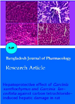
Histopathological observation of liver sections of different groups supported the hepatoprotective activity of both extracts. The control group animal exhibited normal hepatic cells with defined cytoplasm, prominent nucleus, and normal hepatic vein (Figure 2A). The carbon tetrachloride-intoxicated group showed loss of the total hepatic architecture with necrosis, crowding the central vein in many areas (Figure 2B). In other groups, liver improvement was observed by minimizing inflammation, less necrosis, and reducing crowding of the central vein. The histopathological observation confirmed that G. xanthocymus rind extract was more potent than G. lanceifolia extract towards the liver protection damage by carbon tetrachloride (Figure 2D-G).
Figure 2: Effect of methanolic fruit rind extract on liver. Liver section, 0.5 micron size, H&E, 40x. Normal group exhibiting the normal liver architecture with normal hepatic cells, with define cytoplasm, prominent nucleus and normal central vein (A); Negative group exhibiting total loss of hepatic architecture with necrosis, inflammation, crowding of central vein (B); Standard group exhibiting normal hepatic architecture with minimal necrosis, normal central vein; (C); G. xanthocymus 200 mg/kg showing a pattern of reduced necrosis, inflammation (D); G. xanthocymus 400 mg/kgs showing normal architecture with reduced inflammation and normal central vein (E); G. lanceifolia 200 mg/kg showing a pattern of reduced necrosis, inflammation (F); G. lanceifolia 400 mg/kg showing normal architecture with reduced inflammation and almost normal central vein (G)
Discussion
In the present study, pretreatment with the extracts and silymarin, the elevation level of serum marker enzymes activity was decreased as compared to the carbon tetrachloride-intoxicated group in a significant level These species showed significant hepatoprotective activity. G. xanthochymus is found to be more potent than G. lanceifolia. Similar findings on G. pedunculata and G. hombroniana supported the present investigation (Mundugaru et al., 2015; Jamila et al., 2017). The present investigation also revealed that these plant extracts can reduce the production of total bilirubin from damaged liver. A similar observation was reported by earlier workers in the G. kola supported the activity of Garcinia species in reducing the activity of total bilirubin in blood serum (Smith and Adanlawo, 2015). Liver sections of the rats were intoxicated with CCl4 and exhibited disarrangement and degeneration of normal hepatic cells with intense centrilobular necrosis with sinusoidal hemorrhages.
Pretreatment with the silymarin and the other plant extract showed the regeneration of the central vein system, mild narcosis in the liver section, and normal hepatic cells. Histopathological observation of the liver sections found that G. xanthochymus had the highest activity than G. lanceifolia.
It is well established that carbon tetrachloride is a potent hepatotoxin, capable of causing liver damage through the cytochrome P450 mediated activation to free radicals, which induce liver necrosis, inflammation and lead to liver fibrosis. The highly reactive free radicals generated from carbon tetrachloride are trichloromethyle (.CCl3) and trichloromethyle peroxyl (.OOCl3) affect the hepatocytes, leads to structural and functional damage of their cellular membranes which elevated the liver function enzymes such as serum aminotransferases, alkaline phosphatase and reduced the total protein in the serum (Sun et al., 2001, Kaplowitz et al., 1986, Kadiiska et al., 2000). A significant (p<0.001, p<0.005) reduction in the level of serum aminotransferases, alkaline phosphatase was observed in the group of animals pretreatment with 200 and 400 mg/kg body weight of both extracts. The pre-vious study showed the significant free radical scavenging activity of both plants, could exert a beneficial action against liver damage induced by CCl4. However, the preliminary phytochemical investigation of both plants shows the presence of both phenolic and flavonoids with antioxidant activity of garcinol compounds, present in G. xanthochymus and G. lanceifolia might be responsible for the hepatoprotective effect induced by free radicals generated by CCl4 (Yamaguchi et al, 2000, Gogoi et al, 2015, Gogoi, 2019, Gogoi et al, 2020).
In the comparative study between the two Garcinia species, it was found that G. xanthocymus was more hepatoprotective than G. lanceifolia, by lowering serum enzymes and bilirubin and increasing protein levels by a significant level. It might be because of high free radical scavenging activity and the high amount of garcinol found in G. xanthochymus than G. lanceifolia (Gogoi, 2019).
Conclusion
The fruits of G. xanthocymus and G. lanceifolia are potential agents to protect the liver. G. xanthocymus is more potent than G. lanceifolia.
Ethical Issue
Experimental protocol was approved by Dibrugarh University animal ethical committee (Approved No. IAEC/DU/98, Regd. No. 1576/GO/a/11/CPCSEA) and animals were cared and handled with prior to the regulation of CPCSEA. Acute toxicity was determined according to the OECD guideline No. 425 (OECD, 2001).
Acknowledgement
Authors are grateful to Dr. Banashree Mech and Prof. A. K. Rai for providing microtome facility.
References
Achliya GS, Kotagale NR, Wadodkar SG, Dorle AK. Hepatoprotective activity of Panchagavya ghrita against carbon tetrachloride-induced hepatotoxicity in rats. Indian J Pharmacol. 2003; 35: 308-11.
Akbar S, Ishtiaq S. Hepatoprotective effect of Misopates orontium in rats. Bangladesh J Pharmacol. 2020; 15: 75-81.
Babu BH, Shylesh BS, Padikkala J. Antioxidant and hepatoprotective effect of Acanthus ilicifolius. Fitoterapia 2001; 72: 272-77.
Biswas M, Karan TK, Kar B, Bhattacharya S, Ghosh AK, Kumar RBS, Haldar PK. Hepatoprotective activity of Terminlia arjuna leaf against paracetamol induced liver damage in rats. Asian J Chem. 2011; 23: 1739-42.
Burkill IH. A dictionary of economic product of the Malay Peninsula. 1st ed. London, Government of straits settlement, 1935.
Charlton MR. Protein metabolism and liver disease. Baillieres Best Pract Res Clin Endocrinol. 1996; 10: 617-35.
Galigher AE, Kozloff EN, Essentials of practical microtechniques. 2nd ed. Lea and Febiger, 1971.
Gogoi N, Gogoi A, Neog B. Free radical scavenging activities of Garcinia xanthochymus Hook. f. and Garcinia lanceaefolia Roxb. using various in vitro assay models. Asian J Pharm Clin Res. 2015; 9: 138-41.
Gogoi N, Gogoi A, Neog B, Baruah D, Singh KD. Evaluation of antioxidant and hepatoprotective activity of fruit rind extract of Garcinia dulcis (Roxburgh) Kurz. Phcog Res. 2017; 9: 266-72.
Gogoi N. Evaluation of hepatoprotective activity of Garcinia species and its genetic diversity in Upper Assam with special reference to Garcinol. Ph.D. thesis, Dibrugarh University, 2019.
Gogoi N, Gogoi A, Neog B. Assessment of Antioxidant Activity of Garcinol Derived from Garcinia xanthochymus Hook. f. Indian J Nat Sci.2020; 63: 28485-91
Gogoi N, Gogoi A, Neog B, Baruah D, Singh KD. Antioxidant and hepatoprotective activity of fruit rind extract of Garcinia morella (Gaertn.) Desr. IJNPR. 2017; 8: 132-39.
Jamila N, Khan N, Khan AA, Khan I, Khan SN, Zakariae ZA, Kim KS. and in vitro cytotoxic activities of Garcinia hombro-niana (seashore 372 mangosteen). Afr J Tradit Complement Altern Med. 2017; 14: 374-82.
Kadiiska MB, Gladen BC, Baird DD, Dikalova AE, Sohal RS, Hatch GE, Jones DP, Mason RP, Barrett JC. Biomarkers of oxidative stress study: Are plasma antioxidants markers of CCl4 poisoning? Free Radic Biol Med. 2000; 28: 838-45.
Kamraj VP. Herbal medicine. Curr Sci. 2000; 78: 35-39.
Kaplowitz N, Aw TY, Simon FR, Stolz A. Drug-induced hepatotoxicity. Ann Intern Med. 1986; 104: 826-39.
King EJ, Kind RP. Alkaline phosphatase activity assay. J Clin Pathol. 1954; 7: 332.
Lowry OH, Rosebrough NJ, Farr AL, Randall RJ. Protein measurement with the Folin phenol reagent. J Biol Chem. 1951; 193: 265-75.
Malloy HT, Evelyn KA. The determination of bilirubin with the photoelectric colorimeter. J Biol Chem. 1937; 119: 481-90.
Mandal SC, Saraswathi B, Ashok Kumar CK, Mohana Lakshmi S, Maiti BC. Protective effect of leaf extract of Ficus hispida Linn. against paracetamolâ€induced hepatotoxicity in rats. Phytother Res. 2000; 14: 457-59.
Mundugaru, R, Chakkravarthy, M, Basavaiah, R. Hepatoprotective activity of fruit extract Garcinia pedunculata. Bangladesh J Pharmacol. 2014; 9: 483-87.
Moss DW , Butterworth PJ. Enzymology and medicine. London, Pitman Medical, 1974, p 39.
Muriel P, Mourelle M. Prevention by silymarin of membrane alterations in acute CCl4 liver damage. J Appl Toxicol. 1990; 10 : 275-79.
Naik SR, Panda VS. Antioxidant and hepatoprotective effects of Ginkgo biloba phytosomes in carbon tetrachlorideâ€induced liver injury in rodents. Liver Int. 2007; 27: 393-99.
OECD, Guidelines for testing of chemicals, acute oral toxicity, environmental health and safety monograph series on testing and adjustment. No. 425, 2011.
Rao GM, Rao CV, Pushpangadan P, Shirwaikar A. Hepatoprotective effects of rubiadin, a major constituent of Rubia cordifolia Linn. J Ethnopharmacol. 2006; 103: 484-90.
Reitman S, Frankel S. A calorimetric method for the determination of serum glutamate oxaloacetate and glutamate pyruvate transaminase. Am J Clin Path .1957; 28: 53-56.
Singh K, Khanna AK, Chander R. Hepatoprotective activity of ellagic acid against carbon tetrachloride-induced hepatotoxicity in rats. Indian J Exp Biol. 1999; 37: 1025-26.
Smith YRA, Adanlawo IG. Protective effect of saponin extract from the root of Garcinia kola (bitter kola) against paracetamol-induced hepatotoxicity in Albino rats. Int J Sci Res Innov. 2015; 9: 130-34.
Sun F, Hamagawa E, Tsutsui C, Ono Y, Ogiri Y, Kojo S. Evaluation of oxidative stress during apoptosis and necrosis caused by carbon tetrachloride in rat liver. BBA-Mol Basis Dis. 2001; 1535: 186-91
Wiseman H, Halliwell B. Damage to DNA by reactive oxygen and nitrogen species: Role in inflammatory disease and progression to cancer. Biochem J. 1996; 313: 17-29.
Van Puyvelde L, Kayonga A, Brioen P, Costa J, Ndimubakunzi A, De Kimpe N, Schamp N. The hepatoprotective principle of Hypoestes triflora leaves. J Ethnopharmacol. 1989; 26: 121-27.
Yamaguchi F, Ariga T, Yoshimura Y, Nakazawa H. Antioxidative and antiglycation activity of garcinol from Garcinia indica fruit rind. J Agric Food Chem. 2000; 48: 180-85.