Protective effects of ethanolic leaf extract of Medicago sativa against complete Freund's adjuvant-induced arthritis in Wistar rats
Abstract
This study evaluated the antiarthritic activity of the ethanolic leaf extract of Medicago sativa through phytochemical screening and a complete Freund's adjuvant-induced arthritis model in Wistar rats. Phytochemical screening revealed the presence of alkaloids, tannins, phenolics, flavonoids, saponin glycosides, phytosterols, terpenoids, and cardiac glycosides. Six groups of six rats each were treated, and administration of the extract at 400 mg/kg significantly reduced paw swelling, arthritis severity scores, rheumatoid factor, and C-reactive protein levels compared to untreated arthritic controls. Motor coordination, as well as thermal and cold pain sensitivity, were restored at this dose, comparable to the effects of diclofenac sodium at 10 mg/kg. Imaging and histological examination confirmed protection of joint structure. These findings indicate that M. sativa extract possesses significant, dose-dependent, multi-parameter antiarthritic activity.
Introduction
Rheumatoid arthritis is a chronic, systemic autoimmune disorder characterized by synovial inflammation, pannus formation, and progressive joint destruction (McInnes and Schett, 2011; Firestein and McInnes, 2017). It affects a substantial proportion of the global population and is associated with considerable disabi-lity and reduced quality of life (Smolen et al., 2018; Finckh et al., 2022). Current pharmacotherapy is limited by significant gastrointestinal, renal, hepatic, and immunosuppressive adverse effects as well as prohibitive cost (Singh et al., 2016; Smolen et al., 2020), sustaining interest in plant-derived multi-component therapeutics with favorable safety margins (Venkatesha et al., 2011; Ahmad et al., 2018). In vivo studies reported antiarthritic efficacy for several plant extracts, including Bombax ceiba (Riaz et al., 2023), Artemisia pallens (Kumar et al., 2024), Cyathocline purpurea (Kausar et al., 2024), Xanthium strumarium (Moazzam et al., 2024), Acacia modesta (Fayyaz et al., 2023), Acorus calamus (Ilyas et al., 2025), Onosma bracteatum (Raza et al., 2025), and Diospyros kaki (Hosseini et al., 2024).
Medicago sativa Linn. (alfalfa; family Fabaceae) has been used in traditional Ayurvedic and Unani medicine for inflammatory conditions (Bora and Sharma, 2011). Its aerial parts contain isoflavonoids, flavones, phytosterols, phenolic acids, triterpenoid saponins, and alpha-tocopherol (Stochmal et al., 2001; Bora and Sharma, 2011; Khairy et al., 2025), which were reported to confer antioxidant, anti-inflammatory, and immunomodulatory activity through suppression of NF-κB and MAPK signaling (Ahranjani et al., 2026; Vahedi-Mazdabadi et al., 2025).
Literature search identified no published reports evaluating antiarthritic activity in other Medicago species to date, underscoring both the novelty of the present investigation and a current evidence gap for the genus. Comprehensive experimental evaluation of M. sativa in the complete Freund's adjuvant (CFA) model, combining phytochemical characterization with functional, nociceptive, biochemical, radiological, and histopathological endpoints, has not previously been reported. This study addresses this gap by: a) characterizing the phytochemical composition of the ethanolic leaf extract of M. sativa and b) evaluating its antiarthritic efficacy in a comprehensive CFA-induced arthritis model in Wistar rats.
Materials and Methods
Plant material and extract preparation
Fresh leaves of M. sativa were collected in November 2025 from the botanical garden of Appasaheb Birnale College of Pharmacy, Sangli, Maharashtra, India. Botanical authentication was conducted by Prof. V. U. Waghmare, Department of Botany, Willingdon College, Sangli (Voucher No.: WC/BOT/2025/MS01).
Shade-dried leaves were coarsely powdered and sieved through a No. 60 mesh screen. The powder (93 g) was extracted using a Soxhlet apparatus with 750 mL of 70% ethanol for 40 hours. The concentrated dark brown, semi-solid extract was stored at 4°C and freshly recons-tituted in 1% carboxymethylcellulose sodium for animal administration.
Qualitative phytochemical screening of the extract
Qualitative phytochemical screening of the extract was conducted using standard reagent-based tests (Har-borne, 1998). For alkaloids, a small quantity of the extract was separately treated with Dragendorff's reagent (potassium bismuth iodide solution), producing an orange-red precipitate, and with Mayer's reagent (potassium mercuric iodide solution), yielding a cream or white precipitate, both indicating the presence of alkaloids. For tannins, the extract was treated with a few drops of Braymer's reagent (10% ferric chloride solution); the development of a blue-green color indicated tannins. For phenolics, a few drops of 5% ferric chloride solution were added to the extract; a blue-black or greenish coloration confirmed the presence of phenolic compounds. For carbohydrates, Barfoed's test was performed by heating the extract with Barfoed's reagent (cupric acetate in dilute acetic acid) in a boiling water bath for 2 min, a red precipitate indicated monosaccharides. Seliwanoff's test was also performed by heating the extract with Seliwanoff's reagent (resorcinol in dilute hydrochloric acid); a rapid red coloration indicated ketoses. For reducing sugars, Benedict's test was conducted by heating the extract with Benedict's reagent in a boiling water bath for 5 min, a brick-red precipitate confirmed reducing sugars. Fehling's test was also performed by heating the extract with equal volumes of Fehling's A and B solutions; a brick-red precipitate indicated a positive result. For cardiac glycosides, the Keller–Killiani test was performed by treating the extract with glacial acetic acid containing a trace of ferric chloride, then overlaying with concentrated sulfuric acid; a reddish-brown ring at the interface indicated deoxy sugars characteristic of cardenolides. For saponin glycosides, the foam test was performed by vigorously shaking a dilute aqueous solution of the extract for 15 min; a persistent honeycomb froth confir-med the presence of saponins. For flavonoids, the alkaline reagent test was conducted by treating the extract with a few drops of 10% sodium hydroxide solution, producing an intense yellow color that turned colorless upon addition of dilute hydrochloric acid, confirming flavonoids. For terpenoids and phytosterols, Salkowski's test was performed by dissolving the extract in chloroform and carefully adding concentrated sulfuric acid along the side of the tube; a reddish-brown interface indicated terpenoids, while a golden-yellow coloration in the chloroform layer indicated phyto-sterols. For proteins and amino acids, the Biuret test was performed by treating the extract with 4% sodium hydroxide solution followed by a few drops of 1% copper sulfate solution; a violet or pink coloration indicated the presence of proteins or amino acids.
Animals
Adult Wistar albino rats (either sex; 150–200 g; n = 6 per group) were acclimatized for 7 days under controlled conditions (22 ± 2°C; 55 ± 5% relative humidity; 12-hour light/dark cycle) with access to a standard pelleted diet and water ad libitum.
Arthritis induction and treatment
Rheumatoid arthritis was induced by a single sub-plantar injection of 0.1 mL of CFA into the right hind paw on Day 0 (Newbould, 1963). The injection was administered under aseptic conditions, with the injection site clipped and disinfected using 70% isopropyl alcohol before injection. Oral treatment began on Day 7 and continued through Day 28.
The normal control group received 1% carboxymethyl cellulose (10 mL/kg) orally without CFA. The disease control group received CFA plus 1% carboxymethyl cellulose orally. The positive control group received CFA plus diclofenac sodium (10 mg/kg) orally. Another group received CFA plus the extract at 100 mg/kg orally. A separate group received CFA plus the extract at 200 mg/kg orally. The final group received CFA plus the extract at 400 mg/kg orally.
Parameters evaluated
Assessments were conducted on Days 0, 7, 14, 21, and 28 as follows: a) Paw volume was measured using a digital plethysmometer. The hind paw was immersed up to the tibiotarsal joint into the sensor cell filled with a conducting solution, and the displaced volume was recorded in milliliters. b) Arthritis severity was evaluated using a validated 0–4 visual scale: 0 indicated no swelling or erythema; 1 indicated mild erythema and slight swelling confined to the tarsal/ankle joint; 2 indicated moderate erythema and swelling extending from the ankle to the tarsal bones; 3 indicated marked erythema and swelling extending from the ankle to the metatarsal joints; and 4 indicated severe erythema and swelling of the entire paw, including involvement of the digits and joint deformity. c) Thermal hyperalgesia was assessed by the hot plate test. Each rat was placed on a hot plate maintained at 55°C, and the latency to a nociceptive response (paw licking or jumping) was recorded, with a cut-off time of 45 sec to prevent tissue damage. d) Cold allodynia was evaluated using the acetone-drop test. A drop of acetone was applied to the plantar surface of the hind paw, and the withdrawal response was scored on a 0–3 scale: 0, no response; 1, quick withdrawal or flick; 2, prolonged withdrawal with paw licking; and 3, repeated flicking and licking. e) Motor coordination and muscle strength were assessed by measuring the rotarod fall-off time. Rats pre-trained to remain on a rotating rod (16 rpm) were placed on the rotarod, and the time to fall was recorded, with a maximum cut-off of 180 sec. On Day 28, terminal blood samples were collected via retro-orbital puncture under light ether anesthesia. Serum was separated by centrifugation (3,000 rpm, 10 min) for biochemical estimation of rheumatoid factor using the latex agglutination method and C-reactive protein by immunoturbidimetry, following the manufacturer’s protocols with commercial diagnostic kits.
Following euthanasia, the right hind limb joints were dissected, and radiological examination was performed using plain X-ray imaging of the paw in the dorso-plantar view to assess joint space narrowing, soft tissue swelling, and bone erosion.
For histopathological examination, joint tissue was fixed in 10% neutral buffered formalin for 48 hours, decalcified, dehydrated through a graded ethanol series, embedded in paraffin, sectioned at 5 μm thickness, stained with hematoxylin and eosin, and examin-ed under a light microscope at 40x magnification for synovial hyperplasia, inflammatory cell infiltration, pannus formation, cartilage destruction, and subchondral bone erosion.
Statistical analysis
Data are expressed as mean ± SEM. Comparisons among groups were performed using one-way or two-way analysis of variance (ANOVA), followed by Dunnett's or Sidak's multiple comparison tests, as appropriate, with GraphPad Prism. A value of p<0.05 was considered statistically significant.
Results
Extract characteristics and phytochemical screening
The ethanolic extract of M. sativa leaves was obtained as a dark brown semi-solid with a yield of 11.3% w/w.
Qualitative phytochemical screening (data not shown) confirmed the presence of 10 phytoconstituent classes, including alkaloids, tannins, phenolics, flavonoids, saponin glycosides, phytosterols, terpenoids, cardiac glycosides, reducing sugars, and carbohydrates. Proteins and amino acids were absent.
Serum biochemical parameters
The serum biochemical parameters showed a significant increase in rheumatoid factor and C-reactive protein in the disease control group compared to the normal control. On Day 28, rheumatoid factor levels rose from 8.0 ± 0.4 IU/mL in the normal control to 23.4 ± 3.0 IU/mL in the disease control group (p<0.001) (Table I). Treatment with diclofenac sodium at 10 mg/kg significantly reduced rheumatoid factor to 10.4 ± 1.5 IU/mL (p<0.0001 vs. disease control). The extract at doses of 100, 200, and 400 mg/kg decreased rheumatoid factor to 15.2 ± 1.8, 12.8 ± 0.9 and 9.8 ± 0.2 IU/mL, respectively, with significant reductions observed at 200 mg/kg (p<0.001) and 400 mg/kg (p<0.0001). Similarly, C-reactive protein increased from 0.3 ± 0.1 mg/L in the normal control to 2.4 ± 0.3 mg/L in the disease control group. Diclofenac reduced C-reactive protein to 0.5 ± 0.1 mg/L (p<0.0001), while the extract lowered C-reactive protein to 1.5 ± 0.4, 0.9 ± 0.3, and 0.4 ± 0.4 mg/L at 100, 200, and 400 mg/kg, respectively. The reductions were significant at 200 mg/kg (p<0.01) and 400 mg/kg (p<0.0001), whereas the 100 mg/kg dose was non-significant.
Table I: Effect of extract on serum biochemical parameters in CFA-induced arthritic rats on Day 28
| Group | Rheumatoid factor (IU/mL) |
C-reactive protein (mg/L) |
|---|---|---|
| Normal control | 8.0 ± 0.4 | 0.3 ± 0.1 |
| Disease control | 23.4 ± 3.0a | 2.4 ± 0.3a |
| Diclofenac 10 mg/kg | 10.4 ± 1.5d | 0.5 ± 0.1d |
| Extract 100 mg/kg | 15.2 ± 1.8b | 1.5 ± 0.4ns |
| Extract 200 mg/kg | 12.8 ± 0.9c | 0.9 ± 0.3b |
| Extract 400 mg/kg | 9.8 ± 0.2d | 0.4 ± 0.4d |
| Data are mean ± SEM; n=6. ap<0.001 vs. normal control; bp<0.01, cp<0.001, dp<0.0001 vs. disease control; ns: non-significant. One-way ANOVA with Dunnett's test. Extract: extract of M. sativa leaf | ||
Motor coordination (rotarod fall-off time)
Arthritis progressively impaired rotarod performance in the disease control group, with fall-off time decreasing from 21.7 ± 0.6 sec on Day 0 to 5.0 ± 0.4 sec on Day 28. Treatment with the extract at 400 mg/kg restored fall-off time to 26.7 ± 2.0 sec by Day 28 (p<0.001 vs. disease control), comparable to diclofenac sodium (25.0 ± 1.0 sec; Table II).
Thermal hyperalgesia and cold allodynia
CFA administration induced significant thermal hyperalgesia and cold allodynia, with an acetone score of 3.0 ± 0.0 from Day 7 onwards in the disease control group. The extract at 400 mg/kg progressively restored hot plate latency (96.7 ± 2.1% on Day 28, p<0.001) and completely abolished cold allodynia by Day 21 (score 0.0 ± 0.0, p<0.001; Table II).
Table II: Effect of extract on in CFA-induced arthritic rat
| Group | Day 0 | Day 7 | Day 14 | Day 21 | Day 28 |
|---|---|---|---|---|---|
| Paw volume (mL) | |||||
| Normal control | - | 0.8 ± 0.0 | 0.8 ± 0.0 | 0.8 ± 0.0 | 0.9 ± 0.0 |
| Disease control | - | 2.0 ± 0.1c | 1.8 ± 0.0c | 2.0 ± 0.0c | 2.2 ± 0.0c |
| Diclofenac 10 mg/kg | - | 1.6 ± 0.1g | 1.2 ± 0.0g | 1.0 ± 0.0073g | 0.9 ± 0.0g |
| Extract 100 mg/kg | - | 2.0 ± 0.1 | 1.5 ± 0.1f | 1.5 ± 0.1g | 1.4 ± 0.1g |
| Extract 200 mg/kg | - | 2.0 ± 0.1 | 2.0 ± 0.0 | 1.8 ± 0.1e | 1.3 ± 0.0g |
| Extract 400 mg/kg | - | 2.5 ± 0.1 | 2.4 ± 0.1g | 1.7 ± 0.0g | 1.1 ± 0.1g |
| Arthritis severity score (0-4) | |||||
| Normal control | - | 0.0 ± 0.0 | 0.0 ± 0.0 | 0.0 ± 0.0 | 0.0 ± 0.0 |
| Disease control | - | 3.0 ± 0.0 | 3.0 ± 0.4c | 3.7 ± 0.2c | 3.3 ± 0.2c |
| Diclofenac 10 mg/kg | - | 2.3 ± 0.2d | 2.0 ± 0.4 | 0.7 ± 0.2g | 0.3 ± 0.2g |
| Extract 100 mg/kg | - | 2.3 ± 0.2 | 2.3 ± 0.2 | 1.7 ± 0.2g | 1.7 ± 0.2g |
| Extract 200 mg/kg | - | 2.7 ± 0.2 | 2.7 ± 0.2 | 1.7 ± 0.2g | 1.3 ± 0.2g |
| Extract 400 mg/kg | - | 3.0 ± 0.0 | 2.7 ± 0.2 | 1.7 ± 0.2g | 0.3 ± 0.2g |
| Hot-plate latency (% normal) | |||||
| Normal control | 0.0 ± 0.0 | 0.0 ± 0.0 | 0.0 ± 0.0 | 0.0 ± 0.0 | 0.0 ± 0.0 |
| Disease control | 0.0 ± 0.0 | -16.8 ± 3.2 | -32.5 ± 4.4 | -50.8 ± 5.0 | -67.0 ± 1.9 |
| Diclofenac 10 mg/kg | 12.0 ± 3.9 | 2.7 ± 8.8 | 36.3 ± 8.8 | 58.3 ± 5.4 | 76.3 ± 3.4 |
| Extract 100 mg/kg | 78.3 ± 7.4 | 88.7 ± 7.2 | 40.2 ± 15.2 | 61.7 ± 11.7 | 63.7 ± 5.8 |
| Extract 200 mg/kg | 93.3 ± 4.2 | 39.5 ± 3.9 | 47.8 ± 3.8 | 72.6 ± 10.5 | 93.3 ± 2.1 |
| Extract 400 mg/kg | 69.9 ± 4.9 | 35.4 ± 8.9 | 61.5 ± 6.1 | 67.9 ± 13.2 | 96.7 ± 2.1 |
| Cold allodynia / acetone-drop score (0-3) | |||||
| Normal control | - | 0.0 ± 0.0 | 0.0 ± 0.0 | 0.0 ± 0.0 | 0.0 ± 0.0 |
| Disease control | - | 3.0 ± 0.0c | 3.0 ± 0.0c | 2.0 ± 0c | 2.7 ± 0.2c |
| Diclofenac 10 mg/kg | - | 1.3 ± 0.2g | 0.3 ± 0.2g | 0.0 ± 0.0g | 0.0 ± 0.0g |
| Extract 100 mg/kg | - | 2.7 ± 0.2ns | 2.0 ± 0.0g | 1.0 ± 0.0g | 0.7 ± 0.2g |
| Extract 200 mg/kg | - | 1.7 ± 0.2g | 1.0 ± 0.0g | 0.7 ± 0.2g | 0.3 ± 0.2g |
| Extract 400 mg/kg | - | 1.7 ± 0.2g | 1.0 ± 0.0g | 0.0 ± 0.0g | 0.0 ± 0.0g |
| Rotarod fall-off time (sec) | |||||
| Normal control | 24.0 ± 0.6 | 22.3 ± 0.9 | 25.7 ± 0.4 | 23.0 ± 1.0 | 27.0 ± 1.0 |
| Disease control | 21.7 ± 0.6 | 17.7 ± 0.9a | 13.0 ± 0.4b | 13.0 ± 0.4c | 5.0 ± 0.4c |
| Diclofenac 10 mg/kg | 25.7 ± 1.5 | 15.3 ± 1.3ns | 20.3 ± 0.6e | 22.7 ± 0.8g | 25.0 ± 1.0g |
| Extract 100 mg/kg | 21.7 ± 2.8 | 11.3 ± 2.2e | 13.7 ± 2.0ns | 14.3 ± 2.1ns | 14.3 ± 1.8f |
| Extract 200 mg/kg | 28.3 ± 2.8 | 10.7 ± 0.4e | 16.7 ± 1.1ns | 25.0 ± 1.8g | 28.3 ± 1.1g |
| Extract 400 mg/kg | 26.0 ± 1.9 | 13.3 ± 0.4ns | 20.7 ± 1.9f | 22.0 ± 1.3g | 26.7 ± 2.0g |
| Data expressed as mean ± SEM (n=6). ns: non-significant; ap<0.05, bp<0.001, cp<0.0001 vs. normal control; dp<0.05, ep<0.01, fp<0.001, gp<0.0001 vs. arthritic/disease control. One-way or two-way ANOVA followed by Dunnett's or Sidak's multiple comparison test, as appropriate. - : not measured on that day. Extract: ethanolic extract of Medicago sativa leaves; CFA: complete Freund's adjuvant. | |||||
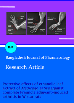
Arthritis severity scoreThe arthritis severity score was 3.3 ± 0.2 in the disease control group on Day 28, compared with 0.0 ± 0.0 in the normal control group. Diclofenac sodium reduced the score to 0.3 ± 0.2. Treatment with the extract decreased the arthritis score to 1.7 ± 0.2, 1.3 ± 0.2, and 0.3 ± 0.2 at doses of 100, 200, and 400 mg/kg, respectively. The 400 mg/kg extract produced an arthritis severity score com-parable to that of the diclofenac-treated group (Table II).Radiological findings Radiographic assessment of CFA-induced disease control rats revealed narrowing of intertarsal joint spaces, periarticular soft tissue swelling, and subchondral bone erosions, confirming progressive joint destruction (Figure 1B). Treatment with the extract at 400 mg/kg markedly reduced bone erosion and soft tissue edema, with radiological findings comparable to those of the diclofenac-treated standard group (Figure 1F). The extract at 200 mg/kg showed moderate structural protection (Figure 1E), while the extract at 100 mg/kg demonstrated limited radiological improvement (Figure 1D).Figure 1: Radiological findings in CFA-induced arthritic Wistar rats on Day 28. Plain X-ray of hind paw jointHistopathological findings Synovial sections from disease control animals exhibited marked synoviocyte hyperplasia, dense inflammatory cell infiltration, pannus formation, cartilage destruction, and subchondral bone erosion—characteristic histological features of CFA-induced rheumatoid arthritis (Figure 2B). Treatment with the extract at 400 mg/kg resulted in near-normal synovial architecture, with substantially reduced inflammatory infiltration and minimal cartilage damage, comparable to diclofenac sodium (Figure 2F). A graded, dose-dependent improvement in synovial histology was observed across the extract-treated groups (Figure 2C–2E).
Figure 2: Histopathological findings in synovial tissue of CFA-induced arthritic rats on Day 28 (H&E stain; 40× magnification). (A) Normal control, (A); Disease control (B); Diclofenac sodium (C); Extract 100 mg/kg (D); Extract 200 mg/kg (E); Extract 400 mg/kg (F). Arrows indicates normal collagen fibers (A), Dense inflammatory cell infiltration (B), Tissue edema and synovial thickening (C), Mild synovial thickening (D), Reduced synovial hyperplasia (E), and Cartilage protection and minimal inflammation (F)
Discussion
The present findings demonstrate that the ethanol extract possesses significant, dose-dependent, multi-parameter antiarthritic activity in the CFA-induced arthritis model, comparable to diclofenac sodium at 10 mg/kg across all assessed parameters. Qualitative phytochemical screening confirmed that the extract is rich in alkaloids, tannins, phenolics, flavonoids, saponin glycosides, phytosterols, terpenoids, and cardiac glycosides. Phytochemical screening was performed specifically on the ethanolic leaf extract of M. sativa used in this study because the qualitative constituent profile and relative abundance of phytochemical classes vary with plant species, extraction solvent, and geographical or seasonal harvest conditions. The present profile, dominated by phytosterols and triterpenoid saponins, is consistent with earlier reports specific to this species (Stochmal et al., 2001; Khairy et al., 2025) and was generated to contextualize and support the mechanistic interpretation of the subsequent pharmacological findings rather than to claim constituent novelty. The richness in anti-inflammatory polyphenols and terpe-noids provides a mechanistic basis for the observed antiarthritic effects. At the highest dose, the extract normalized rheumatoid factor and reduced C-reactive protein relative to untreated arthritic controls; improved hematological indices, reversing anemia and normalizing leukocytosis and thrombocytosis; restored hot plate latency and abolished cold allodynia; recovered rotarod fall-off time; and produced near-normalization of joint architecture on radiological and histopathological examination, with minimal residual cartilage damage.
These results extend prior research on M. sativa, which has largely been limited to single-assay in vitro antioxidant and anti-inflammatory studies (Bora and Sharma, 2011; Ahranjani et al., 2026), by demonstrating efficacy across a comprehensive battery of functional, biochemical, radiological, and histopathological assessments. The findings related to anti-nociceptive effects, motor function, and structural protection are consistent with those reported for Bombax ceiba and Coix lachryma-jobi extracts in the same model (Riaz et al., 2023; Zhang et al., 2019).
Flavonoids and phenolics are known to suppress NF-κB-mediated transcription of COX-2, TNF-α, and IL-6 genes and exert broader anti-inflammatory actions (Serafini et al., 2010; Yahfoufi et al., 2018). Phytosterols modulate osteoclast activation and prostaglandin biosynthesis, providing a plausible mechanistic basis for the radiologically observed protection against bone erosion. Saponin glycosides contribute membrane-stabilizing and immunomodulatory effects, while alkaloids and tannins offer additional analgesic and antioxidant support (Bora and Sharma, 2011; Patel and Patel, 2008; Kaur et al., 2004). Normalization of rheumatoid factor indicates attenuation of B-cell-mediated autoimmunity, whereas normalization of CRP reflects suppression of hepatic acute-phase synthesis driven by the IL-1β/IL-6/NF-κB axis. The restoration of hot plate latency and abolition of cold allodynia are attributable to flavonoid-mediated suppression of prostaglandin- and bradykinin-driven sensitization of peripheral nociceptors. Recovery of rotarod performance reflects concurrent analgesic and anti-edema effects, reducing joint pain, stiffness, and periarticular swelling. The complementary actions of these phytoconstituent classes likely confer pharmacological synergy across multiple inflammatory targets.
Limitations of the present study include: a) the use of a crude extract, which limits the ability to attribute efficacy to individual phytoconstituents; b) LC-MS/HPLC-based identification of specific bioactive markers, in vitro mechanistic assays, and cytokine quantification (TNF-α, IL-1β, IL-6); and c) long-term toxicity and in vivo pharmacokinetic studies before clinical translation (Smolen et al., 2018).
Conclusion
The ethanolic extract of M. sativa leaves exhibits significant, dose-dependent, multi-parameter antiarthritic activity in a rat arthritis model. These findings support its potential as a herbal candidate for the treatment of rheumatoid arthritis.
Ethical Issue
All animal experimental procedures were approved by the Institutional Animal Ethics Committee (IAEC Approval No.: IAEC/ABCP/12/2025–26) under Committee for the Purpose of Control and Supervision of Experiments on Animals, Government of India, and were conducted in compliance with ARRIVE 2.0 guidelines.
References
Ahmad SS, Batool M, Choi S. Structural insights of phytocompounds as potent antiarthritic candidates targeting TNF-α, IL-1β and NF-κB signalling. Front. Pharmacol. 2018; 9: 1129.
Ahranjani PJ, Fathi Achachlouei B, Azadmard-Damirchi S. Bioactive phytochemical compounds in Medicago sativa L. (alfalfa): A systematic review of biological properties and therapeutic relevance in nutraceutical and functional food implications. Food Front. 2026; 7: 1-24.
Bora KS, Sharma A. Phytochemical and pharmacological potential of Medicago sativa: A review. Pharm Biol. 2011; 49: 211-20.
Fayyaz N, Aslam B, Muhammad F. Antiarthritic and immunomodulatory potential of methanolic, n-hexane, and ethyl acetate fractions of bark of Acacia modesta on complete Freund’s adjuvant-induced arthritis in rats. Front Pharmacol. 2023; 14: 1195463.
Finckh A, Gilbert B, Hodkinson B. Global epidemiology of rheumatoid arthritis. Nat Rev Rheumatol. 2022; 18: 591-602.
Firestein GS, McInnes IB. Immunopathogenesis of rheumatoid arthritis. Immunity 2017; 46: 183-96.
Harborne JB. Phytochemical methods: A guide to modern techniques of plant analysis. 3rd ed. Chapman and Hall, London, 1998.
Hosseini SA, Karimi G, Roshan NM. Diospyros kaki fruit extract produces antiarthritic and antinociceptive effects in rats with complete Freund’s adjuvant-induced arthritis. Phytother Res. 2024; 38: 2110-22.
Ilyas U, Mahmood T, Aftab R. Antiarthritic appraisal of Acorus calamus L. extracts in complete Freund’s adjuvant-induced arthritic Wistar rats via regulating inflammatory cytokines and OPG/RANKL pathway. Inflammopharmacology 2025; 33: 1-18.
Kaur G, Hamid H, Ali A, Alam MS, Athar M. Anti-inflammatory evaluation of alcoholic extract of galls of Quercus infectoria. J Ethnopharmacol. 2004; 90: 285-92.
Kausar S, Rehman K, Akash MSH. Antiarthritic activity of methanol extract of Cyathocline purpurea (whole plant) in Freund’s complete adjuvant-induced arthritis in rats. J Ayurveda Integr Med. 2024; 15: 100845.
Khairy A, Elshamy A, Saleh N. Alfalfa (Medicago sativa L.): A review of bioactive components and pharmacological properties. J Adv Pharm Sci. 2025; 2: 1-14.
Kumar V, Bhatt PC, Rahman M. Artemisia pallens W. attenuates inflammation and oxidative stress in Freund’s complete adjuvant-induced rheumatoid arthritis in Wistar rats. Pharmaceuticals 2024; 17: 1361.
McInnes IB, Schett G. The pathogenesis of rheumatoid arthritis. N Engl J Med. 2011; 365: 2205-19.
Moazzam A, Mustafa G, Ahmad M. Antiarthritic and anti-inflammatory activity of Xanthium strumarium L. ethanolic extract in Freund’s complete adjuvant-induced arthritis. Inflammopharmacology 2024; 32: 613-27.
Newbould BB. Chemotherapy of arthritis induced in rats by mycobacterial adjuvant. Br J Pharmacol Chemother. 1963; 21: 127-36.
Patel VR, Patel PR. Therapeutic potential of Medicago sativa in inflammatory disorders. Int J Green Pharm. 2008; 2: 72-75.
Raza SM, Ashraf M, Ahmad S. Onosma bracteatum Wall aqueous–ethanolic extract suppresses complete Freund’s adjuvant-induced arthritis in rats via regulation of TNF-α, IL-6, and C-reactive protein. Molecules 2025; 29: 1830.
Riaz T, Abbasi MA, Shahzadi T. Antiarthritic efficacy of Bom-bax ceiba ethanolic extract in a murine model for rheumatoid arthritis using in vivo, in vitro and radiological analysis. BMC Complement Med Ther. 2023; 23: 356.
Serafini M, Peluso I, Raguzzini A. Flavonoids as anti-inflammatory agents. Proc Nutr Soc. 2010; 69: 273-78.
Singh JA, Saag KG, Bridges SL Jr. 2015 American College of Rheumatology guideline for the treatment of rheumatoid arthritis. Arthritis Care Res. 2016; 68: 1-25.
Smolen JS, Aletaha D, McInnes IB. Rheumatoid arthritis. Lancet 2016; 388: 2023-38.
Smolen JS, Aletaha D, Barton A. Rheumatoid arthritis. Nat Rev Dis Primers. 2018; 4: 18001.
Smolen JS, Landewé RBM, Bijlsma JWJ. EULAR recommendations for the management of rheumatoid arthritis with synthetic and biological disease-modifying antirheumatic drugs: 2019 update. Ann Rheum Dis. 2020; 79: 685-99.
Stochmal A, Piacente S, Pizza C. Alfalfa (Medicago sativa L.) flavonoids. 1. Apigenin and luteolin glycosides from aerial parts. J Agric Food Chem. 2001; 49: 753-58.
Vahedi-Mazdabadi Y, Karimpour-Razkenari E, Akbarzadeh T. Alfalfa (Medicago sativa) and neurodegeneration: Mechanistic insights into oxidative stress, inflammation, and neuronal survival pathways. Nutr Neurosci. 2025; 28: 1-19.
Venkatesha SH, Berman BM, Moudgil KD. Herbal medicinal products target defined biochemical and molecular mediators of inflammatory autoimmune arthritis. Bioorg Med Chem. 2011; 19: 21-29.
Yahfoufi N, Alsadi N, Jambi M, Matar C. The immunomodulatory and anti-inflammatory role of polyphenols. Nutrients 2018; 10: 1618.
Zhang C, Zhang W, Shi R, Tang B, Xie S. Coix lachryma-jobi extract ameliorates inflammation and oxidative stress in a complete Freund’s adjuvant-induced rheumatoid arthritis model. Pharm Biol. 2019; 57: 792-98.
Apply citation style format of Bangladesh Journal of Pharmacology
Copyright (c) 2026 Utkarsha S. Shinde, Tabssum Patwegar, Padma Ladda

This work is licensed under a Creative Commons Attribution-NonCommercial 4.0 International License.